PAVF refers to the condition where the pulmonary artery and pulmonary vein communicate directly through enlarged tortuous blood vessels or hemangioma, resulting in a low incidence. PAVF can be categorized into three types: single, complex, and diffuse. More than 80% of PAVF patients belong to the single type, characterized by a connection between one artery and one vein without division of the cystic tumor. The complex type refers that the lesion is not limited to one artery and one vein, and the cystic tumor is usually separated. The diffuse type refers to the presence of extensive small pulmonary arteriovenous fistulas without cystic tumor formation [4, 5]. The severity of PAVF is directly related to fistula size. A single fistula less than 2cm in diameter usually does not cause symptoms. When the right-to-left shunt volume increases with the enlargement of the fistula, the symptoms such as dyspnea and cyanosis may gradually appear, and the patient's exercise tolerance gradually decreases. About 25% of patients also experience neurological symptoms such as cerebral embolism, transient ischemic attack, migraine, and brain abscess. Pulmonary artery CTA is the preferred diagnostic method for PAVF, providing an overview of the lesion and measuring the diameter of affected vessels. Pulmonary vascular DSA is the gold standard for the diagnosis of PAVF, which can observe the fistulas, feeding arteries and draining veins of PAVF.
After diagnosis, most PAVF patients require treatment. The mortality rate of untreated PAVF patients could be as high as 50%, whereas it could be reduced to 3% after treatment [6]. Current treatments for PAVF include surgery and interventional therapy. The recurrence rate of surgical treatment is low, but it is limited to the treatment of single lesion, and the trauma is large. Interventional therapy could retain more lung tissue and has less trauma, but it is not suitable for more lesions and diffuse lesions. Interventional therapy achieved its first success in treating PAVF in 1978. With the continuous improvement of surgical techniques and interventional materials, interventional treatment of PAVF has been proved to be safe and effective, and has become the first choice in clinical practice [7,8,9,10]. Interventional therapy for PAVF is mainly divided into coil occlusion and occluder occlusion. For PAVF patients with feeding artery diameter < 5 mm, coil closure is recommended. For PAVF with the diameter of feeding artery ≥ 5 mm, the use of coil will increase the risk of its detachment and displacement or even ectopic embolism, and the use of occluder is more effective and safer [11]. At present, the main conservative treatment of diffuse PAVF is improving the symptoms of hypoxia. In this case, the the narrowest diameter of the feeding artery was about 10mm through pulmonary vascular DSA. Therefore the Shenzhen Xianjiang patent ductus arteriosus occluder (XJFD1618) was used for occlusion. After occlusion, the shunt was significantly reduced and the patient's blood oxygen saturation increased rapidly. In the end, the patient's symptoms were significantly improved.
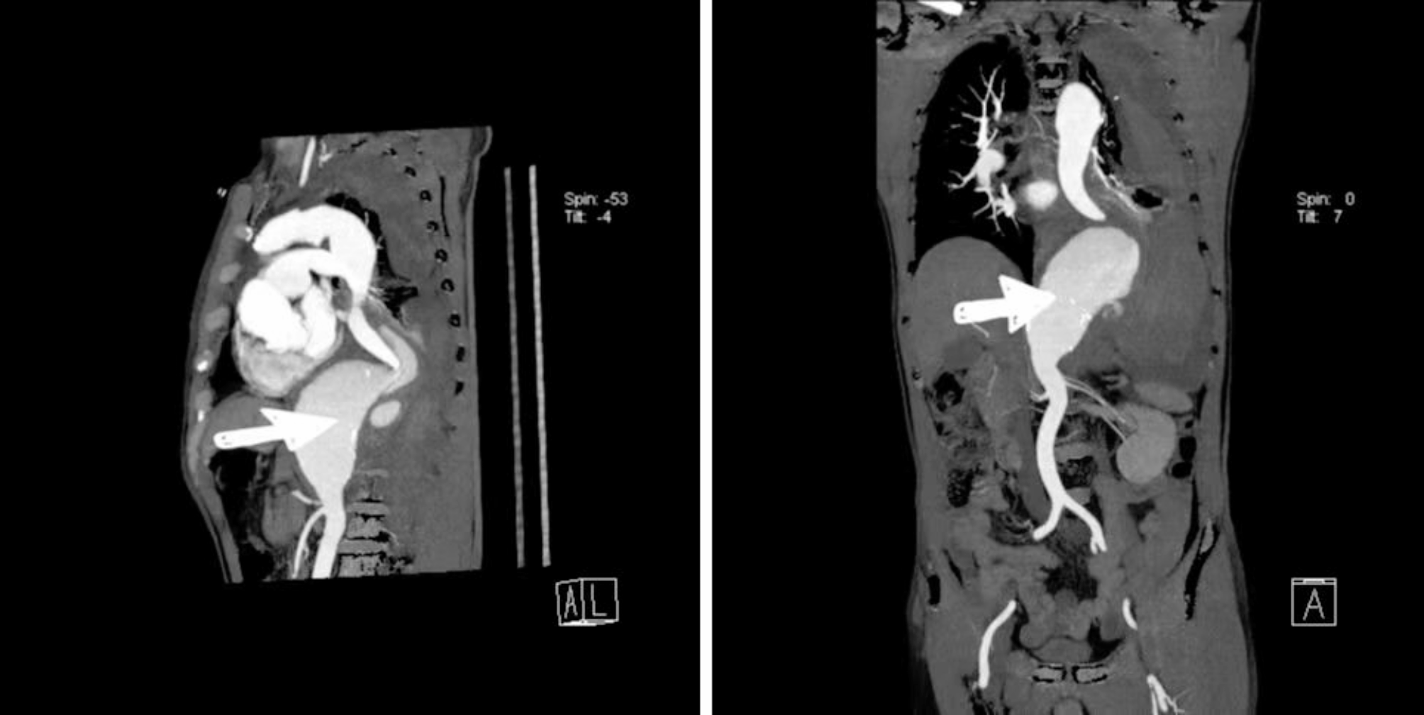
The patient's primary clinical manifestations were chest tightness and shortness of breath following activities, consistent with common symptoms of CHD. Coronary angiography showed that the patient had severe stenosis of the anterior descending artery and the right coronary artery which were consistent with the symptoms, and interventional treatment was given. However, the patient was also complicated with type I respiratory failure, and pulmonary artery CTA showed arteriovenous malformation in the right lower lobe of the lung, which was considered to have PAVF. Pulmonary vascular DSA was performed to confirm the diagnosis of right PAVF, and the patient was treated with occlusion. After the operation, the patient's hypoxemia and symptoms were significantly improved.
PAVF is a rare disease, and its symptoms lack specificity. For elderly patients with CHD, hypertension and other common cardiovascular diseases, PAVF often leads to oversight. Therefore, in patients with unexplained hypoxemia, considering the possibility of PAVF is crucial, necessitating comprehensive auxiliary examinations to prevent missed diagnosis. On this basis, intervention treatment should be carried out to improve the prognosis of patients as much as possible.


留言 (0)