
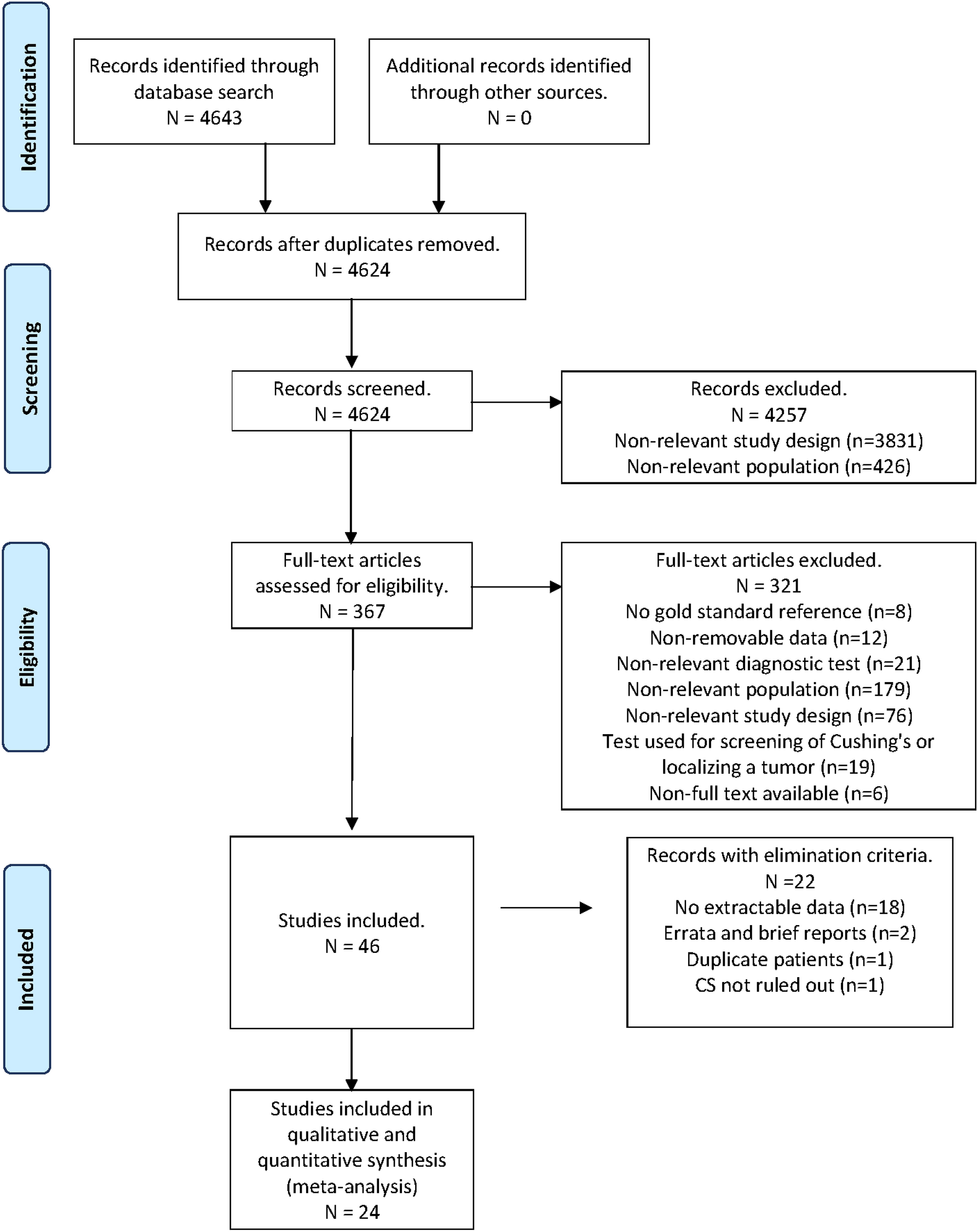
記住我
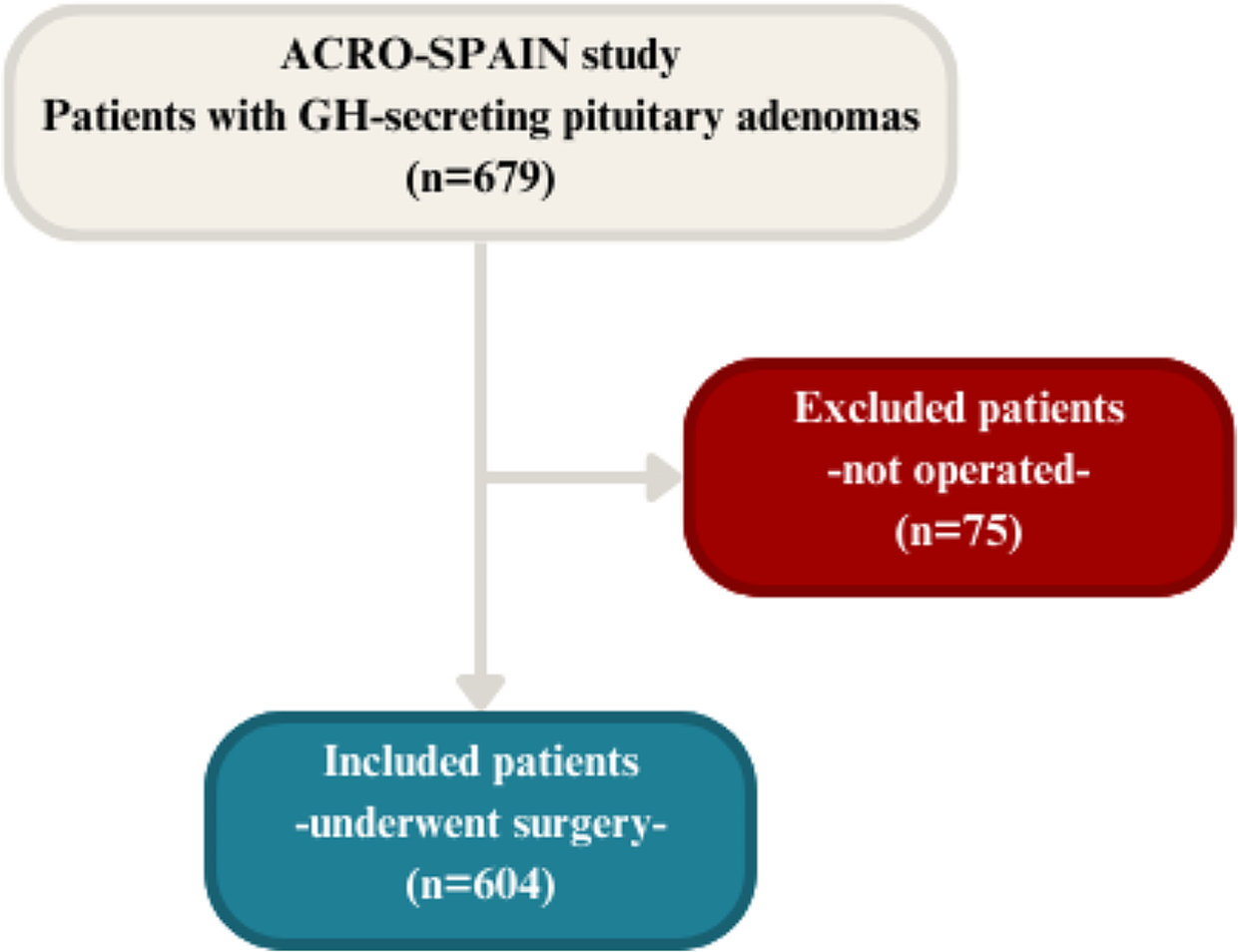
Twenty ACRO (7 females) with a mean age of 50 years (range 31–75 years) entered the study. The median time from diagnosis was 10 years (1–33). At the time of the original diagnosis, mean GH levels were 17.5 ± 12.1 and median IGF1xULN was 2.46 [1.08–4.94]. Eighteen (90%) had previously undergone trans-sphenoidal surgery. At the time of our evaluation, 6 patients (30%) were cured with surgery and 14 (60%) had active disease requiring medical treatment, including 5 patients on first-generation SSAs (25%), 4 on combined therapy with first-generation SSAs and PEG (20%), and 5 on pasireotide therapy (25%). At the time of study enrolment, 11 active patients (55%) exhibited disease control under medical treatment, only 3 (15%) presented with persistent biochemically uncontrolled disease. Median time between initial diagnosis and the achievement of disease control was 56.0 months (5-0-148.0).
Biochemical and clinical evaluationTable 1 compares the biochemical and clinical parameters (comorbidities and their therapies) of ACRO and CTRLs. As expected, sex, age, and BMI were similar between the two groups. We found no significant differences in fasting plasma glucose, HbA1c, lipid profile, blood pressure levels or the prevalence of cardiometabolic complications (hypertension, obesity, OSAS, dyslipidaemia, and glucose metabolism impairment) between ACRO and CTRLs, thus the two groups resulted homogeneous for cardiovascular and metabolic risk factors.
Table 1 Anthropometric, biochemical, clinical, and cardiac parameters at cardiac magnetic resonance (CMR) in patients with acromegaly and controlsCardiac evaluationTable 1; Fig. 1 summarize the cardiac parameters of ACRO and CTRLs. ACRO showed higher left ventricular (LV)-end diastolic volume index (EDVi) (79.6 ± 13.8 vs. 61.7 ± 11.5, p < 0.001), LV- end systolic volume index (ESVi) (33.9 ± 7.8 vs. 23.8 ± 7.0, p < 0.001) as well as left ventricular mass index (LVMi) (57.1 ± 14.2 vs. 43.5 ± 8.5, p = 0.001). Consequently, LV stroke volume index (LV-SVi) was higher in ACRO than in CTRLs (45.7 ± 9.2 vs. 37.9 ± 7.1, p = 0.007), whereas a trend was observed towards lower LV- ejection fraction (EF) in ACRO (57.5 ± 6.1 vs. 61.6 ± 6.7, p = 0.069). Concentricity index (LVMi/LV-EDVi) and interventricular septum (IVS) thickness did not significantly differ between the two groups (p = 0.782 and p = 0.128, respectively). Only one ACRO had LV hypertrophy (LVH) according to conventional CMR thresholds [22, 23]. Specifically, said patient was diagnosed with acromegaly at age 32; following marked changes in facial and body features, the diagnostic workup revealed severe acromegaly (IGF1xULN at diagnosis: 3.94) caused by a GH-secreting, locally invasive pituitary macroadenoma (maximum lesion diameter: 19 mm). The patient underwent pituitary adenomectomy and subsequently received SSA treatment due to persistent disease activity. Of note, the patient was treated with increasing doses of SSAs, but had not yet achieved disease control at the time of study enrolment, two years after medical treatment initiation (GH 2.1 ng/ml, IGF1xULN 1.6). Differences in right-ventricular (RV) parameters between groups resembled the ones observed in LV parameters. RV-EDVi and RV-ESVi were higher in ACRO than CTRLs (89.7 ± 19.7 vs. 63.4 ± 9.7 and 44.8 ± 13.0 vs. 25.8 ± 6.4, respectively; p < 0.001 for both). RV-SVi was also higher in ACRO than in CTRLs (44.9 ± 9.3vs 37.6 ± 7.6, p = 0.021). Moreover, ACRO showed a lower RV-EF than CTRLs (50.6 ± 6.0 vs. 59.3 ± 7.7, p = 0.001). Evaluation with T1 mapping technique did not reveal any significant difference between ACRO and CTRLs prior to administration of intravenous contrast; conversely, we found post contrast T1 intensity to be higher in ACRO than in CTRLs (444.3 ± 51.3 vs. 406.9 ± 50.0, p = 0.038). Three patients (2 with controlled disease under medical treatment and 1 surgically cured) had ECV greater than 30%, indicating the presence of myocardial fibrosis [24]; however, ECV did not significantly differ between the two groups (p = 0.979).
Fig. 1
Cardiac morpho-structural and functional parameters in patients with acromegaly (grey bars, n = 20) and age, sex, and BMI-matched controls (black bars, n = 17). Data are expressed as mean ± SD. * = p < 0.05; ** = p < 0.01; *** = p < 0.001. ACRO = Acromegaly; CTRL = Controls; LV-EDVi = Left Ventricle End-Diastolic Volume index; LV-ESVi = Left Ventricle End-Systolic Volume index; LV-SVi = Left Ventricle Stroke Volume index; LVMi = Left Ventricular Mass index; LV-EF = Left Ventricle Ejection Fraction; RV-EDVi = Right Ventricle End-Diastolic Volume index; RV-ESVi = Right Ventricle End-Systolic Volume index; RV-SVi = Right Ventricle Stroke Volume index; RV-EF = Right Ventricle Ejection Fraction
We found a significant correlation between IGF1 x ULN and LVMi (r = 0.600; p = 0.005), and concentricity index (r = 0.454; p = 0.044), and linear regression analysis confirmed IGF1 x ULN as a predictor for LVMi (B = 0.575, p = 0.008) in the ACRO group, see Fig. 2. Conversely, neither hormone levels at diagnosis (i.e., mean GH, IGF1xULN) nor duration of biochemical uncontrolled disease showed significant associations with any of the cardiac parameters at baseline.
Fig. 2
Scatterplot depicting the correlations between IGF1 levels times the upper limit of normal and cardiac parameters. IGF-1 x ULN = insulin-like growth factor-1 times the upper limit of normal; LVMi = Left Ventricular Mass index. The cut-off used to define left hypertrophy was LVMi higher than 83 g/m2 in men and 67 g/m2 in women. The upper limit of normal for concentricity index is defined as 0.9 g/mL in men and 0.8 g/mL in women
Subgroup analysis according to cardiometabolic comorbidities, disease duration and sexTo investigate the potential impact of cardiometabolic impairment on cardiac structure and function, we compared ACRO based on the presence or absence of several comorbidities and risk factors, including hypertension, obesity, diabetes mellitus, impaired fasting glucose, impaired glucose tolerance, OSAS, dyslipidaemia, and smoking. Notably, the 3 patients with diabetes (2 on pasireotide, 1 on combined SSA and PEG treatment) showed lower LV-EF compared to those without diabetes (50.2 ± 2.0 vs. 58.8 ± 5.7, p = 0.021), whereas we observed no significant differences according to the other factors.
Furthermore, we tried to ascertain the impact of disease duration on cardiac dysfunction. The median disease duration, measured in years passed since the initial diagnosis, was 10 years (1–33). Of note, the duration of disease was similar between active and cured patients, and it was not associated with any of the main cardiac parameters.
Lastly, a comparison according to sex revealed that male patients with acromegaly exhibited higher BMI compared to females (23.0 [20.7–27.8] vs. 29.0 [24.0-38.4], p = 0.007) without other significant differences in cardiometabolic risk factors. Male patients with acromegaly presented with higher LV-ESVi (37.0 ± 7.0 vs. 28.0 ± 5.9, p = 0.011), LVMi (65.1 ± 9.1 vs. 42.1 ± 8.3, p < 0.001), IVS thickness (13.0 [9.0–17.0] vs. 9.0 [7.0–13.0], p = 0.002), RV-EDVi (96.1 ± 20.2 vs. 77.8 ± 12.5, p = 0.044), RV-ESVi (49.8 ± 12.6 vs. 35.3 ± 7.3, p = 0.013), postcontrast T1 intensity (471.2 ± 46.5 vs. 394.0 ± 28.1, p = 0.002) and lower RV-EF (48.4 ± 5.9 vs. 54.6 ± 4.1, p = 0.023) compared to females. After adjusting cardiac parameters for population age- and sex- reference ranges, male patients confirmed higher LVMi (p = 0.025) and IVS thickness (p = 0.003) than females.
Subgroup analysis according to disease statusTo evaluate the impact of disease status on cardiac dysfunction, we compared cardiac parameters between controls, patients with active disease on medical treatment and those in long-term biochemical remission following surgery.
Table 2 and Fig. 3 summarize the results of post-hoc analyses.
Table 2 Subgroup analysis of morpho-structural and functional cardiac parameters at cardiac magnetic resonance according to disease statusFig. 3
The main cardiac morpho-structural and functional parameters in patients with acromegaly after stratification for disease status. Comparisons were made among patients with active acromegaly (white bars, n = 14), patients in remission from acromegaly after surgery (grey bars, n = 6) and age, sex, and BMI-matched controls (black bars, n = 17). Data are expressed as mean ± SD. * = p < 0.05; ** = p < 0.01; *** = p < 0.001. LV-EDVi = Left Ventricle End-Diastolic Volume index; LV-ESVi = Left Ventricle End-Systolic Volume index; LV-SVi = Left Ventricle Stroke Volume index; LVMi = Left Ventricular Mass index; LV-EF = Left Ventricle Ejection Fraction; RV-EDVi = Right Ventricle End-Diastolic Volume index; RV-ESVi = Right Ventricle End-Systolic Volume index; RV-SVi = Right Ventricle Stroke Volume index; RV-EF = Right Ventricle Ejection Fraction
The results showed higher LVMi in patients with active disease compared to the ones who had been cured with surgery (62.0 ± 12.0 vs. 45.5 ± 12.5, p = 0.013), even after correcting for sex (p = 0.011), whereas no differences in the remaining cardiac parameters were detected.
Patients with active disease also presented with higher LV-EDVi (82.0 ± 14.9 vs. 61.7 ± 11.5, p < 0.001), LV-ESVi (35.6 ± 7.4 vs. 23.8 ± 7.0, p < 0.001), LV-SVi (46.3 ± 10.5 vs. 37.9 ± 7.1, p = 0.023), LVMi (62.0 ± 12.1 vs. 43.5 ± 8.5, p < 0.001) compared to healthy controls, even after correcting for sex (p < 0.001).
On the other hand, left ventricle morphologic parameters were similar between controls and patients surgically cured from acromegaly (Table 2).
Regarding the RV, controls exhibited lower RV-EDVi compared to acromegaly patients, both with active disease (63.4 ± 9.7 vs. 90.8 ± 21.5, p < 0.001) and in biochemical remission after surgery (63.4 ± 9.7 vs. 87.1 ± 16.1, p = 0.012), even after correcting for sex (p < 0.001, p = 0.010). Furthermore, controls showed lower RV-ESVi compared to patients with active disease (25.8 ± 6.4, vs. 45.5 ± 12.6 p < 0.001), even after correcting for sex (p < 0.001); higher RV-EF compared to patients with active acromegaly (59.3 ± 7.7 vs. 50.2 ± 5.3, p = 0.003) and cured patients (59.3 ± 7.7 vs. 51.4 ± 7.9, p = 0.058), even after correcting for sex (p = 0.005; p = 0.040).
Lastly, as shown in Table 2, active and cured patients showed a comparable hormonal profile at diagnosis regarding mean GH levels (p = 0.509), with only a trend towards higher IGFxULN levels (p = 0.076) in patients with active disease at the time of enrolment. Time between initial diagnosis and the achievement of disease control (either via medical therapy or persistent remission following surgery) was also comparable between groups (p = 0.754).
Subgroup analysis according to type of medical treatmentLastly, to assess potential differences in cardiac morphology and function induced by various medical treatments, we categorized patients with active disease based on their medication regimen at time of study enrolment. Specifically, we compared patients receiving first-generation SSAs (n = 5), second-generation SSA pasireotide (n = 5), and combined treatment with first-generation SSAs and PEG (n = 4). Table 3 detailed the results of this subgroup analysis. In summary, we did not detect any significant differences in the main disease-related and cardiac variables among the three treatment groups, except for a lower RV-EF in patients on combined medical treatment (p = 0.023).
Table 3 Subgroup analysis of morpho-structural and functional cardiac parameters at cardiac magnetic resonance according to type of medical treatment
留言 (0)