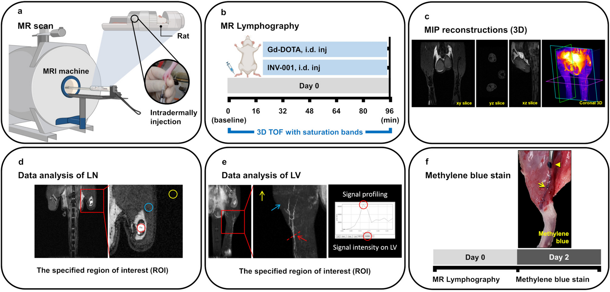
Notohamiprodjo M, Baumeister RGH, Jakobs TF et al (2009) MR-lymphangiography at 3.0T-a feasibility study. Eur Radiol 19:2771–2778
Article
PubMed
Google Scholar
Notohamiprodjo M, Weiss M, Baumeister RG et al (2012) MR lymphangiography at 3.0 T: correlation with lymphoscintigraphy. Radiology 264:78–87
Article
PubMed
Google Scholar
White RD, Weir-McCall JR, Budak MJ, Waugh SA, Munnoch DA, Sudarshan TAP (2014) Contrast-enhanced magnetic resonance lymphography in the assessment of lower limb lymphoedema. Clin Radiol 69:E435–E444
Article
CAS
PubMed
Google Scholar
Mitsumori LM, McDonald ES, Wilson GJ, Neligan PC, Minoshima S, Maki JH (2015) MR lymphangiography: how i do it. J Magn Reson Imaging 42:1465–1477
Article
PubMed
Google Scholar
Kim EY, Hwang HS, Lee HY et al (2016) Anatomic and functional evaluation of central lymphatics with noninvasive magnetic resonance lymphangiography. Medicine 95:e3109
Article
PubMed
PubMed Central
Google Scholar
Salehi BP, Sibley RC, Friedman R et al (2023) MRI of lymphedema. J Magn Reson Imaging 57:977–991
Article
PubMed
Google Scholar
Xiong L, Engel H, Gazyakan E et al (2014) Current techniques for lymphatic imaging: State of the art and future perspectives. Ejso-Eur J Surg Onc 40:270–276
Article
CAS
Google Scholar
Guerrini S, Gentili F, Mazzei FG, Gennaro P, Volterrani L, Mazzei MA (2020) Magnetic resonance lymphangiography: with or without contrast? Diagn Interv Radiol 26:587–595
Article
PubMed
PubMed Central
Google Scholar
Mills M, van Zanten M, Borri M et al (2021) Systematic review of magnetic resonance lymphangiography from a technical perspective. J Magn Reson Imaging 53:1766–1790
Article
PubMed
PubMed Central
Google Scholar
Ruehm SG, Schroeder T, Debatin JF (2001) Interstitial MR lymphography with gadoterate meglumine: Initial experience in humans. Radiology 220:816–821
Article
CAS
PubMed
Google Scholar
Lohrmann C, Foeldi E, Speck O, Langer M (2006) High-resolution MR lymphangiography in patients with primary and secondary lymphedema. AJR Am J Roentgenol 187:556–561
Article
PubMed
Google Scholar
Lohrmann C, Foeldi E, Langer M (2006) Indirect magnetic resonance lymphangiography in patients with lymphedema preliminary results in humans. Eur J Radiol 59:401–406
Article
PubMed
Google Scholar
Felmerer G, Sattler T, Lohrmann C, Tobbia D (2012) Treatment of various secondary lymphedemas by microsurgical lymph vessel transplantation. Microsurg 32:171–177
Article
Google Scholar
Mitsumori LM, McDonald ES, Neligan PC, Maki JH (2016) Peripheral magnetic resonance lymphangiography: techniques and applications. Tech Vasc Interv Radiol 19:262–272
Article
PubMed
Google Scholar
Mitsumori LM (2016) Response: Magnetic resonance lymphangiography: How to prove it? J Magn Reson Imaging 44:1368–1369
Article
PubMed
Google Scholar
Maki JH, Neligan PC, Briller N, Mitsumori LM, Wilson GJ (2016) Dark blood magnetic resonance lymphangiography using dual-agent relaxivity contrast (DARC-MRL): a novel method combining gadolinium and iron contrast agents. Curr Probl Diagn Radiol 45:174–179
Article
PubMed
Google Scholar
Kobayashi H, Kawamoto S, Bernardo M, Brechbiel MW, Knopp MV, Choyke PL (2006) Delivery of gadolinium-labeled nanoparticles to the sentinel lymph node: Comparison of the sentinel node visualization and estimations of intra-nodal gadolinium concentration by the magnetic resonance imaging. J Control Release 111:343–351
Article
CAS
PubMed
Google Scholar
Kobayashi H, Kawamoto S, Choyke PL et al (2003) Comparison of dendrimer-based macromolecular contrast agents for dynamic micro-magnetic resonance lymphangiography. Magn Reson Med 50:758–766
Article
CAS
PubMed
Google Scholar
Müller A, Fries P, Jelvani B et al (2017) Magnetic Resonance lymphography at 9.4 T using a gadolinium-based nanoparticle in rats. Invest Radiol 52:725–733
Article
PubMed
Google Scholar
Kuo PH, Kanal E, Abu-Alfa AK, Cowper SE (2007) Gadolinium-based MR contrast agents and nephrogenic systemic fibrosis. Radiology 242:647–649
Article
PubMed
Google Scholar
Weinreb JC, Rodby RA, Yee J et al (2021) Use of intravenous gadolinium-based contrast media in patients with kidney disease: consensus statements from the American College of Radiology and the National Kidney Foundation. Radiology 298:28–35
Article
PubMed
Google Scholar
Cheong BYC, Wilson JM, Preventza OA, Muthupillai R (2022) Gadolinium-based contrast agents: updates and answers to typical questions regarding gadolinium use. Tex Heart I J 49(3):e217680
Shin TH, Kim PK, Kang S et al (2021) High-resolution T(1) MRI via renally clearable dextran nanoparticles with an iron oxide shell. Nat Biomed Eng 5:252–263
Article
CAS
PubMed
Google Scholar
Mounzer R, Shkarin P, Papademetris X, Constable T, Ruddle NH, Fahmy TM (2007) Dynamic imaging of lymphatic vessels and lymph nodes using a bimodal nanoparticulate contrast agent. Lymphat Res Biol 5:151–158
Article
PubMed
Google Scholar
Malhotra N, Lee JS, Liman RAD et al (2020) Potential toxicity of iron oxide magnetic nanoparticles: a review. Molecules 25:3159
Article
CAS
PubMed
PubMed Central
Google Scholar
Turkbey B, Czarniecki M, Shih JH et al (2020) Ferumoxytol-enhanced MR lymphography for detection of metastatic lymph nodes in genitourinary malignancies: a prospective study. AJR Am J Roentgenol 214:105–113
Article
PubMed
Google Scholar
Hamilton BE (2020) Ferumoxytol-enhanced MRI is not inferior to gadolinium-enhanced MRI in detecting intracranial metastatic disease and metastasis size. Am J Roentgenol 215:1551–1551
Article
Google Scholar
Daldrup-Link HE, Theruvath AJ, Rashidi A et al (2022) How to stop using gadolinium chelates for magnetic resonance imaging: clinical-translational experiences with ferumoxytol. Pediatr Radiol 52:354–366
Article
PubMed
Google Scholar
Werner P, Taupitz M, Schröder L, Schuenke P (2021) An NMR relaxometry approach for quantitative investigation of the transchelation of gadolinium ions from GBCAs to a competing macromolecular chelator. Sci Rep-Uk 11


留言 (0)