Chemicals and reagents
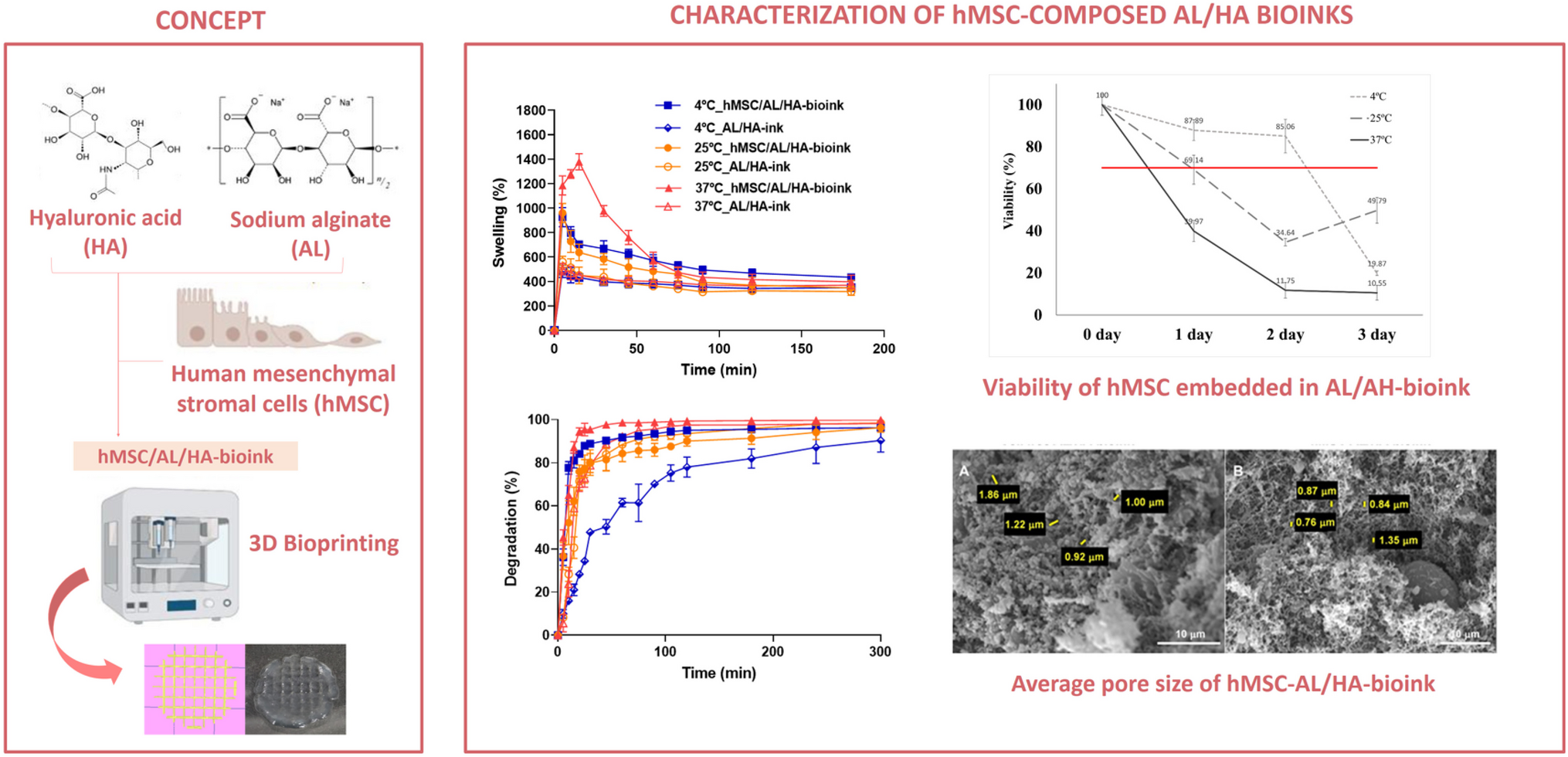
All reagents and chemicals were of analytical grade and purchased from Sigma-Aldrich (Madrid, Spain) unless otherwise stated, and used without prior purification. Hyaluronic acid (HA, 1000 kDa), was kindly gifted by Bioibérica S.A.U. (Barcelona, Spain). Low viscosity sodium alginate (AL, 4-12 cP; CAS number 9005-38-3; molecular weight 216.12) was obtained from Sigma-Aldrich (Madrid, Spain). A Milli-Q® Gradient A10 system apparatus (Millipore Iberica, Madrid, Spain) was used for the filtration of double distilled water prior to its use.
hMSC isolation and culture
Subcutaneous adipose tissue was collected, upon informed consent, from patients that underwent liposuction procedure. This study was approved by the Ethics Committee of Clinical University Hospital of Málaga (Spain) (number: 02/022010). The isolation and culture protocols of hMSC were performed following the work by López-Ruiz et al. [38]. hMSC were characterized following the established criteria of the International Society for Cellular Therapy (ISCT) [39], and following the protocols described by Martínez-Moreno et al. [40]. Adipose tissue collected from lipoaspiration was minced and treated on a shaker by enzymatic digestion solution of 1 mg/mL of collagenase type IA, at 37 °C for 1 h. After digestion, the enzyme collagenase was removed by a single wash in sterile phosphate-buffered saline (PBS), and washed twice with Dulbecco’s Modified Eagle Medium (DMEM) supplemented with 10% fetal bovine serum (FBS) (Invitrogen Inc., New York, USA). The cell pellet was suspended in DMEM containing 10% FBS and 1% streptomycin/penicillin, and cultured at 37 °C in 5% CO2. After 48 h, non-adherent cells were discarded from the removed medium. When 80% level of confluence was reached, cells were released with TrypLE (Invitrogen Inc., New York, USA) and sub-cultured. For all the experiments, hMSC were used between passages 4 and 6.
Preparation of AL/HA-bioink loaded with hMSC
The formulation of the bioink was carried out in three stages. First, the hydrogel was prepared by dissolving AL and HA in PBS at concentrations of 6% (w/v) and 1.33% (w/v), respectively, obtaining a non-crosslinked AL/HA-based hydrogel (AL/HA-hydrogel) [41]. Previously, both materials were sterilized by short cycle autoclaving. This techniques has been previously shown to be effective in the sterilization of materials such as HA against some types of bacteria without affecting the rheology, physicochemical prop-erties and printability [42] and it has also been used to sterilisation of the AL in others previous works [28, 43, 44]. Second, non-crosslinked AL/HA-hydrogel was combined with 100 mM CaCl2 (3:1) to form the semi-crosslinked hydrogel, being the final concentrations 4.5% (w/v) and 1% (w/v) for AL and HA, respectively. The semi-crosslinked hydrogel was mixed homogeneously using a 3 cc Luer-Lock syringe with a connector and stored at 4 °C until use. Briefly, hMSC pellet were suspended in the semi-crosslinked AL/HA-hydrogel at the concentration of 1 × 10⁶ cells/mL resulting the hMSC/AL/HA-based bioink (hMSC/AL/HA-bioink). Then, hMSC/AL/HA-bioink packed in 3 cc syringes was gentle stirred to get homogeneous hMSC suspensions. The bioinks were then stored at different temperatures (4 °C, 25 °C, or 37 °C) for further studies. Identical process of formulation, without hMSC, was performed to obtain a blank AL/HA-ink to be used as a control. Finally, the hMSC/AL/HA-bioink was placed in the bioprinter (REG4LIFE by Regemat; Regemat 3D, S.L., Granada, Spain) and extruded at room temperature (RT) (Flow velocity: 4.5 mm/s; nozzle diameter: 0.4 mm). After bioprinting, the crosslinking process was completed by bath in 180 mM CaCl2 for 30 min at RT. Then, the constructs were placed in culture medium (DMEM containing, 10% FBS and 1% streptomycin/penicillin) and stored for 7 days in an incubator with 5% CO2 at 37 °C.
Sterility treatment and testing
The sterility assay was performed by direct inoculation of 1 mL AL/HA-bioink in two different microbiology media according to the European Pharmacopoeia 9th Edition, by using Thioglycollate Penase Broth (TPB; VWR Int., Radnor, PA, USA) to detect anaerobic and aerobic microorganisms, and Tryptic Soy Penase Broth (TSPB, VWR Int., Radnor, PA, USA) to detect fungi and aerobic microorganisms. Each hydrogel in inoculated media was incubated for 14 days at 22 °C and 35 °C for TSPB and TPB, respectively. All samples were daily inspected, by visually checking for any signs of turbility. If after 14 days microbial growth had taken place, the medium would show turbidity. Aseptic conditions were implemented for this assay, inside a safety cabinet in a clean room. For each media (TSPB and TPB), sterility test and growth promotion test of aerobes, anaerobes and fungi, were previously performed. The formulation and bioprinting processes were carried out under aseptic conditions to maintain the sterility of the final bioink and construct.
Physical and macroscopic appearance
The gelation of AL/HA-hydrogel was assayed by tube inversion test, at three different crosslinking states, namely, non-crosslinked hydrogel, semi-crosslinked hydrogel (adding 100 mM CaCl2), and fully crosslinked hydrogel (with 180 mM CaCl2). The test tube inversion method allows determining the sol or gel state of a hydrogel depending on the flow of the sample. Briefly, the hydrogel was deposited on small-sized glass jars and immersed in a water bath at 37 ± 0.5 °C. To determine the sol-to-gel transition of the hydrogel, the jars were then inverted and the time to stop flowing freely was then recorded. The physical and macroscopic appearance of hMSC/AL/HA-bioink and AL/HA-ink were checked.
Osmolality
Osmolality was determined in a micro-osmometer model 3320 (Advanced Instruments, Inc., Norwood, MA, USA) at 25 ± 0.5 °C. The freezing point depression (FPD), which refers to the direct proportional concentration of osmotically active compounds in aqueous solution, was determined. All measurements were performed in triplicate, and results are given as the mean ± standard deviation (SD).
pH values
The pH values of bioinks were determined using a calibrated pH meter LAQUA pH1100-S (HORIBA Advanced Techno Co., Lille, France) at 4 ± 0.5 °C, 25 ± 0.5 °C and 37 ± 0.5 °C. Each measurement was conducted by direct immersion of the electrode in the samples contained in a glass. All measurements were run in triplicate, and results are given as the mean ± standard deviation (SD).
Degradation test
The degradation test was undertaken by immersion procedure. Accurate weights of hMSC/AL/HA-bioink and AL/HA-ink (0.1 ± 0.02 g) were immersed in an Eppendorf tube with 1 mL PBS (pH 7.4). The test was performed at three distint temperatures, i.e., 4 ± 0.5 °C, 25 ± 0.5 °C and 37 ± 0.5 °C and at two different relative humidity (RH) values, 35% and 70% in a climatic chamber during 300 min. Eppendorf tubes tube were left close during the incubation. After incubation, samples were centrifuged at 5000 rpm for 1 min. The PBS excess was removed and the samples were weighed. Upon each analysis, 1 mL of PBS was added to keep original conditions constant. The degradation percentage was determined in triplicate as a measure of weight loss (WL) using the following equation [45]:
$$WL\;\left(\%\right)=\frac\;\times\;100$$
where W0 is the initial weight of sample and Wt the mass of degraded sample measured at different times.
Swelling test
The water uptake capacity of hMSC/AL/HA-bioink and AL/HA-ink was determined by swelling of freeze dried samples in an Eppendorf tube with 500 µL PBS (pH = 7.4). The test was carried out at 4 ± 0.5 °C, 25 ± 0.5 °C and 37 ± 0.5 °C, and two different RH values, 35% and 70%, in a climatic chamber during 180 min. Eppendorf tubes tube were left close during the incubation. At selected time intervals the swollen samples were centrifuged at 5000 rpm for 1 min, and after eliminating the excess of PBS, they were weighed [45]. Then, samples were replenished with fresh PBS. The swelling (Sw) percentage was calculated in triplicate applying the following equation:
$$Swelling\;ratio\;\left(\%\right)=\frac\;\times\;100$$
where Ws is the weight of the samples at the swelling state and Wd is the initial mass of the freeze dried samples.
Porosity
The porosity of hMSC/AL/HA-bioink and AL/HA-ink was calculated according to the Archimedes’ principle [46]. Analysis of percentage of porosity was performed by immersing the freeze-dried bioinks (0.06 ± 0.01 g) in 200 mL PBS pH 7.4 at 25 ± 0.5 °C. The submerged mass of samples was recorded, and then samples were removed, and the wet mass was recorded. Samples were analysed in triplicate. Porosity of bioinks was calculated applying the following equation:
$$Porosity\;\left(\%\right)=\frac}$$
where Mw is the water saturated wet mass of samples, MD is the freeze dried mass of the samples, and MSUB is the submerged mass of samples.
In addition, AL/HA-ink and hMSC/AL/HA-bioink images obtained by environmental scanning electron microscopy (SEM; ESEM Quanta 200 FEI) were analyzed. The images were processed using ImageJ software following the procedure described by Monticeli et al. [47]. Briefly, the measurement scale was adjusted taking into account the values provided by the microscope. Then, the images were transformed to an 8-bit format and the threshold adjustment was carried out in the range 0-255 to be able to measure gaps in the sample and to set the appropriate threshold. In this case, the optimal range used was 0-115.
Zeta potential and electrical conductivity
The zeta potential (ζ) of hMSC/AL/HA-bioink and AL/HA-ink was determined at 25 ± 0.5 °C from the electrophoretic mobility using a Zetasizer Nano ZS (Malvern Instruments Ltd., Malvern, UK). The electrical properties of samples were tested as a function of pH variation (from 4 to 8, at constant concentration 10−3 M KNO3), and as a function of ionic strength variation (from 10−1 to 10−5 M KNO3, at pH = 6). Before measuring, the samples were dispersed in double distilled water to form a 0.1% (w/v) solution, kept under mechanical stirring (50 rpm) for 24 h [48]. Zeta potential values were recorded as the mean ± SD of three replicates. Electrical conductivity of the samples was recorded in a Crison EC-Meter BASIC 30+ (Crison Instruments, Alella, Spain) conductivity meter. The samples were diluted in water at room temperature (1:20). The device electrode was dipped into a glass vial containing samples. Data are expressed as the mean ± SD of three replicates.
Rheological studies
Samples of hMSC/AL/HA-bioink and AL/HA-ink were submitted to dynamic (oscillatory) test, as well as rotational test by using Haake RheoStress 1 rheometer (Thermo Fischer Scientific, Kalsruhe, Germany) connected to a temperature controller (Thermo Haake Phoenix II + Haake C25P). The initial rotational testing consisted of a stress sweep test followed by a frequency sweep test were carried out for the hMSC/AL/HA sample to determine the appropriate values of shear stress and frequency for the subsequent temperature sweep test. Each sample was set to equilibrate by placing it between the plate-plate sensor systems with a Haake PP60Ti mobile plate (1 mm gap, 60 mm diameter) for 5 minutes to reach the starting temperature. The oscillatory stress sweep tests were conducted at a constant frequency of 1 Hz and a temperature of 25 ± 0.5 °C, in a stress range of 0.04 to 200 Pa. A value of 0.5 Pa, located within the linear viscoelastic region, was selected for the subsequent frequency sweep study ranging from 0.1 to 10 Hz. Consequently, the effect of temperature on the rheology of the bioinks was investigated by performing a temperature sweep test from 10 ± 0.5 °C to 40 ± 0.5 °C at 1 Hz and 0.5 Pa. The temperature increment was controlled at a ramp speed over 2000 seconds, during which the storage modulus (G’), loss modulus (G’’) and the complex viscosity (η*) were measured.
In the second case, rotational tests were run in a Haake C60/2Ti mobile cone (60 mm diameter, 0.102 mm gap and 2 ° angle). Each sample was set to equilibrate by placing it between the cone-plate sensor systems (0.102 mm gap) for 5 min to attain the running temperature. The shear rate was increased from 1 to 100 s-1, then maintained for 1 min at 100 s-1, and finally decreased from 100 to 1 s-1. The shear stress was measured at 25 ± 0.5 °C and 37 ± 0.5 °C.
Data from the flow curve (τ = f (γ̇)) were fitted to selected mathematical models, namely, Bingham, Casson, Cross, Herschel-Bulkley, Newton and Ostwald-De-Waele, to determine the flow type. Viscosity mean values (Pa·s) were determined from the constant share range at 100 s-1 of the viscosity curve (η = f (γ) in response to the temperature (ºC). During the test, the microstructure disturbance or apparent thixotropy (Pa/s) was evaluated by calculating the area of hysteresis loop formed when the shear program is run, i.e., from 1 to 100 s-1 (ramp-up), constant shear at 100 s-1, and from 100 to 1 s-1 (ramp-down).
Stability tests
Freshly prepared samples (n = 3) were submitted to heating-cooling cycles over a temperature range between 4 ± 0.5 °C and 45 ± 0.5 °C by storing them 24 h under each temperature for 6 days, and then checked for signs of physical instability (gel collapse, homogeneity, etc.). Freshly prepared samples (n = 3) were centrifuged (Centrifuge 5804 Eppendorf, Hamburg, Germany) at 3500 rpm for 30 min at room temperature (n = 3), and then examined for signs of physical instability. Freshly prepared samples went through freeze-thaw cycles, by keeping them between − 21 ± 0.5 °C and + 25 ± 0.5 °C, for three cycles of 24 h for 6 days, and then examined for sings of physical instability. Finally, the influence of the temperature on hMSC/AL/HA-ink was studied to define its appropriate temperature of storage and its shelf-life. Bioinks loaded with hMSC were packed in 3 cc Luer-Lock syringes at a concentration of 1 × 10⁶ cells/mL with 0.5 mL of AL/HA-bioink. Different temperatures were assayed 4 °C, 25 °C and 37 °C (three syringes each temperature). Cell viability was tested every 24 h for 3 days using the alamarBlue® (aB) assay following the manufacturer’s protocol (Thermo Fisher Scientific Inc., Carlsbad, CA, USA).
Scanning electron microscopy (SEM)
Cell-free and hMSC/AL/HA-bioinks were maintained under standard cell culture conditions at 0, 1, 2 and 3 days. Bioinks were fixed in a bath of 2.5% glutaraldehyde buffer in cacodylate for 12 h followed by a dehydration process through a graded series of ethanol (30–100%). Then, bioinks were critical-point dried in an Emscope CPD 750 critical point dryer (Emscope Lab., Ashford, UK). Bioinks were placed onto aluminum SEM specimen mounting stubs (Electron Microscopy Sciences, Hatfield, PA, USA) on a carbon tab (Agar Scientific Ltd., Essex, UK) and coated with carbon to improve their electrical conductivity using an Emitech® K950X Carbon Evaporator (QuorumTechnologies Ltd, East Grinstead, UK). Finally, samples were examined using a field emission scanning electron microscope JSM-7001 F (JEOL Ltd., Tokyo, Japan). Morphology and porosity of critical-point dried hMSC/AL/HA-ink were observed and measured respectively by SEM.
Optical microscopy
To determine whether the hMSC were properly embedded and distributed along the bioinks, cells were first examined by optical microscopy at 0 and 7 days. Images were taken using a microscope (Olympus IX71, Tokyo, Japan) at 100× magnification.
Cell viability
The LIVE/DEAD® Viability/Cytotoxicity kit (Thermo Fisher Scientific Inc., Carlsbad, CA, USA) was used following the manufacturer’s instructions to assess cell viability in hMSC/AL/HA-bioinks. Briefly, samples were firstly washed with 1X PBS wash buffer. Subsequently, they were incubated in the dark for 30 min with 8 µL of 4 µM EthD-I (red staining) and 4 µL of 2 µM calcein AM (green staining), both diluted in 4 mL sterile PBS. Following another wash with 1X PBS, samples were observed using a confocal microscope Nikon Eclipse Ti-E A1 (Nikon Instruments Europe B.V., Amsterdam Netherlands) and imaged on days 1, 4 and 7. Green fluorescence was indicative of live cells while red fluorescence indicated dead cells using two distinct filters. The images were then analyzed using ImageJ software v. 1.52i (NIH, Bethesda, MD, USA). Six regions were assessed for each cell type (live or dead) to derive an average value of the percentage of viable cells (n = 3).
Cell proliferation assay
The proliferation rate of cells embedded in hMSC/AL/HA-ink was determined by colorimetric aB assay (Thermo Fisher Scientific Inc., Carlsbad, CA, USA) on day 1, 4 and 7. Appropriate control without cells was used for data normalization. Briefly, samples were incubated with 10 µL of aB solution per each 100 µL of medium at each time point, and incubated for 3 h. The fluorescence intensity was analysed using a plate reader Synergy HT (Bio-Tek Instruments, Inc., Winooski, VT, USA) with excitation and emission wavelengths of 600 nm and 570 nm. Experiments were performed in triplicate (n = 3), and the absorbance data represent as fold increase to day 1.
3D bioprintability
An extrudability test was performed using hMSC/AL/HA-ink in three different crosslinking states (0 mM, 100 mM and 180 mM CaCl2) to determine the appropriate degree of crosslinking for use in bioprinting. It was done by varying bioprinting properties such as flow rate or printing speed [5, 49,50,51].
The selected hMSC/AL/HA-ink was packed in a 3 cc syringe and loaded into the bioprinter. The bioprintability assessment of the bioink was performed using a REG4LIFE by Regemat bioprinter (Regemat 3D, S.L., Granada, Spain). The bioprinting process consisted of the deposition of the selected bioink in a layer-by-layer manner adding 180 mM CaCl2 to generate a linear 3D printed fully-crosslinked construct (porous rectangular-type structure; 10 mm high ×10 mm wide structure) by using the Regemat 3D designer software [52]. Filament after extrusion and nozzle diameter had a width of 0.4 mm. The adequate flow speed was established at 4.5 mm/s. Furthermore, to determine whether the bioprinting process of the cell-loaded bioink affected cell proliferation capacity, an aB assay was performed post-bioprinting at 0 h and 24 h.
Statistical analysis
The obtained results were statistically validated by one-way analysis of variance (ANOVA) or Student’s t-test using GraphPad Prism® v. 5.00 software (GraphPad Software Inc., San Diego, CA, USA). Differences between independent groups were determined by applying Tukey’s test, the significance level was set at 0.05, and adopting a 95% confidence level.


留言 (0)