
記住我


RWPE-1, DU-145, PC-3, 22Rv1, LNCaP, C4-2B, VCaP and HEK293T cell lines were cultured according to the ENCODE cell culture standards. IGF2BPs KO and RBM15 KO cells were generated using the CRISPR-Cas9 technology following the manufacturer’s instructions. The guided RNAs (sgRNAs) were cloned into pLentiCRSPIR V2 (Genscript, USA). Transfections were carried out with Polyplus (Franch) for plasmids and siRNAs. The sgRNA sequences used are listed in Supplementary Table S1.
Clinical tissue samplesThirty-six paired prostate cancer tissues and adjacent tissues were obtained from patients. The clinical tissues were all collected at the First Affiliated Hospital, School of Medicine, Zhejiang University after the approval of the Ethics Committee of Zhejiang University and informed consent. Clinicopathological characteristics of the PCa patients are listed in Supplementary Table S2.
Antibodies, siRNAs, plasmids and RNA oligosAntibodies used in this study were: anti-Myc tag (ab32, Abcam), anti-IGF2BP1 (ab290736, Abcam), anti-IGF2BP2 (ab128175, Abcam), anti-IGF2BP3 (ab177477, Abcam), anti-RBM15 (10587-1-AP, Proteintech), anti-DNMT1 (ab92314, Abcam), anti-DNMT3A (ab307503, Abcam), anti-DNMT3B(67,259, Cell Signaling Technology), anti-SEMA3F (SAB2107196, Sigma), anti-S9.6 (MABE1095, Sigma), anti-5-mC (ab214727, Abcam), anti-m6A (SAB5600251, Sigma), anti-YAP1(phosphor S127, ab76252, Abcam), anti-YAP1(66900-1-Ig, Proteintech), anti-LATS1(66569-1-Ig, Proteintech), anti-LATS2(20276-1-AP, Proteintech), anti-β-Actin(4970, Cell Signaling Technology). siRNAs against DNMT1 used were purchased from Ribobio Co.,Ltd (Guangzhou, China). pcDNA3-based vectors encoding wild-type, RRM domain mutant, KH domain mutant Myc-tagged IGF2BP1, IGF2BP2, IGF2BP3 were produced by Shanghai Yoche Biotechnology Co.,Ltd (Shanghai, China). The plasmids encoding RBM15 and SEMA3F (h-RBM15-pcDNA3.1-c-HA, M35-FLAG-SEMA3F) were obtained from Guangzhou FulenGen Co., Ltd (Guangzhou, China). DNA and RNA oligos synthesized by TsingKe Biotech Co., Ltd (Beijing, China) are listed in Supplementary Table S1.
Lentiviruses transfectionThe lentiviruses vectors, pReceiver-Lv242-FLAG, encoding IGF2BP1, IGF2BP2, IGF2BP3 and RBM15 were obtained from Guangzhou FulenGen Co., Ltd (Guangzhou, China). Lentivirus infection was performed following the manufacturer’s manuals.
Immunoprecipitation and immunoblotingAfter extracting proteins from cultured cells with a modified buffer, immunoprecipitation and following immunoblotting were performed with antibodies as described previously [21]. Briefly, 5 µg corresponding antibodies were added to PC-3 cell lysates and incubated at room temperature, followed by incubation with Protein A/G Magnetic beads (MCE) at 4 °C overnight. After washing with modified wash buffer, protein complexes were eluted by elution buffer and analyzed by immunoblotting and LC-MS/MS. All western blot experiments in Figs. 1, 2, 3, 4, 5, 6, 7, 8, 9 and 10 were repeated once; typical images from a single repeat are shown.
Fig. 1
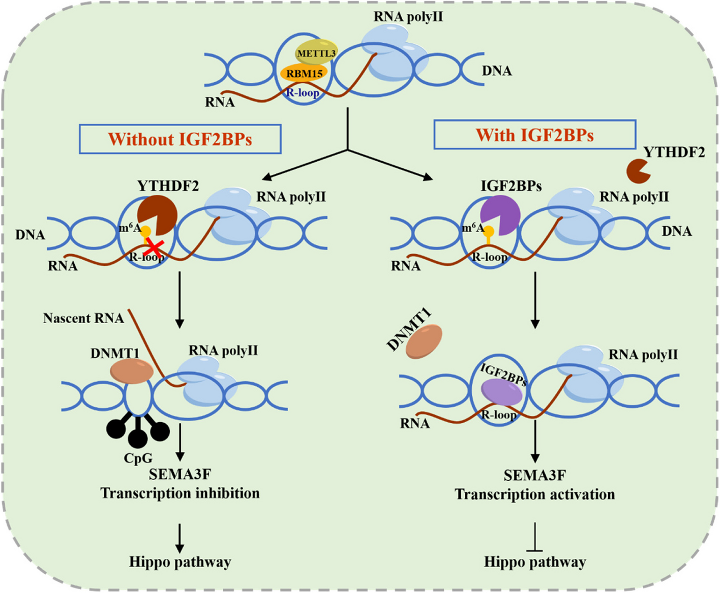
R-loops are reduced in PCa and recruit IGF2BP proteins. A Isolated R-loops from PCa and paired normal tissues were analyzed by Dot-blot. Methylene blue staining served as a loading control. (Left) representative dot-blot results; (Right) quantification of dot blot (normalized by the density of methylene blue staining). B Regulators were identified by LC-MS/MS analysis using S9.6 antibody in PC-3 cells. C Validation of S9.6 IP LC-MS/MS results by western blot assay. Experiments were performed with Myc-purified IGF2BP proteins and S9.6 antibody in 293T cells. (Left): Representative western blot results. (Right): Quantification of western blot assay. D PC-3 cells were immunostained for IGF2BP1/2/3 and S9.6; representative images are shown (scale bar: 60 μm). E Quantification of cellular immunofluorescence (n = 10 per group). Data are presented as means ± 95CI, two-tailed unpaired t-test. *p-value < 0.05, **p-value < 0.01, ***p-value < 0.001, ****p-value < 0.0001
Purification of IGF2BP1/2/3, METTL3 and RBM15Full-lengths IGF2BP1/2/3 were cloned into pcDNA3.1 with Myc-tag, and full-lengths METTL3 was cloned into M35 with Flag-tag (Guangzhou FulenGen Co., Ltd). Full-lengths RBM15 was cloned into pcDNA3.1 with HA-tag (Guangzhou FulenGen Co., Ltd).The plasmid was expressed in 293T cells and incubated with DMEM medium at 37 °C. Cells were lysated in IP lysis buffer and incubated on ice. After incubation, the extract was centrifuged at 13,000 g for 20 min at 4 °C. The tag-agarose was added to supernatant and incubated at 4° C with rotation for 60 min. After centrifugation at 3000 g for 1 min, the tag-agarose-bound proteins were washed with the buffer containing 50 mM Tris pH 7.4, 150 mM NaCl. Recombinant IGF2BPs, METTL3 and RBM15 were eluted with elution buffer (50 mM Tris pH 7.4, 150 mM NaCl, 500 µg/ml peptides).
S9.6 IPRecombinant proteins co-immunoprecipitation with S9.6 antibody was performed from cultured PC-3 and DU-145 cells as previously described [22].
In vitro RNA-protein pulldown assays3’-biotin-labeled ssRNA (5’-CGUCUCGGACUCGGACUGCU-3’) and complementary ssDNA (5’-AGCAGTCCGAGTCCGAGACG-3’) were synthesized from TsingKe Biotech Co., Ltd (Beijing, China). We generated m6A ssRNA by substituting all two adenines with m6A-modified adenines. The ssRNA with or without m6A modification was annealed with complementary ssDNA in a 1:1 ratio. As directed by the manufacturer’s instructions, proteins in cell lysate were pulled down with the ssRNA and DNA: RNA hybrids using the Pierce magnetic RNA-Protein pull-down kit (20,164, ThermoFisher). The final elution and supernatant were analyzed by western blot and LC-MS/MS.
LC-MS/MSElution samples were prepared for LC-MS/MS analysis as described previously [22]. After in-gel digestion, the peptides were dissolved on an EASY-nLC 1000 UPLC system according to the manufacturer’s protocol. And then peptides were subjected to NSI source followed by tandem mass spectrometry (MS/MS) in Q ExactiveTM Plus (Thermo) coupled online to the UPLC. The final MS/MS data were processed using Proteome Discoverer 2.4.
Dot-blotTotal genomic DNA from prostate cells were isolated according to standard procedures. The same amount of DNA from divergent groups was diluted to the same concentration with NaOH/TE solution and denatured at 99 °C. Afterward, DNA was dot-blotted on Hybond N + membrane (GE health) via a dot-blot apparatus (Bio-rad), and linked by a UV crosslink at 1500 µJ. The membrane was stained by 0.1% methylene blue (Sigma-Aldrich). Primary antibody was diluted 1:1000 in universal antibody diluent (NCM Biotech). After incubation at 4 °C, the membrane was washed twice gently in 0.02% TBST for 10 min. Secondary antibody was diluted 1:5000 in 5% BSA in 0.02% TBST and then washed three times. All dot blot experiments in Figs. 1, 2, 3, 4, 5, 6, 7, 8, 9 and 10 were repeated once; typical images from a single repeat are shown.
ImmunofluorescenceCells for immunofluorescence were fixed in ice-cold methanol for 15 min at -20 °C and further treated with 0.1% Triton X-100 for 30 min. Cells were then blocked by 5% BSA for 30 min at room temperature. Primary antibodies were diluted in universal antibody diluent (NCM Biotech) and incubated with prostate cells overnight at 4 °C. The cells were then washed three times with 0.02% TBST, and were incubated with secondary antibodies for 50 min at room temperature followed by wash with 0.02% TBST twice. DAPI was incubated with cells at room temperature for 5 min. Images were acquired with a STEDYCON confocal microscope and processed using Image J and Adobe Photoshop. Quantification of the m6A, RBM15, IGF2BP1/2/3 and S9.6 signal intensities was performed according to the previously described method [7].
DRIP-seq and m6A DIPGenomic DNA was isolated from prostate cells by Proteinase K and RNase A treatment in lysis buffer, followed by phenol-chloroform extraction and ethanol precipitation. The DNA was fragmented to 200–500 bp by sonication. 20 µg of genomic DNA was used for each immunoprecipitation. S9.6 DRIP was performed as described previously using S9.6 antibody [23]. The DNA libraries were sequenced on the Illumina sequencing platform by Genedenovo Biotechnology Co., Ltd (Guangzhou, China). M6A DIP was carried out as described in a previously published study [13] using anti-m6A antibody. With respect to the data analysis, sequencing reads were aligned to genome reference sequences using HISAT2 software (v2.1.0). The DRIP enriched regions (peaks) were visualized by Integrative Genomics Viewer (IGV).
RNA-seqTotal RNA was isolated from different groups of PC-3 cells by standard protocol. RNA-seq libraries were constructed using standard Illumina RNA-seq protocols by Genedenovo Biotechnology Co., Ltd (Guangzhou, China). The paired-end clean reads were mapped to the reference genome using HISAT2. 2.4. Principal component analysis (PCA) was performed with R package gmodels (http://www.r-project.org/) in this experience. RNAs differential expression analysis was performed by DESeq2 software between two different groups (and by edgeR between two samples). The genes/transcripts with the parameter of false discovery rate (FDR) below 0.05 and absolute fold change ≥ 2 were considered differentially expressed genes/transcripts.
ChIPDU-145 and PC-3 cells cultured in 10 cm dishes were fixed with 1% paraformaldehyde for 15 min at room temperature. The ChIP assay was carried out using the ChIP kit (Merck and Millipore). Briefly, cells were lysed using lysis buffer on ice. Nuclei was collected and fragmented by ultrasound. The chromatin was isolated and added to DNMT1 beads in ChIP buffer. After incubation overnight, the beads were washed and eluted. The eluted chromatin was combined and crosslinks reversed, followed by DNA purification by PCR purification columns (Thermo Fisher). The eluted ChIP-DNA was used for RT-PCR analysis.
For ChIP-re-ChIP, the chromatin was isolated and added to RBM15 beads in ChIP buffer. Immunoprecipitates were then eluted with 30 µL 10mM DTT at 37 °C for 30 min. Then they were diluted 20x with Re-ChIP buffer (1% Triton X-100, 150 Mm NaCl, 2 mM EDTA, 20 mM Tris-HCl, and 1x cocktail) on ice. Next, they were incubated with anti-m6A antibody at 4 °C overnight with mixing. Washes and IP elution were performed according to ChIP kit protocols, followed by DNA purification by PCR purification columns (Thermo Fisher). The eluted ChIP-DNA was used for RT-PCR analysis. All the primers used are shown in Supplementary Table S1.
DNMT1 activity assayThe DNMT1 activity assay (Abcam) was used to quantify DNMT1 binding activity. The procedures to measure the DNMT1 activity were directed by manufacturer’s manual.
MethylationEPIC (850 K) BeadChipThe MethylationEPIC BeadChip experiments and data analysis of the PCa samples were conducted by OE Biotechnology Co.Ltd. (Shanghai, China). DNA concentration and integrity were assessed by a NanoDrop 2000 spectrophotometer (Thermo Fisher Scientific, Waltham, MA, USA) and agarose gel electrophoresis, respectively. DNA was bisulfite treated using the Zymo Research EZ DNA methylaiton-Glod Kits (Zymo Research, Irvine, CA, USA). Bisulfite-converted DNA was analysed on an Illumina Infinium MethylationEPIC(850 K) BeadChip (Illumina). Finally, Illumina iSCAN was used to scan the chip to get the Idat files. Idat files were imported and then preprocessed with ChAMP(version 2.12.4) package in R to get raw data. Next, the raw data was normalized with BMIQ method. Statistical differences in continuing variables between two groups were compared by t-test. The significantly DMS(differential methylation Sites) were identified by a threshold of |deltaBeta| > 0.1 and P.Value < 0.05.
Cell migration and proliferation assaysAll specific procedures were performed as previously described [18].
Animal models and in vivo imagingSubcutaneous transplantation models were prepared as described previously [24]. All the animal studies and protocols followed the institutional guidelines of the First Affiliated Hospital, School of Medicine, Zhejiang University.
RNA isolation and quantitative PCRTotal RNA was extracted from prostate cell lines and PCa tissues by RNAiso plus (Takara, Japan) and analyzed by RT-qPCR as previously described [25]. All the primers used in the study are shown in Supplementary Table S1.
Databases usedSeveral user-friendly databases were utilized to download data, analyze or refer to in this study. TCGA database (https://portal.gdc.cancer.gov), starBase online database (https://rnasysu.com/encori/), GEPIA online database (http://gepia2021.cancer-pku.cn/), LinkedOmics online database (http://www.linkedomics.org/), SRAMP (http://www.cuilab.cn/sramp), and Venn diagram (http://bioinfogp.cnb.csic.es/tools/venny/index.html).
StatisticsData are presented as mean ± SD. Differences between two groups were evaluated using the two-tailed, unpaired t test. Survival curves were constructed using the Kaplan-Meier method and analyzed by the log-rank test. All statistical analysis was performed using the GraphPad Prism 9.0 software. Statistical significance was defined as P value of < 0.05.
留言 (0)