
記住我


As reported previously, no neurological signs and pathological changes were found in control animals; VB2− chickens showed progressive weakness and paresis from PH8d until end of this study [15]. The paresis in VB2−/+ chickens became stable at PH16d (4 days after VB2 repletion), and gradually recovered thereafter. Routine pathological examination (Table 1) revealed predominant demyelination in all VB2− groups and predominant remyelination in VB2−/+ chickens at PH16d and PH21d. Endoneurial oedema, hypertrophic Schwann cells with lipid deposition, complex myelin foldings with varying stages of degeneration and myelin fragments in Schwann cell cytoplasm, as noticed in VB2− chickens [15], were also found in VB2−/+ chickens at less severity. But there were no obvious difference of hypertrophic fibroblasts and onion bulb formation [14] between VB2− and VB2−/+ chickens at the same time points. Macrophage-mediated myelin stripping, as described in both human and animal inflammatory demyelinating neuropathies [3, 21], was not found here. Sciatic and brachial nerves showed same pathological changes.
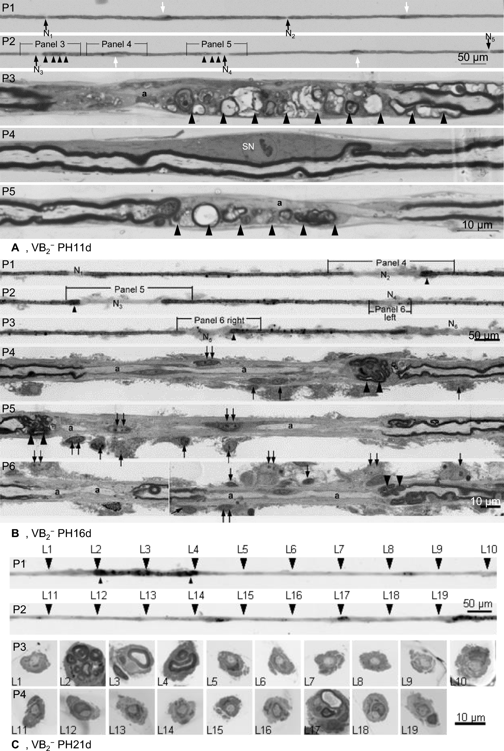
Table 1 Plastic section study of the peripheral nervesaTF studies in VB2− chickensFifty TFs were prepared from each animal. In control animals, at least 5 consecutive internodes in each fibre were examined. Pathological nerve fibres with similar length were examined. Demyelination was the predominant change in TF preparations from all VB2− groups. At PH11d (Fig. 1A), demyelination was mainly confined to the paranodal region, covering not more than 10 percent internodal length, paranodal demyelination. Normal internodes and demyelination were often noticed in the same fibres. At PH16d (Fig. 1B), paranodal demyelination was present in some internodes. In majority of affected internodes, demyelination involved both paranodal and adjoining internodal region, covering more than 10 percent of internodal length and the demyelinated segment not longer than that of neighbouring myelin sheath retained segment, partial internodal demyelination. In some affected internodes, demyelination involved whole internode or the demyelinated segment longer than that of neighbouring myelin retained segment, internodal demyelination. Demyelination of different types were often noticed in the same fibres. At PH21d (Fig. 1C), internodal demyelination was the major type. Quantitative analyses (Table 2) confirmed: the average percentage of paranodal demyelination decreased significantly with age, whereas the percentage of internodal demyelination increased significantly with age; the percentage of partial internodal demyelination increased significantly from P11d to P16d and then decreased significantly at P21d. These results suggest the evolution of demyelination in VB2− chickens: starting from paranodal region, progressing into internodal region, and finally involving whole internode.
Fig. 1
Evolution of demyelination in VB2− chickens. Light micrographs. A Panel 1 (P1) and P2 show part of a TF from a VB2−PH11d chicken. The nodes of Ranvier are indicated by arrows (N1 to N5). Paranodal demyelination is noted at 2 sites (N3 and N4) and normal node of Ranvier at 3 sites (N1, N2 and N5). Schwann cell nuclei (white arrowheads) are located in the middle of each internode. P3 to P5 are longitudinal sections through designated segments. P3 is through the first paranodal demyelinating area. No discernible myelin sheath is seen in the demyelinated segment. Myelin degeneration (large arrowheads in P3) is seen in the internodal region adjoining the paranodal demyelination (small arrowheads in P2). P4 is through the central part of the internode with paranodal demyelination. The myelin sheath is well preserved in this segment and a Schwann cell nucleus (SN) is present. P5 is through the second paranodal demyelinating area. No discernible myelin sheath is seen in the demyelinated segment. Myelin degeneration (large arrowheads in P5) is seen in longitudinal section through the segment (small arrowheads in P2) adjoining the paranodal demyelination. The axon is attenuated in demyelinating regions. B (adapted from Cai et al., 2007 [14] with permission from the author and publisher): P1 to P3 show a TF from a VB2−PH16d chicken displaying paranodal demyelination (N1 and N4) and partial internodal demyelination (N2, and N3, N5 and N6). Paranodal swellings (small arrowheads) and surrounding pale-osmicated material representing the fibroblastic onion bulb like proliferation [14]. P4 to P6 are longitudinal sections through designated segments in the three upper panels. In the demyelinated segments, there is no discernible myelin sheath and axon (a) is attenuated. Redundant myelin foldings with varying degrees of myelin splitting and degeneration (large arrowheads) are found in longitudinal sections through paranodal swellings while the myelin sheaths in the neighbouring non-demyelinated internodal regions are intact. The fibre is surrounded by a variably thick fibroblastic proliferation consisting of fibroblast processes and collagen fibres. Numerous fibroblast nuclei (arrows), some with multiple distinct nucleoli (double arrows), are noted at both demyelinated and non-demyelinated segments. C P1 and P2 show part of a TF from a VB2−PH21d chicken displaying internodal demyelination. P3 and P4 are cross sections through designated sites of the TF, showing denuded axon in the demyelinated segments (L1, 5–19), redundant myelin foldings with myelin splitting and degeneration at both ends of the myelin-maintained segment (L2 and 4). The axon size in the region with intact myelin (L3) is larger than that of demyelinated regions. Fibroblast processes and collagen ensheath the TF at all levels to some degree
Table 2 Frequency of different types of demyelination in VB2− chickensSectional studies were performed in 50 TFs with different types of demyelination (Fig. 1). “Naked” axon, axon without surrounding myelin sheath, was present in the demyelinated regions of various types. It was better demonstrated in longitudinal sections of TFs (Fig. 1A and B): active myelin degeneration in myelin retained segments, more severe close to the denuded site; and well-preserved myelin and Schwann cell nucleus in the middle. Myelin fragments was often noticed in the cytoplasm of the Schwann cell enwrapping the internode, which was better demonstrated in transverse sections of TFs (Fig. 1C). Macrophage stripping myelin, as reported in inflammatory demyelinating neuropathies, was not found here by electron microscopy in both transverse and longitudinal sections of TFs. The axon in the demyelinated sites were preserved but often attenuated. These findings further confirmed the evolution of demyelination, proceeding from paranodal gradually into internodal region, and suggest the “myelinating” Schwann cells play a pivotal in myelin degeneration and clearance.
Like previously reported, enveloping fibroblastic reaction [14] was noticed in all groups, but more prominent at PH16d and PH21d.
TF studies in VB2−/+ chickensAt PH14d (2 days after riboflavin repletion), paranodal and partial internodal demyelination were the predominant change according to the surface appearances (Figs. 2 and 3A). But sectional studies revealed the following features in 14/20 examined such “demyelinating” fibres: (1) a Schwann cell nucleus in the “denuded” region, even a very short segment (50 µm), e.g., panels 2 and 3 right in Fig. 2 and panels 4 and 5 in Fig. 3A; (2) a few numbers of myelin lamellae around the axon with inner mesaxon connecting the axon and outer mesaxon connecting the Schwann cell basal membrane, indicating this Schwann cell was myelinating the axon, e.g., panel 4 in Fig. 2; (3) active myelin degeneration at the peripheries of myelin retained segments (here named original internodes), e.g. panel 3 right in Fig. 2 and panels 4 and 5 in panel 3A. These results are interpreted as co-existence of remyelination and active demyelination within affected internodes.
Fig. 2
Light micrographs, P1 and P3; Electron micrographs, P2 and P4. P1 is part of a TF from a VB2−/+PH14d chicken displaying paranodal and partial internodal demyelination according to the surface appearance. P2 is a longitudinal section through the paranodal demyelination segment. A Schwann cell nucleus (SN) is present. This Schwann cell envelops the axon under basal lamella of the nerve fibre (inserts). P3 left is a longitudinal section through the middle of the neighbouring myelin-maintained segment showing a Schwann cell nucleus (SN), 2 attached fibroblasts (Fi) and myelin debris (white arrows). P3 right is a longitudinal section through the partial internodal demyelination segment. A centrally located Schwann cell nucleus (SN) is present. Redundant myelin foldings with myelin breakdown (white arrows) is present in the paranodal region of the neighbouring myelin-maintained segment. P4 is a longitudinal section (rotate 90°) through the partial internodal demyelination region (arrowhead in panel 1) showing 4 layers of non-compacted myelin lamellae surrounding the axon under the basal lamella of the nerve fibre: an inner mesaxon (white arrowhead) connecting the axolemma and outer mesaxon (double white arrowheads) connecting Schwann cell basement membrane
Fig. 3
Evolution of demyelination and remyelination in VB2−/+ chickens. Light micrographs except for electron micrograph at P6 in A. A P1 and P2 are part of a TF from a VB2−/+PH14d chicken with partial internodal demyelination. P3 to P5 are longitudinal sections through designated sites of the TF. Redundant myelin foldings are seen in externally normal paranodal regions (P3 left) and paranodal regions adjoining partial internodal demyelination (P4 and P5). A centrally located Schwann cell nucleus (SN) is noticed in the demyelinated regions (P4 and P5) and neighbouring myelin-maintained segment (P3 right). Supernumerary fibroblasts (Fi) are seen attached to the TF preparation at paranodal and internodal regions. Electron microscopy (P6) further demonstrates the fibroblast with enriched rough reticulum (white rectangle in P3 left). B P1 and P2 are part of a TF from a VB2−/+PH16d chicken, showing remyelination. P3 to P5 are longitudinal sections of designated segments. The nodes of Ranvier (N1–N13 arrows) are hardly seen from the surface appearance of the TF, but are clearly identified in longitudinal sections. The 1st (N1–N2), 4th (N4–N5), 8th (N8–N9) and 11th (N11–N12) internodes are original internodes with thick myelin sheaths. The 2nd (N2–N3), 3rd (N3–N4), 5th (N5–N6), 6th (N6–N7), 7th (N7–N8), 9th (N9–N10), 10th (N10–N11) and 12th (N12–N13) internodes are remyelinating internodes with thin myelin sheaths. Substantial variation of the internodal length is seen in both original and remyelinating internodes. The length of some remyelinating internodes, such as the 2nd (N2–N3), 3rd (N3–N4), 7th (N7–N8) and 10th (N10–N11) internodes, are similar or even longer than some original internodes, such as the 4th (N4–N5) and 8th (N8–N9) internodes. Focal myelin swellings (arrowheads) are present at the paranodal regions of original internodes. The nodal gap is not extra ordinarily large, implicating that the length of the remyelinating internode is already fixed at this stage. A Schwann cell nucleus (SN) and lipid deposition (asterisk) are present in the middle of an original internode. Myelin debris is seen in the paranodal Schwann cell cytoplasm of the original internode (double arrows). The TF is surrounded by a variably thick fibroblastic proliferation consisting of fibroblast processes and collagen fibres. Numerous fibroblast nuclei (white arrows) are noted in the longitudinal section at both original and remyelinating internodes. C P1 is part of a TF from a VB2−/+PH21d chicken, showing remyelination. The nodes of Ranvier are indicated by arrows (N1–N6). There is no considerable variation of the internodal length between the original (N2–N3 and N4–N5) and remyelinating internodes (N1–N2, N3–N4 and N5–N6). One of the remyelinating internode (N1–N2, 194 µm in length) is even longer than that of original internodes (N2–N3, 176 µm; N4–N5, 159 µm). P2 and P3 are longitudinal sections through the full length of a remyelinating and a neighbouring original internode in the first panel. Schwann cell nucleus (SN) is present in the middle of both the remyelinating and original internodes. Degenerating myelin of varying stages (double arrows) is noticed in Schwann cell cytoplasm of both original and remyelinating internodes. White arrows indicate fibroblast nuclei
At PH16d and PH21d (4 and 9 days after riboflavin repletion), remyelination (thin myelin sheath) was present in majority of TFs. In these remyelinating fibres, the characteristic feature was the short original internodes (with thick myelin sheaths), especially at PH21d: they were usually irregular, the shortest less than half of the longest and even some remyelinating internodes (Fig. 3B); sometimes regular, similar to the length of adjacent remyelinating internodes (Fig. 3C). Sectional studies of 38 such fibres revealed: (1) new nodes of Ranvier, which were hardly visible from the surface appearance especially at PH16d (Fig. 3B); (2) active myelin degeneration at the peripheries of original internodes at PH16d (Fig. 3B), but much less in frequency and severity at PH21d (Fig. 3C); (3) myelin debris in the cytoplasm of myelinating Schwann cell encompassing the original (e.g., panels 4 and 5 middle in Fig. 3B and panel 3 in Fig. 3C) and sometimes remyelinating internodes (e.g., panel 3 in Fig. 3C). These findings are interpreted as: the progression of demyelination within individual internodes was interrupted, at least alleviated; both the original myelinating Schwann cell and remyelinating Schwann cell involve in myelin clearance.
Enveloping fibroblasts as seen in VB2− animals were also present in VB2−/+ animals (Figs. 2 and 3).
留言 (0)