
記住我


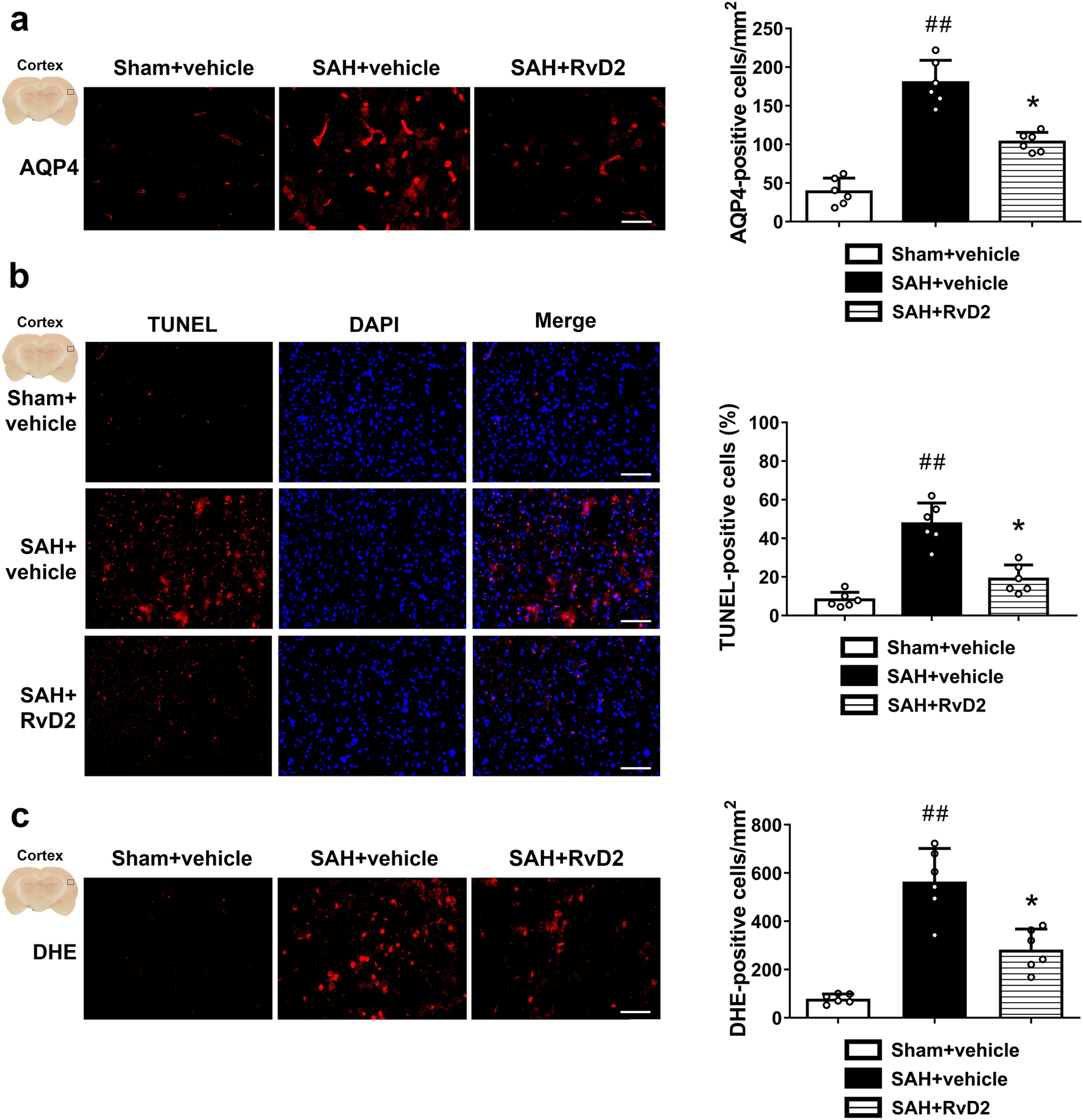


Figure 1 illustrates the study design. We meticulously examined the CDHR1 transcriptome using a publicly accessible dataset (GSE104687). This dataset included 55 participants with TBI who reported loss of consciousness and were meticulously matched with 55 control patients based on age, sex, and year of death. The participants provided brain samples from various regions, including the hippocampus, white matter of the forebrain, parietal neocortex, and temporal neocortex, totaling 376 samples. Our differential analysis of CDHR1 expression across different brain tissues revealed significantly elevated levels compared to those in the control group (white matter of the forebrain, p = 0.0359; hippocampus, p = 0.0072; parietal neocortex, p = 0.0072; and temporal neocortex, p = 0.0093, Fig. 2A–D).
Fig. 1 Fig. 2
Comprehensive analysis was conducted using the public database GSE104687, encompassing distinct brain regions and detection of pro-inflammatory and anti-inflammatory cytokines through enzyme-linked immunosorbent assay (ELISA): A white matter of forebrain, B hippocampus, C parietal neocortex, D temporal neocortex, E the left side illustrates correlation analyses between immune cells and CDHR1, employing xCell methodology, while the right side presents correlation analyses between inflammatory/metabolic pathways and CDHR1. Pathway enrichment was assessed through Gene Set Variation Analysis, as described in the “Methods” section. F In cortical tissue, pro-inflammatory factors (TNF-β1 and IL-10) and anti-inflammatory factors (IFN-γ and IL-6) were detected by ELISA, and G hippocampal tissue, pro-inflammatory factors (TNF-β1 and IL-10), and the expression levels of anti-inflammatory factors (IFN-γ and IL-6). The bar graph is presented as mean ± standard deviation (n = 3, number of animals); *p < 0.05 vs. sham group
Moreover, when examining the correlation between CDHR1 expression and pathways related to immune cells and inflammation, we observed a robust association between CDHR1 expression and endothelial cells. Notably, acute and chronic inflammatory processes have emerged as pivotal explanations for TBI pathogenesis of TBI, underscoring the significance of CDHR1 in these mechanisms (Fig. 2E).
We examined the levels of pro-inflammatory factors (TNF-β1 and IL-10) and anti-inflammatory factors (IFN-γ and IL-6). In cortical tissues, the concentrations of TNF-β1 (unpaired t-test, t = 13.30, df = 4, p = 0.0002) and IL-10 (unpaired t-test, t = 10.73, df = 4, p = 0.0004) were significantly higher in the TBI group compared to the sham group. For the anti-inflammatory factors, the concentrations of IFN-γ and IL-6 were both correspondingly decreased (sham vs. TBI, unpaired t-test, t = 11.88, df = 4, p = 0.0003; sham vs. TBI, unpaired t-test, t = 11.47, df = 4, p = 0.0003, Fig. 2F). In mouse hippocampal tissues, pro-inflammatory factors (TNF-β1, sham vs. TBI, unpaired t-test, t = 19.45, df = 4, p < 0.0001; IL-10, sham vs. TBI, unpaired t-test, t = 19.41, df = 4, p < 0.0001) and anti-inflammatory factors (IFN-γ, sham vs. TBI, unpaired t-test, t = 17.79, df = 4, p < 0.0001; IL-6, sham vs. TBI, unpaired t-test, t = 13.77, df = 4, p = 0.0002) showed similar performance (Fig. 2G). This suggests that TBI occurs throughout the course of inflammation.
Incorporating Associations Between Multi-omics Data from Blood and TBIBased on the aforementioned finding of differential CDHR1 expression in TBI, we hypothesized that CDHR1 expression could explain the causal presumption of the disease. Our objective was to delineate the gene regulation of CDHR1 in blood during TBI and explore the potential underlying epigenetic regulatory mechanisms. To achieve this, we employed a three-step SMR approach. Instances where HEIDI > 0.05 were considered to lack significant heterogeneity. This study integrated cis-eQTL and cis-mQL data for CDHR1 and amalgamated them with GWAS summary-level statistics from the largest TBI dataset (Fig. 3A).
Fig. 3
Association of CDHR1 with traumatic brain injury (TBI) in blood. A eMeta dataset. Solid diamonds represent probes that passed the HEIDI test, while hollow diamonds indicate probes that failed the HEIDI test. Probes highlighted in maroon passed the Summary-data-based Mendelian Randomization (SMR) threshold; B similar to the eMeta dataset, the LBC_BSGS dataset shows associations consistent with the eMeta dataset, and C single nucleotide polymorphisms (SNP) and SMR association results of methylation quantitative trait loci (mQTL), expressed quantitative trait loci (eQTL), and genome-wide association study (GWAS). The first graph illustrates the SNP-log10 (p-value) of TBI from GWAS. Red diamonds and blue diamonds depict − log10 (p-value) in the SMR test for the association of CDHR1 expression and methylation probes with TBI, respectively. Solid diamonds denote probes that passed the HEIDI test, whereas hollow diamonds represent probes that did not pass the HEIDI test. Yellow asterisks indicate the top SNP
Specifically, in the GWAS summary statistics analysis from BrainMeta (n = 1194) and TBI, we integrated the results of eQTL for CDHR1, focusing on probes ENSG00000148600 for CDHR1 (β-SMR = 0.0603821, p-SMR = 0.003934558, p-HEIDI = 0.7282966, and FDR = 0.00393). Similarly, we incorporated summary-level data of mQTL from the LBC-BSGS (n = 1980) to identify probes related to the epigenetic inheritance of CDHR1. Our analysis identified eight methylation sites, of which cg24196693, cg08892899, and cg03541835 were the most prominent (p-SMR < 0.05, p-HEIDI > 0.05). Following FDR correction, we selected cg03541835 as the prime CpG site candidate (β-SMR = 0.231634, p-SMR = 0.006528648, p-HEIDI = 0.2205731, and FDR = 0.052229184) despite an FDR exceeding 0.05 (Fig. 3B).
These results underscored the association between SNPs linked to CDHR1 in GWAS and eQTL as well as GWAS and mQTL of TBI (Fig. 3C). The expression level of CDHR1 (β-SMR = 0.0603821) exhibited a positive correlation with the onset of TBI, mirroring the analogous relationship observed in the level of methylation at the cg03541835 site (β-SMR = 0.0603821). In conclusion, our study elucidated a mechanism explaining the causal effect of CDHR1 in the presumptive occurrence of TBI in the blood, which is potentially influenced by both CDHR1 transcriptional expression and epigenetic modifications.
The second graph shows the − log10 (p-value) of the SNP association of the CDHR1 gene probe ENSG00000148600 from the eMeta dataset. The third graph presents the association of SNPs [− log10 (p-value)] with methylation probes from the LBC_BSGS dataset.
In the lower panel, 127 chromatin state annotations (in color) for 14 samples from the Roadmap Epigenomics Mapping Consortium (REMC) are shown for different primary cell and tissue types (rows). REMC = Roadmap Epigenomics Mapping Consortium, TSSA = active Transcription Start Site, Prom = promoter, and Tx. for active transcription; TxWk for weak transcription; TxEn for transcribed and regulatory promoters/enhancers; EnhA for active enhancers; and EnhW for weak enhancers. Other annotations include DNase (primary DNase), ZNF/Rpts (ZNF genes and repeats), Het (heterochromatin), PromP (poised promoter), PromBiv (bivalent promoter), ReprPC (repressed PolyComb), and Quies (quiescent/low).
Integration of GWAS and eQTL Data from Brain TissueGene expression patterns vary significantly between blood and tissue locations, with the genetic heterogeneity of TBI contributing to these differences. We hypothesized that understanding the interplay between eQTL expression in the brain tissue and TBI could help elucidate causal inferences related to TBI. To test this, we selected cis-eQTL data from the cortex, hippocampus, and frontal cortex of the GTEx Consortium. Subsequently, these datasets were subjected to SMR analysis within the cortex (p-SMR = 0.005003246, p-HEIDI = 0.5233138, and FDR = 0.005003246, Fig. 4A), frontal cortex (p-SMR = 0.00500549, p-HEIDI = 0.5401322, and FDR = 0.005005, Fig. 4B), and hippocampus (p-SMR = 0.007399831, p-HEIDI = 0.1626091, and FDR = 0.007399831, Fig. 4C).
Fig. 4
Association between CDHR1 and traumatic brain injury in different tissues. A Cortex, B frontal cortex, and C hippocampus. Solid diamonds indicate probes that passed the HEIDI test and hollow diamonds indicate probes that failed the HEIDI test. Labels highlighted in maroon indicate probes that passed the Summary-data-based Mendelian Randomization threshold
The SMR analysis yields compelling results. Notably, CDHR1 exhibited a significant absence of association within the cortex (β-SMR = 0.0812947), frontal cortex (β-SMR = 0.0895947), and hippocampus (β-SMR = 0.140338). However, the expression of CDHR1 in the frontal cortex, hippocampus, and cortex may play a causal role in TBI (Fig. 4).
Validation of CDHR1 in an In Vivo Model of TBIIn this study, we utilized a TBI model induced by gravity strikes and examined the cortical and hippocampal tissues. Through rigorous analyses using western blotting and immunohistochemistry, we elucidated the expression patterns of CDHR1. Our findings revealed a significant upregulation of CDHR1 protein expression in both the cortex (unpaired t-test, t = 15.25, df = 8, p < 0.0001) and the hippocampus (unpaired t-test, t = 8.485, df = 8, p < 0.0001) when compared with the sham group, demonstrating a consistent trend (Fig. 5A, B). This pattern was further corroborated by immunohistochemical analysis, underscoring the robustness of our results (cortex: TBI vs. sham, unpaired t-test, t = 8.929, df = 4, p = 0.0009; hippocampus: TBI vs. sham, unpaired t-test, t = 8.332, df = 4, p = 0.0011, Fig. 5C, D). We also examined the contralateral side and the expression of TBI of different severities. Thus, our data unequivocally established a direct association between CDHR1 expression and TBI.
Fig. 5
CDHR1 expression levels detected in an in vivo traumatic brain injury (TBI) model. A, B Comparative analysis of CDHR1 expression levels in distinct brain tissues (TBI vs. sham group), encompassing the cortex and hippocampus, and C, D Furthermore, fluorescence microscopy was employed to examine the fluorescence expression and quantification of CDHR1 in different brain tissues (TBI vs. sham group). The bar graph is presented as mean ± SD (n = 5, number of animals); *p < 0.05 vs. sham group
These results lead us to posit that heightened CDHR1 expression observed in the context of TBI may serve as a pivotal target for potential therapeutic interventions or diagnostic strategies.
留言 (0)