Cell culture
HDFa were cultured in FibroLife Basal Medium (LIFELINE Cell Technology cat.no LL-0011) supplemented with rh FGF basic (5 ng/mL), rh insulin (5 µg/mL), Ascorbic Acid (50 µg/mL), L-Glutamine (7.5 mM), hydrocortisone hemi-succinate (1 µg/mL), FBS 2%. Gentamicin (30 µg/mL) and Amphotericin B (15 ng/mL). All these supplements were provided with the FibroLife S2 LifeFactors Kit (LIFELINE Cell Technology cat.no LL-0011). The HDFa supplemented medium was changed every other day and the cells culture was incubated at 37 °C providing with 5% CO2.
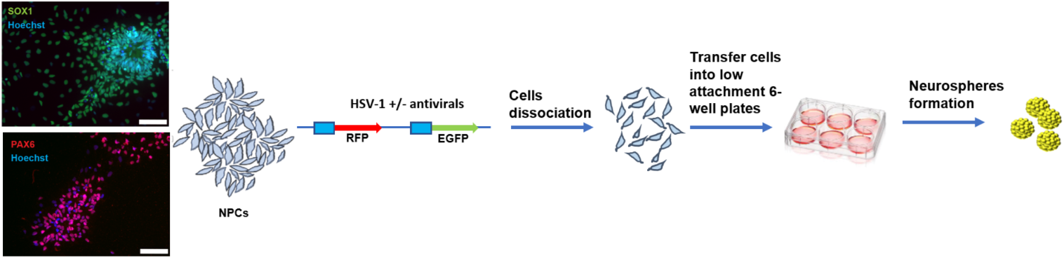
Generation of hiPSCs
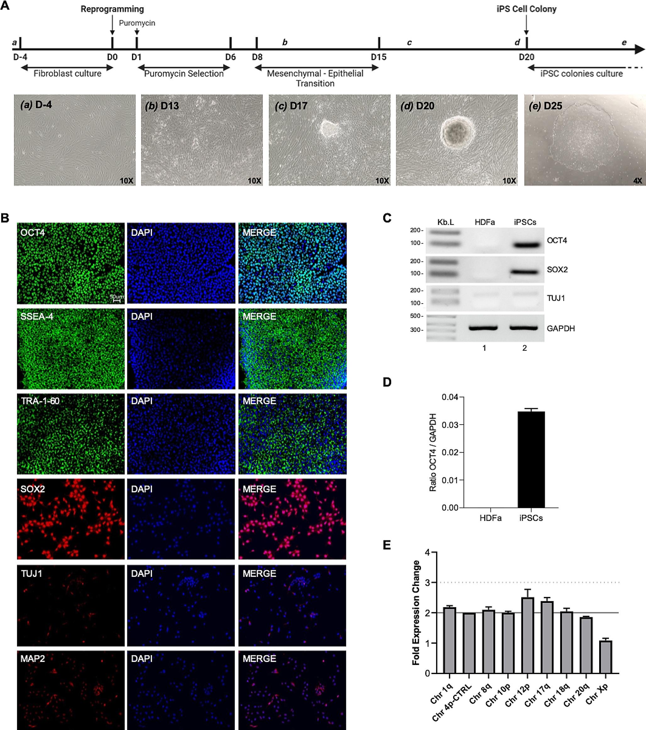
HDFa from different donors (LIFELINE Cell Technology cat.no FC-0001) were used to generate hiPSC. HDFa were reprogrammed to hiPSCs with ReproRNA Kit (Stem Cell Technologies cat.no 05930) following the manufacturer’s instructions. Briefly, hiPSC HDFa were plate in a 6-wells plate, previously coated with Matrigel® (Corning® cat.no 354,277), at the confluency of 100,000 HDFa per wells. The day after the HDFa were transfected with the ReproRNA™-OKSGM, a non-integrating, self-replicating RNA-based reprogramming vector which expresses Oct-3/4, Klf-4, Sox2, Glis1, c-Myc, and a puromycin-resistant cassette. The so transfected HDFa were cultured in a selective growth medium made of Advanced DMEM (Thermo Fisher cat.no 12,491,015) supplemented with 10% FBS, 200 mM L-Glutamine (STEMCELL Technology cat.no 07100), 0.5 mg/mL Recombinant B18R Protein at the final concentration of 175 ng/mL (STEMCELL Technology cat.no 78,075) and in presence of 1 mg/mL solution of puromycin (STEMCELL Technology cat.no 73,342) at the final concentration 0.8 µg/mL. The selective growth medium was changed every day for 1 week and then it was replaced by complete ReproTeSR™ medium (STEMCELL Technology cat.no 05926) without puromycin and supplemented with 0.5 mg/mL Recombinant B18R Protein at the final concentration of 175 ng/mL for an additional week. Two weeks after transfection, ReproTeSR™ medium without B18R was daily change until hiPSC colonies formed and were ready to be manually isolated and cut into small fragments with a 22-gauge needle. Then the colony fragments were scraped and aspirated using a 200 µL pipettor with a filtered tip. The hiPSC colony fragments were plated on culture-ware coated with Matrigel® and containing hiPSC maintenance mTeSR™ Plus complete medium (STEMCELL Technology cat.no 100–0276) supplemented with Y-27,632 (STEMCELL Technology cat.no 72,302) at a final concentration of 10 µM. After 24 h, the maintenance medium was replaced to remove the Y-27,632 and the hiPSC were incubated at 37 °C providing with 5% CO2. The hiPSC maintenance medium was changed every other day until the hiPSC colonies reach the confluency of 70%.
Generation of human cerebral organoids (hCOs)
Newly generated hiPSCs (Fig. 1) were used for creating hCOs by using STEMdiff™ Cerebral Organoid Kit (STEMCELL, 08570), and following manufacturer’s instructions with few modifications. Culture media were freshly prepared for each stage of embryoid body (EB) formation, induction, expansion, and maturation and used at room temperature (RT). On day 0, hiPSCs were detached using the Gentle Cell Dissociation Reagent (STEMCELL, 100–0485) at 37˚C and cells were resuspended in mTeSR™ Plus media (STEMCELL, 100–0276). After counting, cell mixture was centrifuged at 300 g for 5 min before EB seeding. 12,000 hiPSCs were used per EBs. The cell pellet was resuspended in EB seeding medium containing Y-27,632 (STEMCELL, 72,302) at a final concentration of 10 µM. 100 µL of cell suspension was seeded in each well of 96-Well Ultra-Low Attachment Round-Bottom plate (Millipore Sigma, CLS7007). The cells were kept in incubator at 37˚C with 5% CO2. On day 2 and day 4, 100 µL of EB Formation Medium was added per well. On day 6, each EB was transferred into a single well of 24-Well Ultra-Low Attachment plates (Millipore Sigma, CLS3473) containing Induction Medium. They were incubated at 37˚C under 5% CO2 condition for 48 h. On day 8, each EB was embedded in 15 µL of Matrigel® (CORNING, 354,277) on the organoid embedding sheets (STEMCELL, 08579). The droplets of embedded EBs were transferred into each well of 6-Well Ultra-Low Adherent plates containing Expansion Medium. A maximum of 16 EBs were transferred into each well. EBs were then incubated at 37˚C under 5% CO2 condition until the EBs exhibit multi-budding structures. On day 12, a full-mediachange took place with the Maturation Medium. EBs were then placed on an orbital shaker and incubated at 37˚C with 5% CO2. A half-media change was performed every 3 to 4 days during and after maturation with identical conditions.
HIV infection of hCOs and cART treatments
To characterize HIV-1 infection in cerebral organoids, 28 hCOs were incubated with 300 ng NL4.3 HIV-1 EGFP BaL reporter virus. First, hCOs were seeded in 96-well-plates and inoculated with 100 µl in OptiMEM containing diluted virus and Polybrene at a final concentration of 5 µg/ml. hCOs were subjected to spinoculation at 1200 g for 2 h at 32 °C, then placed in incubators at 37 °C overnight. The following day, inoculum was removed, hCOs were washed twice in PBS, plated in ultra-low attachment 24 well-plates, and incubated at 37 °C in complete hCO media on a continuous shaker. Fourteen of hCOs were left untreated while fourteen hCOs were treated with a cART regimen (Raltegravir-10 µg/ml, Tenofovir disoproxil- 2 µg/ml, Emtricitabine-2 µg/ml). cART treatments and half media changes were performed daily during the duration of the experiments. HIV-infected hCOs were collected at day 4 based on the cytopathic effects observed. HIV-infected and cART-treated hCOs were collected at day 7. Supernatants were collected daily, and Gag p24 ELISA was performed using supernatants at day 1, 4 and day 7. For gDNA analysis, four individual hCOs were used per each condition. For immunofluorescence, four individual hCOs were used per condition. For RNA analysis, six hCOs per condition were used. Fourteen hCOs were left uninfected and used as controls.
Immunocytochemistry (ICC)
250,000 hiPSC were plated in 2-well chamber slides previously coated with Matrigel®. The cells were washed with D-PBS (without Ca + + and Mg++) and fixed with cold 4% PFA in PBS solution for 10 min at RT, permeabilized with 0.25% Tx-100 in PBS for 5 min at RT and blocked with 5% Normal Donkey Serum (NDS) in 0.1% Bovine Serum Albumin in PBS (BSA/PBS). The primary antibody was diluted in 0.1% BSA/PBS individually and incubated at 4˚C overnight with gentle rocking. Primary antibodies used are listed in Table 1. The corresponding secondary antibodies of Alexa Fluor 488 (Donkey Anti-Mouse), Alexa Fluor 488 (Donkey Anti-Rabbit), Alexa Fluor 594 (Donkey Anti-Mouse), and Alexa Fluor 594 (Donkey Anti-Rabbit) were diluted in 0.1% BSA/PBS solution at dilution 1:400 and incubated for 2 h in the dark container at RT with gentle rocking. The chambers were disassembled from the slides before cover-slipping with DAPI mounting medium. The coverslips were secured with clear nail polish. The slides were imaged with a Keyence microscope after 30 min.
Table 1 List of primary antibodies usedRT-PCR analysis of pluripotency and differentiation markers
Total RNA was extracted from HDFa, iPSCs and hCOs using an RNA extraction kit (New England Biolabs) according to the manufacturer’s instructions. RT–PCR reactions were performed as previously described (Donadoni et al. 2019). Briefly, cDNAs were synthesized by using M-MLV Reverse Transcriptase (Invitrogen) followed by removal of RNA templates by RNase H digestion. A total of 50 ng cDNA was used as template for PCR reactions. Pluripotency markers OCT4 and SOX2, and differentiation markers, such as astrocytic structural marker, GFAP, neuronal differentiation markers, TUJ1 and MAP2, and oligodendroglia marker, Olig2, were amplified by PCR. GAPDH mRNA was amplified from the same set as internal controls. List of primers used is available in Table 2. Amplified products were resolved on 1% DNA agarose gels and visualized by ethidium bromide staining.
Table 2 List of primers and probes usedhiPSC genetic analysis
The hiPSCs genetic analysis was performed to detect karyotypic abnormalities using hPSC Genetic Analysis Kit (STEMCELL Technology), following manufacturer’s instruction. Briefly, 300 ng of gDNA was analyzed using a qPCR-based technology to calculate copies number of chromosomes 1q, 4p, 8q, 10p,12p,17q,18q, 20q and Xp, in a LightCycler 96 instrument (Roche, Indianapolis, IN, USA). Results obtained were analyzing using application provided by the company (www.stemcell.com/geneticanalysisapp).
Real-time RT-qPCR
RT-qPCR assays for HIV receptors CD4, CCR5 and CXCR were performed using RNA from hiPSCs from three different donors and eight different hCOs, with hCOs combined in two. RT-qPCR assays for characterization of HIV receptors following HIV infection were performed using RNA from uninfected and infected hCOs. RNA was extracted using an RNA extraction kit (New England Biolabs) according to the manufacturer’s instructions. RT-qPCR was performed using Luna® Universal One-Step RT-qPCR Kit (New England Biolabs) in a LightCycler 96 instrument (Roche, Indianapolis, IN, USA). The reaction mixtures contained: 1 × Luna Universal One-Step Reaction Mix, 1 × Luna WarmStart® RT Enzyme Mix, 0.4 µM of forward primer, 0.4 µM of reverse primer, 100 ng of template RNA, and nuclease-free water to a final volume of 20 µL. The protocol was reverse transcription at 55 °C for 10 min, initial denaturation at 95 °C for 60 s, followed by 45 cycles of denaturation at 95 °C for 10 s, and extensions at 60 °C for 30 s, with single acquisition. The amplification steps were then followed by melting steps: initial denaturation at 95 °C for 10 s, followed by 60 s at 65 °C, and temperature increase with continuous readings for 1 s to reach 97 °C. RT-qPCR assays for HIV infection characterization were performed on uninfected or infected hCOs, with or without cART treatments. RNA was extracted using an RNA extraction kit (New England Biolabs) according to the manufacturer’s instructions. RT-qPCR was performed using Luna® Universal Probe One-Step RT-qPCR Kit (New England Biolabs) in a LightCycler 96 instrument (Roche, Indianapolis, IN, USA). The reaction mixtures contained: 1 × Luna Universal Probe One-Step Reaction Mix, 1 × Luna WarmStart® RT Enzyme Mix, 0.4 µM of forward primer, 0.4 µM of reverse primer, 0.2 µM of probe, 25 ng of template RNA, and nuclease-free water to a final volume of 20 µL. The protocol was reverse transcription at 55 °C for 10 min, initial denaturation at 95 °C for 60 s, followed by 45 cycles of denaturation at 95 °C for 10 s, and extensions at 60 °C for 30 s, with single acquisition. The primers used in all the RT-qPCR assay are listed in Table 2.
NL4.3 HIV-1 EGFP BaL viral preparation
NL4.3 HIV-1 EGFP BaL (M-tropic HIV-1 strain) was prepared transfecting the pNL4.3-EGFP-BaL plasmid (containing the HIV-1 NL4.3 strain with the env of BaL strain and the gene encoding EGFP between env and nef without affecting expression of any HIV gene) in HEK 293 T cells. Briefly, 10ug of pNL4.3-EGFP-BaL plasmid were transfected in HEK 293 T cells at the confluency of 60% in 100 mm dish, using Lipofectamine™ 3000 Transfection Reagent (InvitrogenTM cat.no L3000015). After 24 h the media with DNA-lipid complex was replaced with DMEM 5% FBS. After 48 h and 72 h the NL4.3 HIV-1 EGFP BaL virus was collected and concentrated according to the pseudotyped HIV-1-based lentiviral vector (Kutner et al. 2009). Briefly, the collected cell supernatant was centrifuged at 500 g for 10 min at 25 °C to remove cells and large cell debris. Then the pooled supernatants were filtered using a 0.45 μm PES filter (Corning, cat.no. 430,768). Until 32mL aliquots of filtered lentivirus containing cell culture supernatant were transferred into each of the 6 Ultra-clear SW28 tubes (Beckman, cat.no 344,058) and 4 mL of 20% sucrose solution (20 g of UltraPure sucrose, 100mM NaCl, 20mM HEPES pH 7.4 and 1mM EDTA) was released all the way to the bottom of the SW28 tube filled with the filtered lentivirus-containing supernatant. The SW28 tubes were centrifuged for 2 h at 82,700 g and 4 °C using an ultracentrifuge. After centrifugation the supernatant was poured off and the pellet at the bottom of the tube was resuspended in 100 µL of PBS without Ca/Mg per tube. The tubes were incubated at 4 °C for 2 h on a shaking platform and then they were spined at 500 g for 1 min at 25 °C to collect the lentivirus-containing liquids. Finally, the lentivirus was aliquoted in screwcap cryo-vials in 30 µL portions, snap-frozen in crushed dry ice and stored at -80 °C.
HIV-1 gag p24 ELISA (enzyme-Linked immunosorbent assay)
NL4.3 HIV-1 EGFP BaL titer was measured by p24 Gag ELISA (Advanced BioScience Laboratories, Inc.), following instructions provided by the manufacturer. After HIV infection and ART treatment of hCOs, supernatants were collected, and levels of HIV-1 viral load were also quantified by p24 Gag ELISA. Proper dilutions of supernatant were made to be within the range of the assay.
Droplet Digital Polymerase Chain Reaction (ddPCR)
To analyze HIV-1 DNA in hCOs, ddPCR amplifying HIV-1 Ψ gene and human TERT as a reference gene was performed, using the QX200™ Droplet Digital™ PCR System (Bio-Rad, Hercules, CA, USA). The ddPCR reaction mixtures was prepared adding the following reagents: 1 × ddPCR™ Supermix for Probes (No dUTP) (Bio-Rad, Pleasanton, CA, USA), 500 nM of HIV-1 Ψ forward primer, 500 nM HIV-1 Ψ reverse primer, 500 nM of HIV-1 Ψ FAM probe, 500 nM of HsTERT forward primer, 500 nM HsTERT reverse primer, 500 nM of HsTERT HEX probe, 50 ng gDNA and water to a final volume of 22 µL. The ddPCR droplet and plate preparation were performed as previously described (Donadoni et al. 2019). The thermocycling protocol was previously described: initial denaturation at 95 °C for 10 min, then 45 cycles of denaturation at 94 °C for 30 s, annealing and extension at 59 °C for 1 min, followed by a final last incubation at 98 °C for 10 min and storage at 4 °C (Bruner et al. 2019). After amplification, positive and negative droplets of each sample were read and analyzed and graphed as HIV-1 Ψ copies per 1 million of cells. List of primers and probes use in Table 2.
Fixation and embedding of human cerebral organoids (hCOs)
Before immunoanalytical assays, hCOs were collected and washed three times with D-PBSfor 10 min. hCOs were fixed with 4% PFA overnight at 4˚C for 16 h, washed with 0.1% PBS-T, and kept in D-PBS for 1 to 7 days at 4˚C. The 30% Sucrose in D-PBS solution was used for dehydration for 1–2 days until the hCOs were submerged. hCOs were then embedded in Optimal Cutting Temperature Embedding Medium (OCT) to obtain 10 μm thick sections at cryostat and air-dried for 20 min at RT before staining or storing at -80˚C.
Immunofluorescence
The hCO sections were washed with 0.05% Tween-20 in PBS (PBS-T) to remove OCT, permeabilized for 15 min with 0.3% Tx-100 in PBS and blocked with 5% NDS for 1 h at RT in a humidifying chamber. The primary antibodies were diluted in the Dako Antibody Diluent (Agency for Science, Technology and Research, S3022) and incubated at 4˚C overnight in a humidifying chamber. Primary antibodies used are listed in Table 1. The secondary antibodies of Alexa Fluor 488 (Donkey Anti-Mouse), Alexa Fluor 488 (Donkey Anti-Rabbit), Alexa Fluor 594 (Donkey Anti-Mouse), and Alexa Fluor 594 (Donkey Anti-Rabbit) were diluted in the ratio of 1:1000 and incubated at RT for 1 h in a lightless humidifying chamber. The slides were cover slipped with DAPI mounting medium (Invitrogen, P36935). After 30 min of solidification at RT, the slides were imaged with a Keyence microscope.
RNAscope
The protocol of the RNAscope® 2.5 HD Detection Reagents-RED assay (ACD™, 322,360) was followed for the frozen sections of hCOs with modifications. The OCT reagent was washed off from cryosections with PBS-T before incubating at 60˚C for 30 min. The slides were fixed with cold 4% PFA in PBS for 15 min at 4˚C, dehydrated with 50%, 70%, 90%, and 100% Ethanol (EtOH), respectively. The sections were incubated with RNAscope® Hydrogen Peroxide for 10 min and rinsed with DI water. For antigen retrieval, the slides were kept in the RNAscope™ Target Retrieval Reagent (ACD, 322,000) for 5 min at 98–102˚C and equilibrated to RT before rinsing. A barrier was created around the sections with a ImmEdge® Pen (Vector Laboratories, H-400), then slides were stored at 4˚C overnight. The sections were blocked for 10 min with Boxall Blocking reagent at RT and treated with RNAscope® Protease Pluss (ACD, 322,330) for 30 min at 40˚C inside a humidifying chamber. The slides were washed with the RNAscope wash buffer between each treatment. Sections were hybridized with HIV probe (ACD, 444,061), Positive probe (ACD, 313,901), and Negative probe (ACD, 310,043) for 2 h at 40˚C in the humidifying chamber. The Amplification 1–6 solutions were applied at their specific conditions. The chromogen development was achieved with the RED B and RED A solution, which had a ratio of 1:60 and incubated for 10 min at RT in the dark. The sections were counter-stained with Hematoxylin QS and dehydrated with the indicated technique above. The slides were cover-slipped with permanent mounting medium (Vector, H-500-60).
Immunohistochemistry (IHC)
After RNAscope, the IHC was performed before counter-staining. The sections were blocked with Serum-Free Protein Block (Dako, X090930-2) reagent for 30 min at RT. The primary antibodies were incubated overnight at 4˚C, after diluting with Dako Antibody Diluent. The secondary antibody of Dako EnVision + System-HRP Labeled Polymer anti ‘host’ was applied for 30 min at RT in the dark. The DAB + Chromogen solution (Dako, K3468) was kept for 1–2 min and deactivated with DI water.
Statistical analysis
All values presented on the graphs are given a mean ± SEM. Analysis of variance and unpaired Student’s t-test were used to analyze the statistical significance. p-values of < 0.05 were considered statistically significant.


留言 (0)