
記住我

A 60-year-old woman was diagnosed with SLE 17 years ago based on clinical features, such as polyarthritis and fever, and serological studies. The patient had a low disease activity state by initial treatment. One year after discontinuation of immunosuppressive therapy because of personal reasons, she experienced a relapse; hence, a daily regimen of immunosuppressive therapy comprising 10 mg of prednisolone (PSL), 50 mg of azathioprine (AZA), and 1.5 mg of tacrolimus (TAC) was resumed. However, the low-grade fever persisted until admission to our hospital.
The patient exhibited various symptoms, including agraphia, amnesic aphasia, acalculia, left–right agnosia, ideational apraxia, hemispatial neglect, and homonymous hemianopsia. Blood tests showed a low white blood cell count (1.56 × 10³/μL) with lymphocytopenia (360/μL; CD4+ lymphocytes 193/μL, CD8+ lymphocytes 164/μL), hypocomplementemia, elevated anti-ds DNA antibodies (37.7 IU/mL), and vitamin B12 deficiency (125 pg/mL). Anemia or thrombocytopenia was not observed. Antibody tests for cytomegalovirus, hepatitis B viruses, human immunodeficiency virus, and intrinsic factor antibodies were negative. Tuberculosis-interferon-gamma release assay results were negative. Gastric endoscopy was not performed. Bone marrow cells showed mild hyperplasia with normal maturation and differentiation without dysplasia. A cerebrospinal fluid (CSF) examination showed no white blood cells or malignant cells and a protein level of 63 mg/dL. The real-time polymerase chain reaction showed 758 copies/mL of JC virus (JCV) DNA in the CSF.
MRI revealed high-intensity signals in the bilateral parieto-occipital lobe white matter on fluid-attenuated inversion recovery (FLAIR) images, as well as multiple punctate lesions in the bilateral frontal lobe white matter. Susceptibility-weighted imaging (SWI) showed hypointense signal rims in a confined lesion in the left occipital lobe cortex adjacent to the white matter lesion. No discernible enhancement was evident on T1-weighted images following gadolinium administration. Based on these diagnostic findings, the patient was diagnosed with PML secondary to SLE. Subsequently, AZA was discontinued after admission.
Despite a low SLE disease activity index (SLEDAI) score of 5, the disease activity may have smoldered (Cook et al. 2000). Therefore, immunosuppressive therapy was administered, including PSL, TAC, and hydroxychloroquine (HCQ), in addition to mirtazapine, risperidone, mefloquine, and intramuscular injections of vitamin B12. Since her lymphocyte counts improved one month after therapy resumed, PSL was subsequently reduced. Follow-up MRI examinations showed large hyperintense and punctate lesions exhibiting varying behaviors on FLAIR images. The hypointense signal rims on SWI became more prominent (Fig. 1). Although cognitive abnormalities persisted, only ideational apraxia showed improvement, and the patient’s condition was stable. She continued to live in her own house with caregiver support for > 13 months.
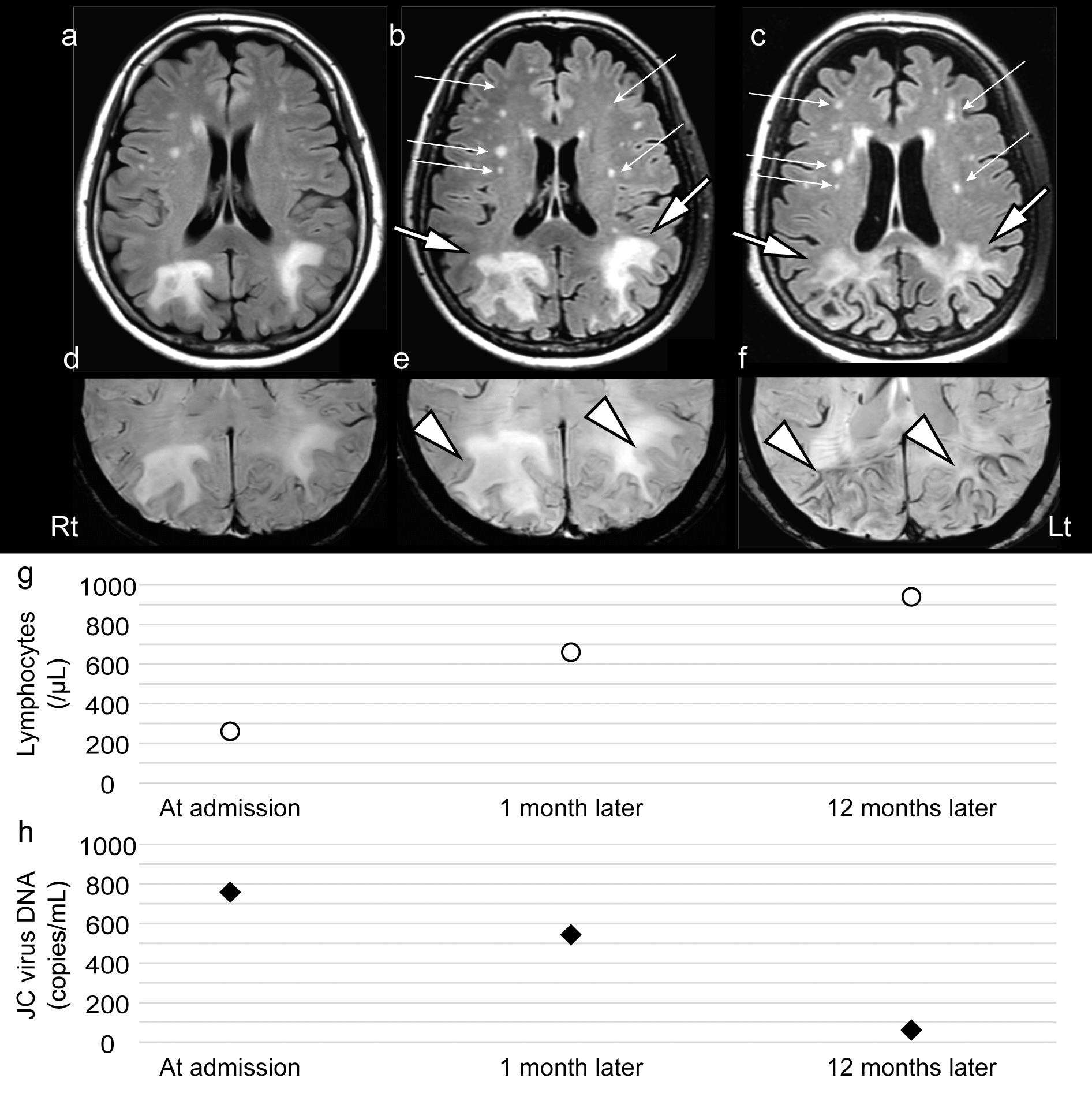
Fig. 1
MRI and JC virus DNA copy numbers in the CSF at each time point are shown. Axial FLAIR images a at admission, b one month later, and c 12 months later are shown. SWI findings d at admission, e one month later, and f 12 months later are shown. Lymphocyte number g and JC virus DNA copy number in the CSF h at each time point are summarized. Both large hyperintense and punctate lesions are enlarged on FLAIR images (one month later). After 12 months, the initially large hyperintense lesions on FLAIR images gradually shrank and became pale (large arrows). The punctate lesions on FLAIR images remained prominent (small arrows). SWI shows hypointense signal rims adjacent to white matter lesions in the bilateral parieto-occipital lobe cortex, becoming more prominent over time (arrowheads). The follow-up lymphocyte counts were 660 and 940/μL at one and 12 months. The JC virus DNA copy number in the CSF significantly decreased from 543 to 61 copies/mL after 12 months. Abbreviations: MRI, magnetic resonance imaging; CSF, cerebrospinal fluid; FLAIR, fluid-attenuated inversion recovery; SWI, susceptibility-weighted imaging
留言 (0)