
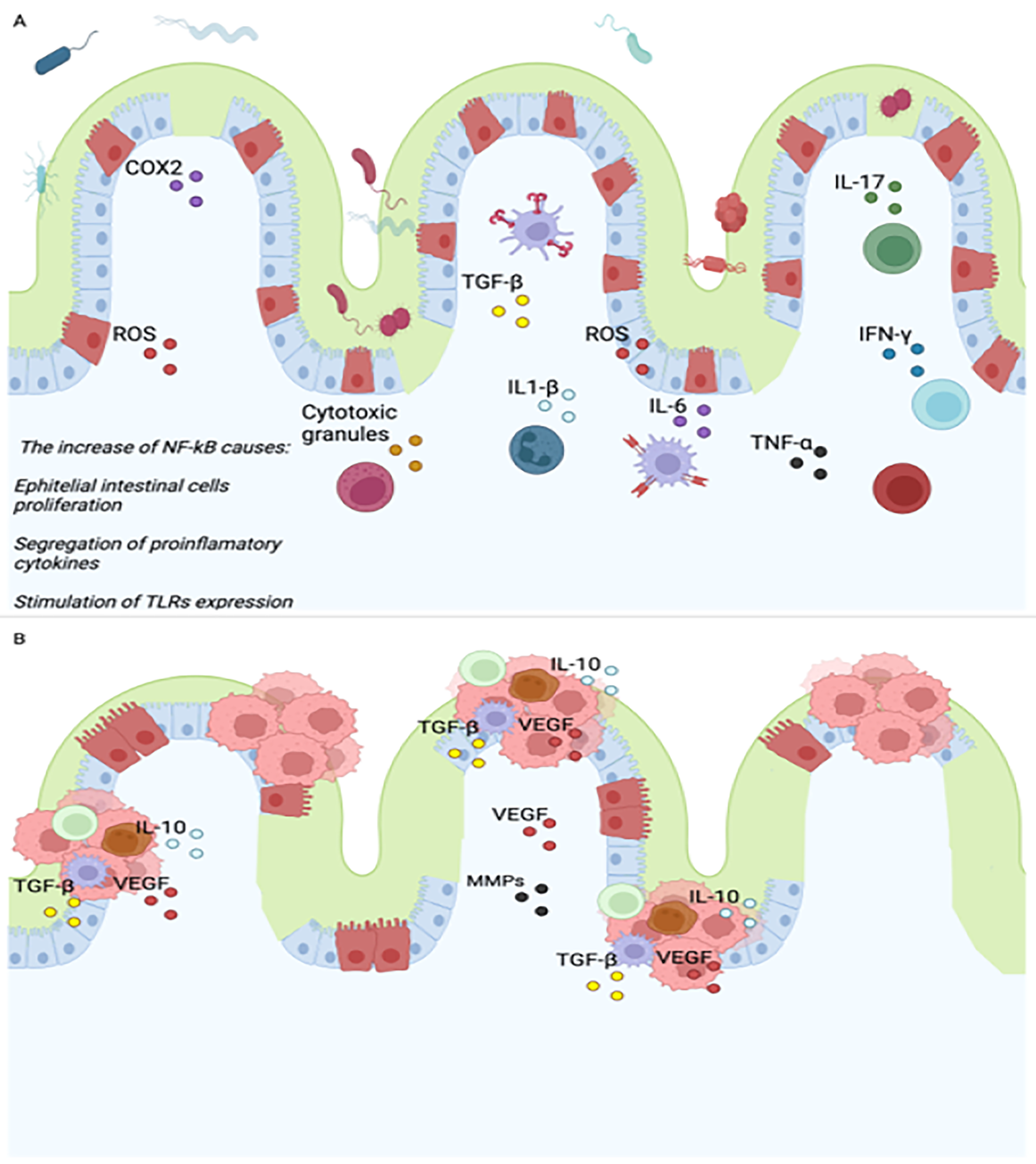
記住我


Since previous studies reported that Pappa2 deletion reduces IGF1 bioavailability and affects postnatal growth [4], here we first analyzed the body and bone length of Pappa2ko/ko mice (Fig. 1). A significant genotype effect on the body length at weaning (PND21) was found (F1,44 = 15.95, p < 0.001), with ko/ko males having an overall decrease in body length compared to wt/wt males (***p < 0.001; Fig. 1A). At puberty (PND40), the significant effect of genotype on body length remained (F1,44 = 56.01, p < 0.001), with ko/ko males and females being shorter than their respective wt/wt groups (***p < 0.001; Fig. 1B). We also observed that wt/wt and ko/ko females were shorter than same genotype males (##/###p < 0.01/0.001; sex effect: F1,44 = 36.95, p < 0.001; Fig. 1B). In adulthood (8 months of age), there was a significant interaction between genotype and sex on body length (F1,44 = 4.05, p < 0.05), suggesting that PAPP-A2 deficiency reduced body length differentially in male and female mice with Pappa2 deletion. In addition, effects of genotype (F1,44 = 90.47, p < 0.001) and sex (F1,41 = 3.99, p = 0.05) were found on body length in adulthood, with body length in ko/ko males and females being reduced compared to their respective wt/wt groups (***p < 0.001; Fig. 1C), and with wt/wt and ko/ko females being shorter than the respective male groups (#p < 0.05; Fig. 1C).
Fig. 1
Auxological parameters of body and bone (femur and tibia) length in Pappa2ko/ko and Pappa2wt/wt mice (males and females) at weaning (A), puberty (B) and adulthood (C-G). Data are represented as mean ± S.E.M (N = 8–10 per experimental group). Two-way ANOVA and Tukey-corrected tests: ***p < 0.001 versus respective wt/wt males and females; #/##/###p < 0.05/0.01/0.001 versus respective wt/wt and ko/ko males
In adulthood (8 months of age), the length of long bones (femurs and tibias) was also affected, with a significant interaction between genotype and sex found in femur length (F1,28 = 6.37, p < 0.05) and femur length relative to body length (F1,28 = 8.35, p < 0.01), suggesting that PAPP-A2 deficiency reduced femur length differentially in male and female mice with Pappa2 deletion. In addition, effects of genotype (F1,28 > 27.87, p < 0.001) and sex (F1,28 > 7.65, p < 0.01) were observed on femur length and femur length relative to body length (Fig. 1D, E), with ko/ko males and females having shorter femurs than their respective wt/wt groups (***p < 0.001), and with wt/wt females having shorter femurs than wt/wt males (##/###p < 0.01/0.001). No significant interaction was observed on tibia length. A significant effect of genotype was found on tibia length (F1,28 = 53.45, p < 0.001) and tibia length relative to body length (F1,28 = 43.52, p < 0.001), with ko/ko males and females having a shorter tibia length than their respective wt/wt groups (***p < 0.001; Fig. 1F, G).
In summary, we observed a sex-specific effect of Pappa2 deletion on body and femur length, with ko/ko females having a more prominent reduction in body length and ko/ko males a more prominent reduction in femur length.
Effects of Pappa2 deletion on circulating and/or pituitary concentrations of IGF1, IGFBP5 and GHSince body and bone length deficits could be consequent to hormonal alterations, we assessed plasma and/or pituitary concentrations of IGF1, IGFBP5 and GH in Pappa2ko/ko mice (Fig. 2). A significant interaction between genotype and sex was found in the circulating levels of IGF-1 (F1,28 = 17.20, p < 0.001) and IGFBP5 (F1,28 = 9.22, p < 0.01), suggesting that the plasma levels of both IGF-1 and IGFBP5 were differentially increased in male and female mice with Pappa2 deletion (Fig. 2A, B). A genotype effect was found on the circulating levels of IGF-1 (F1,28 = 29.14, p < 0.0001), with an overall increase in IGF-1 levels in ko/ko females compared to wt/wt females (***p < 0.001; Fig. 2A). We also found sex effects on the circulating levels of IGF1 (F1,28 = 25.85, p < 0.0001) and IGFBP5 (F1,28 = 11.65; p < 0.01), with the ko/ko female group having significantly more IGF1 and IGFBP5 that the ko/ko male group (###p < 0.001; Fig. 2A, B). No effects or interaction were found on the plasma levels of GH (Fig. 2C). Additional hormonal factors were also evaluated in plasma (Supplementary Figures S5A-F).
Fig. 2
Hormone concentrations in the plasma and pituitary gland of Pappa2ko/ko and Pappa2wt/wt mice (males and females) in adulthood. Concentrations of total IGF1 (A), IGFBP5 (B) and GH in plasma (C), and GH in pituitary gland (D) are represented as mean ± S.E.M (n = 8–10 per experimental group). Two-way ANOVA and Tukey-corrected tests: */***p < 0.05/0.001 versus respective wt/wt males and females; ###p < 0.001 versus respective wt/wt and ko/ko males
Since GH is secreted in a pulsatile fashion and a single plasma measurement does not necessarily reflect its total integrated levels, we also analyzed GH concentrations in Pappa2ko/ko mice at the pituitary level (Fig. 2D). An effect of genotype was found on pituitary GH content (F1,28 = 3.99, p < 0.05), which was most likely due to increased levels of GH in the pituitary gland of the ko/ko males and females (Fig. 2D). Additional hormonal factors were also evaluated in pituitary gland (Supplementary Figures S5G, H).
In summary, there was a sex-specific effect of Pappa2 deletion on total IGF1 and IGFBP5 concentrations in plasma, with a significant increase in ko/ko females. Pappa2 deficiency was also accompanied by increased pituitary GH concentrations in both sexes.
Effects of Pappa2 deletion on gene expression of the hypothalamic IGF1 systemWe analyze whether Pappa2 insufficiency affects gene expression of the main components of the IGF1 system in the hypothalamus (Fig. 3A-I). An effect of genotype was found on the mRNA levels of Pappa2 (F1,28 = 123.2, p < 0.001), with an overall decrease in Pappa2 mRNA in ko/ko males and females compared to their respective wt/wt groups (***p < 0.001; Fig. 3A). There was a sex effect on the mRNA levels of Ghrh (F1,28 = 6.649, p < 0.05), with females having lower Ghrh mRNA levels than males (Fig. 3B). No interaction or main effects were found in Ghih expression (Fig. 3C).
Fig. 3
IGF1 system gene expression in the hypothalamus, the pituitary gland and the liver of Pappa2ko/ko and Pappa2wt/wt mice (males and females) in adulthood. Hypothalamus: relative Pappa2 (A), Ghrh (B), Ghih-Sst (C), Igf1 (D), Igf1r (E), Igfbp3 (F), Igfbp5 (G), Igfals (H) and Stc1 (I) mRNA levels. Pituitary gland: relative Pappa2 (J), Gh (K), Ghrhr (L), Igf1 (M), Igf1r (N), Igfbp3 (O), Igfbp5 (P), Igfals (Q) and Stc1 (R) mRNA levels. Liver: relative Ghr (S), Igf1 (T), Igf1r (U), Igf2 (V), Igfbp3 (W), Igfbp5 (X), Igfals (Y) and Stc1 (Z) mRNA levels. Data are represented as mean ± S.E.M (n = 8–10 per experimental group). See Table S1 for additional information. Two-way ANOVA and Tukey-corrected tests: */***p < 0.05/0.001 versus respective wt/wt males and females; ##/###p < 0.01/0.001 versus respective wt/wt and ko/ko males
No interaction or main effects were found in Igf1 expression (Fig. 3D), but a sex effect was found on the mRNA levels of Igf1r (F1,28 = 7.547, p < 0.05), with females having lower Igf1r mRNA levels compared to males (Fig. 3E). No interaction or main effects were found in Igfbp3 expression (Fig. 3F). An effect of genotype (F1,28 = 5.681, p < 0.05) was found on the mRNA levels of Igfbp5, with ko/ko males having higher levels compared to wt/wt males (*p < 0.05; Fig. 3G). There was a sex effect on the mRNA levels of Igfals (F1,28 = 4.445, p < 0.05), with females having higher mRNA levels compared to males (Fig. 3H). No main effects or interaction between factors were found in the Stc1 mRNA levels (Fig. 3I).
Thus, there were sex specific effects of Pappa2 deletion on the hypothalamic IGF system with an increase in Igfbp5 and Igfals mRNA levels in ko/ko males and decreased Ghrh and Igf1r mRNA levels in females.
Effects of Pappa2 deletion on gene expression of the pituitary IGF1 systemWe also analyzed the gene expression of the main components of the IGF1 system in the pituitary gland of Pappa2ko/ko mice (Fig. 3J-R). A significant interaction between sex and genotype (F1,28 = 34.22; p < 0.001), and main effects of sex (F1,28 = 48.96; p < 0.001) and genotype (F1,28 = 219.0, p < 0.001) were found on the mRNA levels of Pappa2, with an overall decrease in ko/ko male and female mice compared to respective wt/wt mice (***p < 0.001; Fig. 3J), and lower levels of Pappa2 mRNA in wt/wt females compared to wt/wt males (###p < 0.001; Fig. 3J). An interaction between sex and genotype (F1,28 > 8.177, p < 0.01) and a sex effect (F1,28 > 31.88, p < 0.001) were found on the mRNA levels of both Gh and Ghrhr. Multiple comparison tests showed lower levels of Gh and Ghrh in wt/wt females than wt/wt males (###p < 0.001; Fig. 3K, L). We also found lower Ghrh levels in the pituitary gland of ko/ko males compared to wt/wt males (*p < 0.05; Fig. 3L).
A significant interaction between sex and genotype (F1,28 = 17.01; p < 0.001), and main effects of sex (F1,28 = 23.47; p < 0.001) and genotype (F1,28 = 23.75; p < 0.001) were found on the mRNA levels of Igf1. Multiple comparison tests indicated overall higher levels if Igf1 mRNA in ko/ko females compared to wt/wt females and ko/ko males (***/###p < 0.001; Fig. 3M). No interaction or effects between factors were found in the pituitary mRNA levels of Igf1r, Igfbp3, Igfbp5, Igfals or Stc1 (Fig. 3N-R).
In summary, we observed a sex-specific effect of Pappa2 deletion on pituitary Gh, Ghrhr and Igf1 mRNA levels, with lower Ghrhr expression in ko/ko males and higher Igf1 expression in ko/ko females.
Effects of Pappa2 deletion on gene expression of the liver IGF1 systemSince changes in PAPP-A2 activity can also affect the liver, we analyzed the gene expression of the main components of the IGF1 system in the liver of Pappa2ko/ko mice (Fig. 3S-Z). Main effects of sex (F1,28 = 8.932, p < 0.01) and genotype (F1,28 = 13.87, p < 0.01) were found on the mRNA levels of Ghr, with an overall increase in Ghr mRNA in ko/ko male and female groups compared to their respective wt/wt groups (*p < 0.05; Fig. 3S).
A significant interaction between sex and genotype was found in the mRNA levels of Igf1 (F1,23 = 9.216, p < 0.01), suggesting that the liver Igf1 mRNA levels were differentially altered in male and female mice with Pappa2 deletion (Fig. 3T). We also found a genotype effect on the mRNA levels of Igf1 (F1,28 = 16.38, p < 0.001). Multiple comparison analysis indicated that ko/ko females showed higher mRNA levels of Igf1 than wt/wt females (***p < 0.001) and ko/ko males (##p < 0.01; Fig. 3T). No interaction or effects were found on the mRNA levels of Ifg1r or Igf2 (Fig. 3U, V).
An interaction between sex and genotype (F1,28 = 5.518, p < 0.05), and an effect of genotype (F1,28 = 6.400, p < 0.05) on the mRNA levels of Igfbp3 were found, with ko/ko females having higher Igfbp3 expression compared to wt/wt females (*p < 0.05; Fig. 3W). An effect of sex was found on the mRNA levels of Igfbp5 (F1,28 = 6.59; p < 0.05), with females having higher expression (Fig. 3X). An effect of genotype was found on the mRNA levels of Igfals (F1,28 = 10.52; p < 0.01), with an increase in Igfals expression in ko/ko females compared to wt/wt females (*p < 0.05; Fig. 3Y). A genotype effect was also found in the mRNA level of Stc1 (F1,28 = 11.00, p < 0.01), with ko/ko males and females expressing significantly more Stc1 than their respective wt/wt groups (*p < 0.05; Fig. 3Z). Pappa2 mRNA was undetected in the liver of all experimental groups analyzed.
Summarizing, we found a sex-specific effect of Pappa2 deletion on liver Igf1 and Igfbp3 mRNA levels, with higher mRNA expressions in ko/ko females. Pappa2 deficiency was also accompanied by increased liver Ghr, Igfals and Stc1 mRNA levels in both sexes.
Effects of Pappa2 deletion on IGF1 signaling pathways in the hypothalamusTo further understand how PAPP-A2 deficiency affects hypothalamic regulatory systems, we analyzed IGF1 signaling pathways in this brain area (Fig. 4A-I). No interaction or effects were found on the phosphorylated form (Tyr607) of PI3K (Fig. 4A). A significant interaction between sex and genotype was found in the protein levels of AKT when it was phosphorylated (p) on serine 473 (F1,20 = 22.25, p < 0.001), mTOR phosphorylated on serine 2448 (F1,20 = 57.51, p < 0.001), GSK3β phosphorylated on serine 9 (F1,20 = 4.57; p < 0.05), ERK1 phosphorylated on threonine 202 (F1,20 = 15.54, p < 0.001), and ERK2 phosphorylated on tyrosine 204 (F1,20 = 4.79, p < 0.05), suggesting that the hypothalamic levels of p(Ser473)-AKT, p(Ser2448)-mTOR, p(Ser9)-GSK3β, p(Thr202)-ERK1 and p(Tyr204)-ERK2 were differentially altered in male and female mice with Pappa2 deletion (Fig. 4B-F). We also found genotype effects on the hypothalamic levels of p(Ser473)-AKT (F1,20 = 7.024; p < 0.05) and p(Thr202)-ERK1 (F1,20 = 5.29, p < 0.05). Effects of sex were found on the protein levels of p(Ser473)-AKT (F1,20 = 5.22, p < 0.05), p(Ser9)-GSK3β (F1,20 = 7.47, p < 0.05) and p(Thr202)-ERK1 (F1,20 = 9.029, p < 0.01). No interaction or effects were found on the protein levels of p(Thr172)-AMPKα or p(Ser641)-GS (Fig. 4G, H).
Fig. 4
Key sensors of IGF1 signaling pathways in the hypothalamus, the pituitary gland and the liver of Pappa2ko/ko and Pappa2wt/wt mice (males and females) in adulthood. Hypothalamus: phosphorylated forms of PI3K (A), AKT (B), mTOR (C), GSK3β (D), ERK1 (E), ERK2 (F), AMPKα (G) and GS (H). Pituitary gland: phosphorylated forms of GSK3β (J), AMPKα (K), ERK1 (L) and ERK2 (M). Liver: phosphorylated forms of PI3K (O), AKT (P), mTOR (Q), GSK3β (R), ERK1 (S), ERK2 (T), AMPKα (U) and GS (V). Data are represented as mean ± S.E.M (n = 6 per experimental group). Representative immunoblots (I, N and W) are also shown. See Table S2 and Figures S1-S3 for additional information. Two-way ANOVA and Tukey-corrected tests: */**/***p < 0.05/0.01/0.001 versus respective wt/wt males and females; #/##/###p < 0.05/0.01/0.001 versus respective wt/wt and ko/ko males
Multiple comparison analysis indicated that protein levels of p(Ser473)-AKT, p(Ser2448)-mTOR and p(Thr202)-ERK1 were higher in the hypothalamus of ko/ko females compared to wt/wt females (**/***p < 0.01/0.001) and ko/ko males (##/###p < 0.01/0.001; Fig. 4B, C, E). However, ko/ko males had lower p(Ser2448)-mTOR than wt/wt males (**p < 0.01; Fig. 4C). Regarding sex differences, ko/ko female had higher levels of p(Ser9)-GSK3β and p(Tyr204)-ERK2 than ko/ko males (#/##p < 0.05/0.01; Fig. 4D, F), whereas wt/wt females had lower p(Ser2448)-mTOR wt/wt males (##p < 0.01; Fig. 4C). Representative immunoblots are shown in Fig. 4I (see also the Supplementary Figure S1 for unedited blots).
These results indicate a sex-specific effect of Pappa2 deletion on the main intracellular regulators of IGF1 signaling pathways in the hypothalamus, with higher protein levels of p(Ser473)-AKT, p(Ser2448)-mTOR and p(Thr202)-ERK1 in ko/ko females.
Effects of Pappa2 deletion on IGF1 signaling pathways in the pituitary glandWe also analyzed IGF1 signaling pathways in the pituitary gland (Fig. 4J-N). An interaction between sex and genotype (F1,20 = 5.9; p < 0.05), and effects of sex (F1,20 = 11.08, p < 0.01) and genotype (F1,20 = 7.42, p < 0.05) were only found in the protein levels of p(Ser9)-GSK3β. Multiple comparison analysis indicated that protein levels of p(Ser9)-GSK3β were higher in ko/ko males compared to wt/wt males and ko/ko females (**/##p < 0.01; Fig. 4J). No interaction or effects were found on the protein levels of p(Thr172)-AMPKα, p(Thr202)-ERK1 or p(Tyr204)-ERK2 (Fig. 4K-M). Representative immunoblots are shown in Fig. 4N (see also the Supplementary Figure S2 for unedited blots).
Thus, there is a sex-specific effect of Pappa2 deletion on p(Ser9)-GSK3β in the pituitary gland, with higher protein levels in ko/ko males.
Effects of Pappa2 deletion on IGF1 signaling pathways in the liverThe IGF1 signaling pathways were also analyzed in the liver of Pappa2ko/ko mice (Fig. 4O-W). An interaction between sex and genotype was observed when protein levels of p(Tyr607)-PI3K were analyzed (F1,20 = 6.55, p < 0.05), suggesting that the protein levels of p(Tyr607)-PI3K were differentially altered in the liver of male and female mice with Pappa2 deletion (Fig. 4O). Effects of genotype were found on the protein levels of p(Tyr607)-PI3K (F1,20 = 10.93, p < 0.01) and p(Thr172)-AMPKα (F1,20 = 6.264, p < 0.05), with higher protein levels in the liver of ko/ko females compared to wt/wt females (*/**p < 0.05/0.01; Fig. 4O, U). Effects of sex were found on the protein levels of p(Ser2448)-mTOR (F1,20 = 4.19, p = 0.05) and p(Thr172)-AMPKα (F1,20 = 8.185, p < 0.05), with females having lower protein levels than males (#p < 0.05; Fig. 4Q, U). No interaction or effects on protein levels of p(Ser473)-AKT, p(Ser9)-GSK3β, p(Thr202)-ERK1, p(Tyr204)-ERK2 or p(Ser641)-GS were found (Fig. 4P, R, S, T, V). Representative immunoblots are shown in Fig. 4W (see also the Supplementary Figure S3 for unedited blots).
These results indicate a sex-specific effect of Pappa2 deletion on p(Tyr607)-PI3K in the liver, with higher protein levels in ko/ko females. Pappa2 deficiency was also accompanied by increased protein levels of p(Thr172)-AMPKα in ko/ko females.
Effects of rhGH, rhIGF1 and rhPAPP-A2 on body and bone lengthGiven that treatment with recombinant human IGF1 (rhIGF1) in patients with PAPP-A2 mutations improved growth velocity and height following recombinant human (rh)IGF1 treatment [5], here we first analyzed body and bone (femur and tibia) length in Pappa2wt/wt and Pappa2ko/ko male and female mice treated with rhGH, rhIGF1 and rhPAPP-A2 for 30 postnatal days (PND) from PND5 to PND35 (Fig. 5A). A significant interaction between treatment and genotype was found in body length, but not bone length, of mice treated with rhGH (F1,64 = 9.94, p < 0.01), suggesting that the effect of rhGH treatment on body length is genotype-dependent. However, interactions between treatment and sex were observed in body and femur lengths of mice treated with rhIGF1 and rhPAPP-A2 (body length: F1,64 = 6.69, p = 0.01, F1,64 = 6.03, p = 0.01, respectively; femur length: F1,64 = 5.72, p < 0.02, F1,64 = 9.28, p < 0.005, respectively), suggesting that the effect of rhIGF1 and rhPAPP-A2 treatments on body and femur length is sex-dependent.
ffect of rhGH treatment was only observed on body length (F1,64 = 11.32, p < 0.002), whereas effects of rhGH (F1,64 = 13.45, p < 0.001), rhIGF1 (F1,64 = 5.64, p = 0.02) and rhPAPP-A2 (F1,64 = 7.64, p < 0.01) were found on femur length. Analyzing the sexes separately, the rhPAPP-A2 effect on body length was specifically significant in females (F1,33 = 13.1, p < 0.001). Genotype and sex effects on body length were observed for all three treatments (genotype: F1,64 > 8.5, p < 0.005; sex: F1,64 > 20.03, p < 0.001), as well as the sex effects on bone length (F1,64 > 4.28, p < 0.05).
Multiple comparisons showed that rhGH treatment increased body and bone length in Pappa2wt/wt mice of both sexes, but not Pappa2ko/ko mice, compared to respective untreated mice (*/**/***p < 0.05/0.01/0.001; Fig. 5B-D). Moreover, body length of rhGH-treated Pappa2ko/ko mice of both sexes and femur length of rhGH-treated Pappa2ko/ko females were shorter than respective rhGH-treated Pappa2wt/wt mice (#/###p < 0.05/0.001; Fig. 5B, C). rhIGF1 and rhPAPP-A2 treatments specifically increased femur length in Pappa2wt/wt and Pappa2ko/ko females, but not males, compared to respective untreated mice (*/***p < 0.05/0.001; Fig. 5C), whereas rhPAPP-A2 also increased body length in Pappa2ko/ko females only, compared to untreated Pappa2ko/ko females (**p < 0.01; Fig. 5B).
Thus, these results indicate a female-specific effect of rhPAPP-A2 treatment on body and femur length.
Fig. 5
Experimental design (A) and auxological parameters of body (B), femur (C) and tibia (D) length in Pappa2ko/ko and Pappa2wt/wt mice (males and females) treated with rhGH, rhIGF1 and rhPAPP-A2 for 30 days, from PND5 to PND35. Data are represented as mean ± S.E.M (N = 8–10 per experimental group). Two-way ANOVA and Tukey-corrected tests: */**/***p < 0.05/0.01/0.001 versus respective saline-treated males and females; #/##/###p < 0.05/0.01/0.001 versus respective wt/wt males and females
Effects of rhGH, rhIGF1 and rhPAPP-A2 on circulating levels of IGF1 and IGFBP5Given that treatment with recombinant human IGF1 (rhIGF1) in patients with PAPP-A2 mutations increased bioactive IGF1 but serum levels of total IGF1 and ternary complexes remained elevated [5], we analyzed circulating concentrations of total IGF1 and IGFBP5 in Pappa2wt/wt and Pappa2ko/ko male and female mice treated with rhGH, rhIGF1 and rhPAPP-A2 for 30 postnatal days (PND) from PND5 to PND35 (Fig. 6A, B). Complete statistical analysis of the data is shown in the Supplementary Table S3. Interaction between treatment, genotype and sex was only observed in plasma levels of total IGF1 in mice treated with rhPAPP-A2 (F1,65 = 5.47, p < 0.05), suggesting that the effect of rhPAPP-A2 treatment on plasma total IGF1 levels is sex- and genotype-dependent. However, interactions between treatment and genotype were found in plasma levels of IGFBP5, but not IGF1, in mice treated with rhGH (F1,68 = 13.3, p = 0.01), rhIGF1 (F1,63 = 33.6, p < 0.001) and rhPAPP-A2 (F1,64 = 5.15, p < 0.05), suggesting a similar genotype-dependent effect of all three treatments on plasma IGFBP5 levels. Analyzing the sexes separately, this interaction between treatment and genotype in plasma levels of IGFBP5 was significant in females treated with rhGH (F1,31 = 10.1, p < 0.01) and rhIGF1 (F1,34 = 12.8, p < 0.01), and in males treated with rhIGF1 (F1,34 = 24.7, p < 0.001). Also, an interaction between treatment and genotype was specifically observed in plasma levels of total IGF1 of males treated with rhPAPP-A2 (F1,35 = 4.9, p < 0.05). Interactions between treatment and sex were only observed in plasma levels of total IGF1 after rhIGF1 (F1,65 = 6.45, p < 0.05) and rhPAPP-A2 (F1,65 = 17.8, p < 0.001) treatments. Interactions between genotype and sex were found in plasma levels of total IGF1 after rhGH (F1,67 = 26.6, p < 0.001), rhIGF1 (F1,65 = 25.0, p < 0.001) and rhPAPP-A2 (F1,65 = 15.0, p < 0.001) treatments (Supplementary Table S3).
Fig. 6
Experimental design (A) and schematic diagram (B) of the analysis of circulating hormone concentrations, and liver IGF1 system gene expression in Pappa2ko/ko and Pappa2wt/wt mice (males and females) treated with rhGH, rhIGF1 and rhPAPP-A2 for 30 days, from PND5 to PND35. Concentrations of total IGF1 (C) and IGFBP5 (D) in plasma, and relative Igf1 (E), Ghr (F), Igf1r (G), Igfbp3 (H), Igfbp5 (I), Igfals (J), Stc1 (K) and Stc1 (L) mRNA levels are represented as mean ± S.E.M (n = 8–10 per experimental group). See Table S1 for additional information. Two-way ANOVA and Tukey-corrected tests: */**/***p < 0.05/0.01/0.001 versus respective saline-treated males and females; #/##/###p < 0.05/0.01/0.001 versus respective wt/wt males and females
Effects of rhGH treatment were observed on plasma levels of total IGF1 (F1,67 = 12.1, p = 0.001) and IGFBP5 (F1,68 = 14.7, p < 0.001), whereas effects of rhIGF1 (F1,65 = 39.8, p < 0.001) and rhPAPP-A2 (F1,65 = 82.4, p < 0.001) were only found on plasma levels of total IGF1. Analyzing the sexes separately, the rhGH effects were significant on IGFBP5 in males (F1,37 = 6.83, p < 0.05), and total IGF1 (F1,30 = 13.8, p < 0.001) and IGFBP5 (F1,31 = 7.52, p = 0.01) in females, whereas the rhIGF1 and rhPAPP-A2 effects were only significant on total IGF1 in both males (F1,34 = 32.2, p < 0.001 and F1,34 = 86.2, p < 0.001, respectively) and females (F1,31 = 9.74, p < 0.01 and F1,34 = 12.9, p = 0.001, respectively). Genotype and sex effects on plasma levels of both total IGF1 and IGFBP5 were observed for all three treatments (F1,63-68 > 4.89, p < 0.05 and F1,62-68 > 24.8, p < 0.001, respectively) (Supplementary Table S3).
Multiple comparisons showed that rhGH treatment increased plasma levels of total IGF1 in Pappa2wt/wt and Pappa2ko/ko females, but not males, compared to respective untreated mice (*p < 0.05; Fig. 6C). In contrast, rhIGF1 treatment decreased plasma levels of total IGF1 in Pappa2wt/wt and Pappa2ko/ko males and females compared to respective untreated mice (**/***p < 0.01/0.001; Fig. 6C). rhPAPP-A2 treatment also decreased plasma levels of total IGF1 in Pappa2wt/wt and Pappa2ko/ko males, and Pappa2ko/ko females compared to respective untreated mice (*/***p < 0.05/0.001; Fig. 6C). rhGH treatment increased plasma levels of IGFBP5 specifically in Pappa2ko/ko males and females compared to respective untreated mice (*/**p < 0.05/0.01; Fig. 6D). rhIGF1 treatment decreased plasma levels of IGFBP5 in Pappa2wt/wt males and females, and increased plasma levels of IGFBP5 in Pappa2ko/ko males only, compared to respective untreated mice (*/***p < 0.05/0.001; Fig. 6D). Interestingly, rhPAPP-A2 treatment decreased plasma levels of IGFBP5 in Pappa2wt/wt females, and increased plasma levels of IGFBP5 in Pappa2ko/ko females, but not males, compared to respective untreated females (*p < 0.05; Fig. 6D). Regarding genotype, lower plasma total IGF1 levels were observed in both untreated (saline) and treated Pappa2ko/ko males, but not females, compared to respective Pappa2wt/wt mice (#/##/###p < 0.05/0.01/0.001; Fig. 6C). In contrast, higher plasma IGFBP5 levels were observed in both untreated and treated Pappa2ko/ko males, and in rhIGF1- and rmPAPPA2-treated Pappa2ko/ko females compared to respective Pappa2wt/wt mice (#/###p < 0.05/0.001; Fig. 6D).
In summary, there is an effect of rhIGF1 and rhPAPP-A2 treatments on circulating total IGF1, resulting in lower plasma levels of total IGF1 in males and females.
Effects of rhGH, rhIGF1 and rhPAPP-A2 on liver gene expression of IGF1 system componentsSince changes in IGF1 bioactivity can be modulated by liver production of IGF1 ternary complexes in response to treatment, we also analyzed liver gene expression of IGF1 system components in Pappa2wt/wt and Pappa2ko/ko male and female mice treated with rhGH, rhIGF1 and rhPAPP-A2 for 30 postnatal days (PND) from PND5 to PND35 (Fig. 6A, B). Complete statistical analysis of the data is shown in the Supplementary Tables S4 and S5. Interactions between treatment, genotype and sex were only observed in Stc1 mRNA expression levels after rhGH treatment (F1,60 = 6.39, p < 0.05), and in Ghr and Stc1 mRNA expression levels after treatment with rhIGF1 (F1,63 = 6.5, p < 0.05 and F1,58 = 9.7, p < 0.01, respectively) and rhPAPP-A2 (F1,59 = 11.9, p = 0.001 and F1,57 = 7.75, p < 0.01, respectively), suggesting that the effect of rhIGF1 and rhPAPP-A2 treatments on liver gene expression of Ghr and Stc1 are sex- and genotype-dependent. In addition, interactions between treatment and genotype were observed in Ghr mRNA expression levels in mice treated with all three treatments (rhGH: F1,62 = 7.12, p = 0.01; rhIGF1: F1,63 = 6.98, p = 0.01; rhPAPP-A2: F1,59 = 20.6, p < 0.001), suggesting a similar genotype-dependent effect of all three treatments on Ghr gene expression. Analyzing the sexes separately, this interaction between treatment and genotype in Ghr mRNA expression levels was observed in males, but not females, treated with all three treatments (rhGH: F1,30 = 12.1, p < 0.01; rhIGF1: F1,31 = 10.7, p < 0.01; rhPAPP-A2: F1,29 = 27.2, p < 0.001), whereas interaction between factors in Stc1 mRNA expression levels was only found in males treated with rhIGF1 and rhPAPP-A2 (F1,27 = 14.5, p = 0.001 and F1,28 = 16.4, p < 0.001, respectively). Interestingly, interactions between treatment and genotype were also found in Igf1r and Igfals mRNA expression levels after rhPAPP-A2 treatment only, being specifically significant in females (F1,29 = 5.53, p < 0.05 and F1,29 = 7.7, p = 0.01, respectively). Interactions between treatment and sex were observed in Ghr, Igf1r and Stc1 mRNA expression levels after all three treatments (rhGH: F1,60-62 > 7.94, p < 0.01; rhIGF1: F1,58-63 > 9.22, p < 0.005; rhPAPP-A2: F1,57-59 > 6.61, p < 0.05). In addition, interactions between treatment and sex were also observed in Igfbp5 mRNA expression levels after rhGH and rhIGF1 treatments (F1,60-61 > 8.7, p < 0.005), and in Stc2 mRNA expression levels after rhGH and rhPAPP-A2 treatments (F1,57-58 > 5.29, p < 0.05). Interactions between genotype and sex were also found in Ghr and Igfals mRNA expression levels in mice treated with rhGH (F1,62 > 4.53, p < 0.05), and in Igfals and Igfbp5 mRNA expression levels in mice treated with rhIGF1 (F1,61-62 > 6.76, p < 0.05) and rhPAPP-A2 (F1,58-60 > 4.65, p < 0.05). Further statistical data can be found in the Supplementary Tables S4 and S5.
There were effects of all three treatments on Igf1 mRNA expression levels (rhGH: F1,62 = 4.26, p < 0.05; rhIGF1: F1,63 = 10.2, p < 0.01; rhPAPP-A2: F1,60 = 34.7, p < 0.001), effects of rhGH and rhIGF1 treatments on Igfals and Igfbp5 mRNA expression levels (rhGH: F1,60-62 > 24.6, p < 0.001; rhIGF1: F1,61-63 > 8.52, p < 0.005), and effects of rhIGF1 and rhPAPP-A2 treatments on Stc2 mRNA expression levels (rhIGF1: F1,57 = 15.4, p < 0.001; rhPAPP-A2: F1,57 = 6.68, p < 0.05). Also, an effect of rhGH treatment on Igf1r mRNA expression levels (F1,62 = 5.46, p < 0.05) and an effect of rhIGF1 treatment on Igfbp3 mRNA expression levels (F1,59 = 6.66, p < 0.05) were specifically observed. Analyzing the sexes separately, the rhGH treatment effects on Ghr and Igfbp5 mRNA expression levels were specifically observed in females (F1,31 > 14.0, p < 0.001), whereas those on Stc1 and Stc2 mRNA expression levels were specifically observed in males (F1,27 > 5.57, p < 0.05). The rhIGF1 treatment effects on Igf1, Igfals, Igfbp3 and Stc2 mRNA expression levels were specifically observed in males (F1,27-31 > 5.18, p < 0.05), whereas those on Igf1r, Igfbp5 and Stc1 mRNA expression levels were specifically observed in females (F1,31 > 8.18, p < 0.008). The rhPAPP-A2 treatment effect on Stc2 mRNA expression levels was specifically observed in males (F1,28 = 13.0, p = 0.001), whereas that on Stc1 mRNA expression levels was specifically observed in females (F1,29 = 5.45, p < 0.05). Genotype effects were observed on Stc1 mRNA expression levels for all three treatments (F1,57-60 > 10.3, p < 0.002), Igfals mRNA expression levels for rhGH and rhIGF1 treatments (F1,62-63 > 4.51, p < 0.05), and Igfbp3 mRNA expression levels for rhIGF1 treatment (F1,59 > 5.24, p < 0.05). Finally, sex effects on Ghr, Igf1r, Igfbp3 and Igfbp5 mRNA expression levels were observed for all three treatments (rhGH: F1,60-62 > 18.9, p < 0.001; rhIGF1: F1,59-62 > 10.5, p < 0.002 rhPAPP-A2: F1,58-59 > 6.05, p < 0.05), whereas sex effects on Igfals mRNA expression levels were specifically found for rhIGF1 (F1,63 = 13.7, p < 0.001) and rhPAPP-A2 (F1,60 = 13.8, p < 0.001) treatments. See the Supplementary Tables S4 and S5 for further statistical data.
Multiple comparisons showed that rhIGF1 treatment increased Igf1 mRNA expression levels only in Pappa2wt/wt males compared to respective untreated males (***p < 0.001; Fig. 6E). In addition, rhPAPP-A2 treatment increased Igf1 mRNA expression levels in Pappa2wt/wt and Pappa2ko/ko males and females compared to respective untreated mice (*/**/***p < 0.05/0.01/0.001; Fig. 6E). All three treatments increased Ghr mRNA expression levels in Pappa2wt/wt males, but not Pappa2ko/ko males, and decreased Ghr mRNA expression levels in Pappa2ko/ko females, but not Pappa2wt/wt females, compared to respective untreated mice (**/***p < 0.01/0.001; Fig. 6F). All three treatments also increased Igf1r mRNA expression levels in Pappa2wt/wt females (**p < 0.01; Fig. 6G). In addition, rhGH treatment, but not rhIGF1 and rhPAPP-A2, specifically increased Igf1r mRNA expression levels in Pappa2ko/ko females compared to untreated females (**p < 0.01; Fig. 6G). In contrast, rhPAPP-A2 treatment, but not rhGH and rhIGF1, specifically decreased Igf1r mRNA expression levels in Pappa2ko/ko males (*p < 0.05; Fig. 6G). rhIGF1 treatment increased Igfbp3 mRNA expression levels in Pappa2wt/wt and Pappa2ko/ko males, whereas rhPAPP-A2 treatment decreased Igfbp3 mRNA expression levels in Pappa2ko/ko females only (*/**p < 0.05/0.01; Fig. 6H). rhGH and rhIGF1 treatments increased Igfbp5 mRNA expression levels in Pappa2wt/wt and Pappa2ko/ko females, but not males, compared to respective untreated mice (*/**p < 0.05/0.01; Fig. 6I). r
留言 (0)