
記住我
Combination antiretroviral therapy (cART) reduces the replication of HIV-1, but HIV-1 proviruses survive in the latent reservoir [1]. The latent reservoir is a population of immune cells that contain an integrated HIV-1 provirus that does not undergo viral replication and as such hides from the immune system. However, the latent reservoir remains replication competent, and therefore, treatment interruption results in viral rebound, resulting in the necessity of life-long therapy adherence to cART. Drug fatigue, side effects, high treatment costs, the emergence of resistant strains and stigma are still associated with HIV-1 infection [2]. In addition, people living with HIV (PLWH) suffer from early aging due to chronic inflammation [3]. This emphasizes the importance of continued efforts to cure HIV-1 infection.
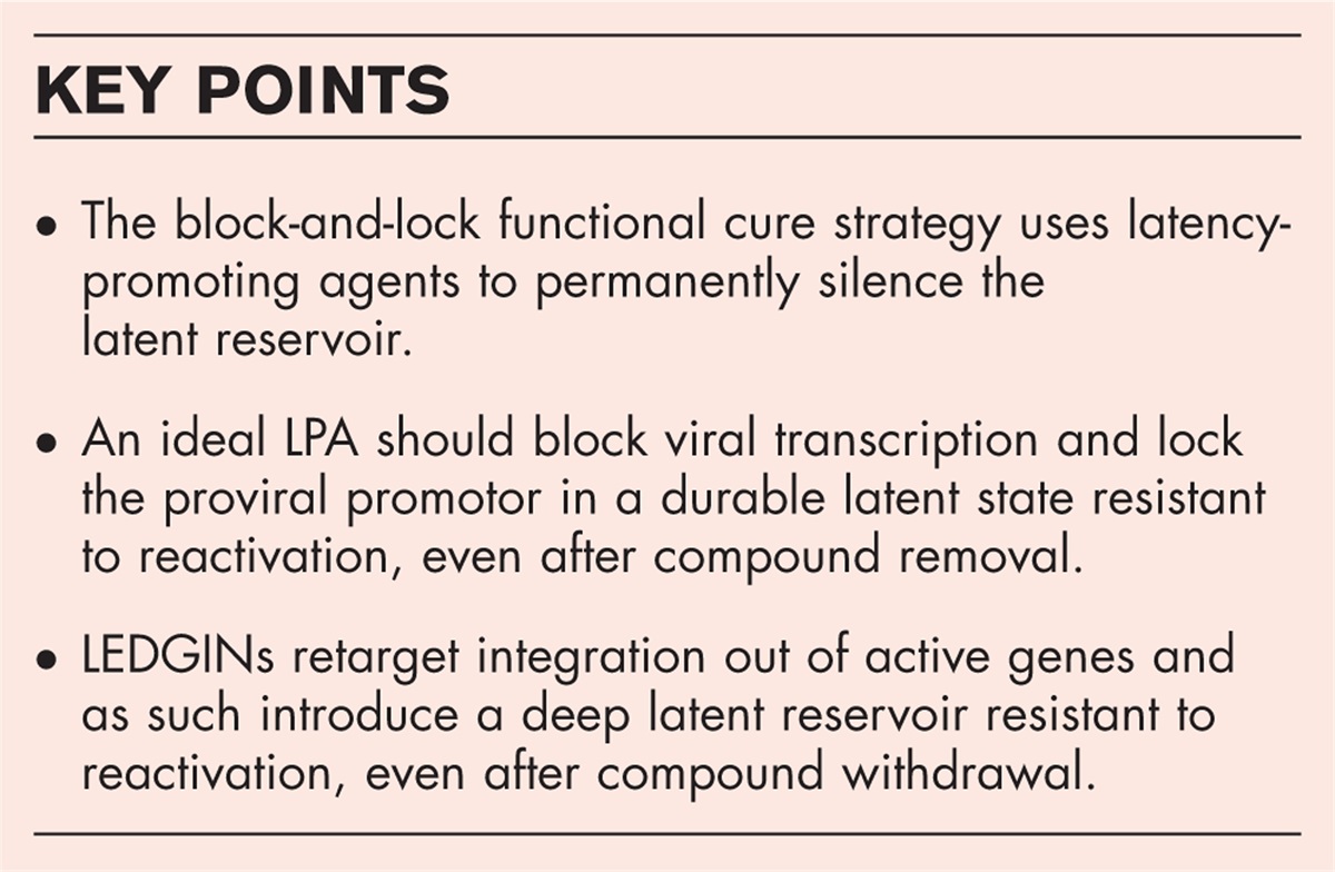
To cure HIV-1 infection, the latent reservoir needs to be eradicated or silenced permanently. Several strategies are under investigation to eradicate the latent reservoir, such as chimeric antigen receptor T-cell (CAR-T) therapies [4] and gene editing strategies [5–8]. However, the shock-and-kill strategy is probably the most investigated eradicational cure approach [9]. This strategy uses latency-reactivating agents (LRAs) to reactivate latently infected cells and as such kill these cells by cytopathic effects of the virus itself or by immune-mediated clearance [9]. Limited success in ex-vivo studies and clinical trials [10] shifted the interest towards a functional cure based on permanent silencing of the latent reservoir [11–14]. A functional cure may be more feasible than eradication, as evolutionary evidence shows that about 8% of the human genome consists of sequences derived from endogenous retroviruses that have been transcriptionally silenced [15]. Secondly, some HIV-infected patients show virological control in the absence of treatment, referred to as elite controllers and posttreatment controllers (PTCs) [16]. The functional cure for HIV-1 infection is called the block-and-lock approach. The block-and-lock approach uses latency-promoting agents (LPAs) to “block” HIV-1 transcription and “lock” the proviral promotor in a durable deep latent state, even after compound withdrawal [11–14]. To induce this permanent lock phenotype, LPAs must induce epigenetic modifications that silence HIV-1 gene expression. Various LPAs have been proposed [17]. One of the most well known LPAs is the Tat inhibitor didehyro-cortistatin A (dCA). This compound was shown to prevent reactivation in CD4+T cells isolated from HIV-1-infected patients for 25 days after compound removal in vitro and for 19 days after treatment interruption in vivo in BLT (bone marrow-liver-thymus) humanized mice [18–27]. However, until now, none of the LPAs under investigation have resulted in a permanent deep latent state. This indicates that persistent research is required to discover new drug targets and identify new LPAs. In this review, we will provide a recent update on the block-and-lock cure approaches.
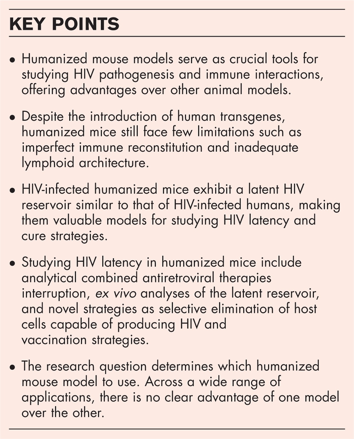
 Box 1:
 no caption available
LATENCY-PROMOTING AGENTSAll LPAs that have been discovered during the last 2 years (2022–2023) will be discussed below.
LEDGIN GS-9822The epigenetic reader lens-epithelium derived growth factor (LEDGF/p75) consists of an integrase binding domain (IBD), binding the HIV-1 integrase and a PWWP domain, interacting with the methylated histone mark H3K36me2/3, which is associated with active transcription [28]. This interaction allows LEDGF/p75 to guide HIV-1 integration into transcriptionally active genes. The small molecules referred to as LEDGINs, designed via structure-based drug design [29], bind the LEDGF/p75-binding pocket of the viral integrase, inhibiting viral integration [14,17,29]. It has been shown that next to their antiviral effect, LEDGINs also contain latency promoting effects by altering the integration preferences of HIV-1 [30▪,31,32▪]. Vansant et al.[30▪] further characterized the potential of LEDGINs for a functional cure by using the barcoded HIV-ensembles (B-HIVE) technology, which tags the HIV genome with a unique barcode to trace insert-specific HIV expression. The authors showed that after addition of the LEDGIN CX014442, integration occurred out of active chromatin regions and more towards intergenic regions and genes that are less transcriptionally active [30▪,31,32▪]. Next, Janssens et al.[32▪] optimized single-cell branched DNA imaging, a fluorescent in-situ hybridization technique that allows the simultaneous detection of viral DNA and RNA, to further characterize the latency promoting effect of LEDGINs. By using this technique, the authors confirmed that LEDGINs reduce integration, transcription and reactivation, both in T cell lines and primary cells infected in vitro[32▪]. Additionally, changes in the three-dimensional localization of the provirus within the nucleus were observed, as integration was shifted towards the core of the nucleus rather than the periphery [30▪,31,32▪]. Gilead developed a preclinical LEDGIN candidate, GS-9822. This compound showed favorable characteristics such as high metabolic stability, high barrier to emergence of resistance, and desirable pharmacokinetic properties for the development of clinical LEDGIN candidates [33▪▪]. In 2023, Bruggemans et al.[33▪▪] did a side-by-side comparison of the two LEDGINs GS-9822 and CX014442. Both GS-9822 and CX014442 hampered HIV-1 replication in multiple round assays [33▪▪]. Both LEDGINs also abrogated the interaction between LEDGF/p75 and the HIV-1 integrase [33▪▪]. Integration site sequencing revealed that both LEDGINs retargeted integration towards repressive chromatin regions. Finally, using a double reporter system both LEDGINs were shown to increase the fraction of latent vs. productive provirus and to hamper viral reactivation [33▪▪]. GS-9822 was significantly more potent than CX104442 at nanomolar concentrations in all assays. This nanomolar activity of GS-9822 supports the high potential for a functional cure strategy based on this class of compounds [33▪▪]. Nevertheless, clinical development of GS-9822 was hindered due to urothelial toxicity in cynomolgus monkeys [33▪▪]. Although proof-of-principle for LEDGIN-mediated retargeting and a functional block-and-lock cure has been obtained, additional lead optimization is necessary to increase the potency of future LEDGIN candidates.
A consensus has been reached that the transcriptional state of the HIV provirus is influenced by the integration site and its chromatin environment. Therefore, LEDGINs are appealing as LPAs. The use of LEDGINs may extend beyond preexposure prophylaxis, as they could be added to drug regimens during acute infection. Early treatment with ART is known to reduce the size of the latent reservoir [34]. The addition of LEDGINs to this early treatment regime could potentially transform the residual reservoir in a deep latent state, increasing the chance of a durable silencing after treatment discontinuation. The effects of LEDGIN on HIV-1 integration sites in patients with chronic HIV-1 infection are yet to be determined. However, in 2019, Swanström's group showed that replication-competent reservoirs primarily arise at the beginning of treatment [35,36]. LEDGINs may thus also serve as LPAs in chronically infected patients diagnosed years after infection, if included in first-line treatment. In chronically infected patients already treated with ART, the extent to which latent reservoirs are activated and modulated after treatment discontinuation remains to be investigated. LEDGINs will only work in this patient category if the reservoir is dynamic. Next research steps include quantitative viral outgrowth assays (QVOA) using samples from HIV-1-infected patients treated with ART. In this setting LEDGIN treatment could hamper reactivation of HIV-1. Additionally, LEDGINs can be tested in vivo in mouse models or macaque monkeys infected with SIV. Primate studies require the availability of a LEDGIN that also targets HIV-2/SIV. Finally, we recommend that during clinical trials with LEDGINs to test antiviral potency, samples are collected for proviral load determination, integration site sequencing and QVOA to further characterize how LEDGINs silence the latent reservoir in patients.
Ponatinib (AP2453)A drug screen of 1701 FDA-approved compounds for their inhibitory effect on PMA-induced reactivation in J-Lat cells, resulted in the discovery of ponatinib (AP2453). Ponatinib is a tyrosine kinase inhibitor, used for the treatment of chronic myeloid leukemia (CML) and acute lymphoid leukemia (ALL) [37–39]. Huang et al.[40▪▪] confirmed the ability of ponatinib to reduce reactivation in J-Lat cells induced by several LRAs with distinct mechanisms of action. Moreover, the authors tested ponatinib's potential as an LPA in multiple J-Lat cell lines with diverse viral integration sites, indicating that the latency promoting activity is independent of the integration pattern. In addition, Bcl-2-transduced primary CD4+T cells and CD4+T cells collected from ART-suppressed HIV-1 infected patients were used to confirm the inhibitory effect of ponatinib on HIV-1 transcription [40▪▪]. An important and challenging aspect of the block-and-lock approach is the ability of LPAs to maintain deep latency after compound withdrawal. Therefore, Huang et al.[40▪▪] showed that pretreatment of J-Lat cells with ponatinib increased the resistance to reactivation for several days. Next, the authors investigated the mechanism by which ponatinib exerted its latency promoting effect. Ponatinib inhibited the phosphorylation of the phosphatidylinositol 3-kinase (PI3K)/protein kinase B (AKT)/mammalian target of rapamycin (mTOR) pathway and p65 [40▪▪]. This is not far-fetched, as the Verdin already linked the mammalian target of rapamycin complex (mTORc) to HIV-1 latency and latency reversal [41,42]. Several inhibitors of the mTORc such as rapamycine, torin and pp242, have been shown to silence HIV-1 gene expression [41,42]. For ponatinib, investigation into the chromatin environment near the LTR promotor remains warranted, as the permanent effect cannot be reached by mTOR inhibition alone. To conclude, screening for FDA-approved drugs is a desirable strategy for discovering new LPAs. However, ponatinib is FDA-approved for the treatment of cancer and has serious side effects, including arterial ischemic events [43]. For other indications such as HIV-1 infection, the risk-benefit ratio must be reconsidered, taking into account that in the context of a functional cure strategy a short treatment is envisaged rather than a long-term dosing.
SulforaphaneEarlier research has demonstrated that sulforaphane inhibits HIV-1 infection of macrophages by activating nuclear erythroid 2-related factor (Nrf2) [44,45]. Nrf2 is a transcription factor regulating the expression of genes involved in oxidative stress [46]. Interestingly, nrf2 has been shown to inhibit NF-κB signaling, a determinant of HIV-1 gene expression [47]. Therefore, Jamal et al.[48▪▪] studied the effect of sulforaphane in latently infected cells. Sulforaphane hampered TNF-α and PMA-induced reactivation in distinct monocyte and T cell lines without showing toxicity [48▪▪]. Mechanistically, qPCR showed that sulforaphane inhibits initiation and elongation of HIV-1 transcription [48▪▪]. In addition, immunoblot analysis indicated sulforaphane-induced accumulation of Nrf-2 in the cells, resulting in decreased NF-κB phosphorylation [48▪▪]. Therefore, sulforaphane is hypothesized to interfere with NF-κB activation through Nrf-2 accumulation. A major strength is that this study demonstrates the consistent pro-latency effect of sulforaphane in monocytes and CD4+ T cell lines. However, it is still of great importance to explore whether sulforaphane alters the epigenetic landscape at the HIV-1 promoter and would be able to permanently prevent reactivation after drug discontinuation.
Senexin a and BRD6989Cyclin-dependent kinase 8/19 (CDK8/19) positively regulates HIV-1 transcription by phosphorylating the CTD of the RNA Pol II [49]. Horvath et al.[50▪▪] showed that inhibitors of CDK8/19, Senexin A and BRD6989, inhibit latency reversal induced by several LRAs, both in latency cell line models and primary cells from infected patients. In line, they showed that CRISPR mediated knock-out of CDK8, impaired latency reversal as well [50▪▪]. Mechanistically, chromatin immunoprecipitation (Ch-IP) showed that RNA Pol II recruitment to the HIV promotor was dependent on CDK8/19, as the addition of CDK8/19 inhibitors hampered RNA Pol II recruitment to the HIV LTR. As such, this study highlights that CDK8/19 represents a valid target for the block-and-lock cure approach and that repurposing of other CDK8/19 inhibitors is of interest.
ARN-3236, YKL-05-099 and HG-9-91-01Organisms’ responses to the environment and pathogens are controlled by endogenous daily fluctuations called circadian rhythms [51]. The HIV-1 promotor is the binding site for many circadian transcription factors, such as REV-ERB and Basic Helix-Loop-Helix ARNT Like 1 (BMAL1) [51]. As such, these circadian transcription factors regulate viral transcription [51]. Interestingly, the activation of REV-ERB through SR9009 was shown to prevent HIV-1 replication in primary T cells and was found to be effective against multiple HIV subtypes [51]. This prompted the investigation into pharmaceutical compounds regulating circadian transcription factors. Because the salt inducible kinases (SIK) have been shown to control circadian rhythms [52], Borrmann et al.[53▪▪] decided to investigate the SIK inhibitors ARN-3236, YKL-05-099 and HG-9-91-01 for their effect on HIV-1 transcription and reactivation. These three SIK inhibitors were shown to hamper HIV-1 replication in circadian synchronized cells and primary cells [53▪▪]. In addition, these SIK inhibitors inhibited TNF-α mediated reactivation in J-Lat cells [53▪▪]. Overall, this study highlights the potential of SIK inhibitors as LPAs. However, general knowledge about the mechanism of action is still necessary, as well as investigation into the persistent effect of SIK inhibitors on HIV-1 latency.
WogoninInstead of screening FDA-approved drugs, Zhang et al.[54▪▪] aimed to screen drugs derived from herbs. The most interesting LPA that emerged out of this screen is called wogonin. Wogonin is a flavone derived from the Chinese herb S. baicalensis, known to play a role in the cancer biology [55,56]. Using flowcytometry and qPCR wogonin was shown to inhibit HIV-1 reactivation in cell models with distinct integration sites and in patient-derived CD4+T cells [54▪▪]. Wogonin was effective against reactivation induced by several LRAs (PMA, vorinostat, panobinostat, ricolinostat, JQ1 and anti CD3/CD28 antibodies) [54▪▪]. Moreover, the latency-promoting effect was translated to the clinic as Wogonin impaired HIV-1 transcription in CD4+T cells collected from ART-suppressed patients [54▪▪]. A necessary quality for a successful LPA is a long-lasting effect induced by epigenetic fingerprints near the HIV LTR [54▪▪]. Remarkably, pretreatment for 24 h with wogonin, promoted the resistance to reactivation in J-Lat cells [54▪▪]. This prompted the investigation into epigenetic modifications induced by wogonin. Crotonylation of histones at the HIV LTR is known to stimulate transcription even more potently than acetylation [57,58]. The upregulation of histone crotonylation is dependent on the activity of the histone acetyltransferase p300 and its downregulation on HDAC1 [57,58]. Western blot analysis proved that wogonin decreases the protein level of p300, but has no effect on HDAC1 [54▪▪]. The p300-specific activator, CTPB, reversed the effects of wogonin on HIV-1 reactivation and histone crotonylation, suggesting that wogonin's primary mechanism of action is histone crotonylation [54▪▪]. Overall, these results seem highly promising, as the effect of wogonin was investigated in vitro in latent cell line models and ex vivo in patient-derived cells, but independent research groups still need to confirm these results. Furthermore, this study highlights that apart from well known epigenetic modifications such as acetylation and methylation, also other more unique chromatin alterations can potently induce latency.
TopotecanThe FDA-approved camptothecin analog topotecan is used to treat cancer by targeting topoisomerase I [59]. Already in 1993, scientists indicated that topotecan has antiviral activity in acutely and chronically HIV-1-infected cells, independently of topoisomerase I inhibition [60,61]. In 2023, Mukim et al.[62▪▪] first reported topotecan as a block-and-lock agent as addition of topotecan to in-vitro infected primary CD4+T cells resulted in impaired HIV-1 transcription and reactivation. Moreover, topotecan appeared to induce a durable silencing as 24 h treatment induced deep latency for 3 additional days after removal of the compound in vitro[62▪▪]. Next, the authors investigated topotecan's mechanism of action. Interestingly, topotecan appeared to alter splicing as an increased number of unspliced transcripts and a decreased number of multiply spliced and singly spliced transcripts were observed after addition of topotecan [62▪▪]. Further mechanistic studies revealed that topotecan promoted intron retention and upregulated SRSF6 expression. However, further mechanistic studies are warranted as Bennet et al.[63] showed that the antiviral effect of another camptothecin analogue was caused by disrupting the multimerization of the Vif protein. Moreover, additional studies on the epigenetic landscape at the HIV promotor after topotecan treatment are necessary. Topotecan inhibited reactivation for 3 days after withdrawal of the compound from cell culture, but long-term experiments are still required to confirm how long topotecan's effects on HIV-1 latency lasts. Additionally, the efficacy of topotecan needs to be validated ex vivo using cells from HIV-1-infected patients. Finally, the toxicity of topotecan seen in clinical trials [64], indicated that further lead optimization is required to produce less toxic and more potent analogues.
CONCLUSIONIn recent years, the concept of a block-and-lock functional cure based on long-term silencing of the latent reservoir as opposed to virus eradication has gained popularity. This motivated scientists to develop and investigate LPAs. During the last 2 years, several research groups screened FDA-approved drugs or herbs to discover new LPAs such as ponatinib [40▪▪], wogonin [54▪▪] and topotecan [62▪▪], while other research groups targeted proteins involved in HIV-1 latency to design LPAs. For example, by targeting the interaction between LEDGF/p75 and the viral integrase, LEDGINs were developed [29,30▪,31,32▪,33▪▪]. In addition, sulforaphane targets Nrf2 [48▪▪], while senexin A and BRD6989 target CDK8/19 [50▪▪]. The LPAs that are currently under investigation are promising but require further optimization. There are two important aspects to latency promoting activity: the block and the lock phenotype. An LPA with a ’block’ phenotype inhibits HIV-1 transcription, whereas LPAs with a ’lock’ phenotype induce a deep latent reservoir resistant to reactivation, even in the absence of treatment. Although many LPAs can inhibit HIV-1 transcription (block), few can permanently put the provirus in a deep latent state (lock). The GS-9822 compound induces a long-lasting effect by retargeting the provirus out of active regions [33▪▪]. In addition, pretreatment with wogonin renders the cells resistant to reactivation, probably by inhibiting crotonylation near the HIV promotor [54▪▪]. Ponatinib [40▪▪] and topotecan [62▪▪] could maintain a deep latent state after compound withdrawal, through unknown mechanisms. However, it is likely that these compounds may induce chromatin modifications as well, which last after compound withdrawal. Still, it will remain challenging to durably silence HIV-1 gene expression in patients. The relationship between permanent suppression of HIV-1 transcription/reactivation, epigenetics and the time to viral rebound is still elusive and requires further investigation. However, during the last 2 years, the block-and-lock functional cure strategy moved significantly forward, increasing the hope for a functional cure for HIV-1 infection.
AcknowledgementsE.P. wrote the manuscript. A.D. and Z.D. critically reviewed the manuscript. All authors have read and agreed to the published version of the manuscript.
Financial support and sponsorshipThis research was funded by the Research Foundation Flanders [FWO] [G0A5316N and SBO-Saphir] and the KU Leuven Research Council [C14/17/095-3M170311].
Conflicts of interestThere are no conflicts of interest.
REFERENCES AND RECOMMENDED READINGPapers of particular interest, published within the annual period of review, have been highlighted as:
▪ of special interest
▪▪ of outstanding interest
REFERENCES 1. Hamlyn E, Ewings FM, Porter K, et al. Plasma HIV viral rebound following protocol-indicated cessation of ART commenced in primary and chronic HIV infection. PLoS One 2012; 7:e43754. 2. Paramesha AE, Chacko LK. Predictors of adherence to antiretroviral therapy among PLHIV. Indian J Public Health 2019; 63:367–376. 3. Desai S, Landay A. Early immune senesence in HIV disease. Curr HIV/AIDS Rep 2010; 7:4–10. 4. Qi J, Ding C, Jiang X, Gao Y. Advances in developing CAR T-cell therapy for HIV cure. Front Immunol 2020; 11:1–13. 5. Peterson CW, Kiem HP. Cell and gene therapy for HIV cure. Curr Topics Microbiol Immunol 2018; 417:211–248. 6. Allen AG, Chung C-H, Atkins A, et al. Gene editing of HIV-1 co-receptors to prevent and/or cure virus infection. Front Microbiol 2018; 9:1–14. 7. Liu Z, Chen S, Jin X, et al. Genome editing of the HIV co-receptors CCR5 and CXCR4 by CRISPR-Cas9 protects CD4+ T cells from HIV-1 infection. Cell Biosci 2017; 7:1–15. 8. Kaminski R, Chen Y, Fischer T, et al. Elimination of HIV-1 genomes from human T-lymphoid cells by CRISPR/Cas9 gene editing. Sci Rep 2016; 6:1–15. 9. Abner E, Jordan A. HIV “shock and kill” therapy: in need of revision. Antiviral Res 2019; 166:19–34. 10. Kim Y, Anderson JL, Lewin SR. Getting the “kill” into “shock and kill”: strategies to eliminate latent HIV. Cell Host Microbe 2018; 1:14–26. 11. Janssens J, Bruggemans A, Christ F, Debyser Z. Towards a functional cure of HIV-1: insight into the chromatin landscape of the provirus. Front Microbiol 2021; 12:636642. 12. Mori L, Valente ST. Key players in HIV-1 transcriptional regulation: targets for a functional cure. Viruses 2020; 12:1–35. 13. Moranguinho I, Valente ST. Block-and-lock: New horizons for a cure for hiv-1. Viruses 2020; 12:E1443. 14. Debyser Z, Vansant G, Bruggemans A, et al. Insight in HIV integration site selection provides a block-and-lock strategy for a functional cure of HIV infection. Viruses 2019; 11:E12. 15. Griffiths DJ. Endogenous retroviruses in the human genome sequence. Genome Biol 2001; 2:1–5. 16. Li JZ, Blankson JN. How elite controllers and posttreatment controllers inform our search for an HIV-1 cure. J Clin Invest 2021; 131:149414. 17. Vansant G, Bruggemans A, Janssens J, Debyser Z. Block-and-lock strategies to cure HIV infection. Viruses 2020; 12:1E84. 18. Mousseau G, Clementz MA, Bakeman WN, et al. An analog of the natural steroidal alkaloid cortistatin A potently suppresses Tat-dependent HIV transcription. Cell Host Microbe 2012; 12:97–108. 19. Mousseau G, Kessing CF, Fromentin R, et al. The tat inhibitor didehydro-cortistatin a prevents HIV-1 reactivation from latency. MBio 2015; 6:e00465. 20. Li C, Mousseau G, Valente ST. Tat inhibition by didehydro-Cortistatin A promotes heterochromatin formation at the HIV-1 long terminal repeat. Epigenetics Chromatin 2019; 12:23. 21. Kessing1 CF, Nixon CC, Li C, et al. In vivo suppression of HIV rebound by didehydro-Cortistatin A, a “block-and-lock” strategy for HIV-1 cure. Cell Rep 2017; 21:600–611. 22. Mediouni S, Jablonski J, Paris J, et al. Didehydro-Cortistatin A inhibits HIV-1 Tat mediated neuroinflammation and prevents potentiation of cocaine reward in Tat transgenic mice. Curr HIV Res 2015; 13:64–79. 23. Mousseau G, Aneja R, Clementz MA, et al. Resistance to the tat inhibitor didehydro-cortistatin a is mediated by heightened basal HIV-1 transcription. MBio 2019; 10:e01750-18. 24. Rice AP. Unexpected mutations in HIV-1 that confer resistance to the tat inhibitor Didehydro-Cortistatin A. MBio 2019; 10:e01547-19. 25. Shi J, Manolikakes G, Yeh CH, et al. Scalable synthesis of cortistatin A and related structures. J Am Chem Soc 2011; 133:8014–8027. 26. Mediouni S, Chinthalapudi K, Ekka MK, et al. Didehydro-cortistatin a inhibits HIV-1 by specifically binding to the unstructured basic region of tat. MBio 2019; 10:e02662-18. 27. Mediouni S, Kessing CF, Jablonski JA, et al. The Tat inhibitor didehydro-cortistatin A suppresses SIV replication and reactivation. FASEB J 2019; 33:8280–8293. 28. Blokken J, De Rijck J, Christ F, Debyser Z. Protein-protein and protein-chromatin interactions of LEDGF/p75 as novel drug targets. Drug Discov Today Technol 2017; 24:25–31. 29. Christ F, Voet A, Marchand A, et al. Rational design of small-molecule inhibitors of the LEDGF/p75-integrase interaction and HIV replication. Nat Chem Biol 2010; 6:442–448. 30▪. Vansant G, Chen HC, Zorita E, et al. The chromatin landscape at the HIV-1 provirus integration site determines viral expression. Nucleic Acids Res 2020; 48:7801–7817. 31. Vranckx LS, Demeulemeester J, Saleh S, et al. LEDGIN-mediated inhibition of Integrase-LEDGF/p75 interaction reduces reactivation of residual latent HIV. EBioMedicine 2016; 8:248–264. 32▪. Janssens J, De Wit F, Parveen N, Debyser Z. Single-cell imaging shows that the transcriptional state of the HIV-1 provirus and its reactivation potential depend on the integration site. MBio 2022; 13:e0000722. 33▪▪. Bruggemans A, Vansant G, Balakrishnan M, et al. GS-9822, a preclinical LEDGIN candidate, displays a block-and- lock phenotype in cell culture. Antimicrob Agents Chemother 2021; 65:1–17. 34. Buzon MJ, Martin-Gayo E, Pereyra F, et al. Long-term antiretroviral treatment initiated at primary HIV-1 infection affects the size, composition, and decay kinetics of the reservoir of HIV-1-infected CD4 T cells. J Virol 2014; 88:10056–10065. 35. Abrahams M, Joseph SB, Garrett N, et al. The replication-competent HIV-1 latent reservoir is primarily established near the time of therapy initiation. Sci Transl Med 2020; 11:eaaw5589. 36. Brodin J, Zanini F, Thebo L, et al. Establishment and stability of the latent HIV-1 DNA reservoir. Elife 2016; 5:e18889. 37. Tan FH, Putoczki TL, Stylli SS, Luwor RB. Ponatinib: a novel multityrosine kinase inhibitor against human malignancies. Onco Targets Ther 2019; 12:635–645. 38. Cortes JE, Kim DW, Pinilla-Ibarz J, et al. Ponatinib efficacy and safety in Philadelphia chromosome-positive leukemia: final 5-year results of the phase 2 PACE trial. Blood 2018; 132:393–404. 39. Kelsey C, Martin Mhatre V, Ho J-AL, Kevin Range DMYAM. Ponatinib in refractory philadelphia chromosome-positive leukemias. N Engl J Med 2012; 367:2075–2088. 40▪▪. Huang T, Cai J, Wang P, et al. Ponatinib represses latent HIV-1 by inhibiting AKT-mTOR. Antimicrob Agents Chemother 2023; 67:1-15. 41. Besnard E, Hakre S, Kampmann M, et al. The mTOR complex controls HIV latency article the mTOR complex controls HIV latency. Cell Host Microbe 2016; 20:785–797. 42. Giacca M. HIV latency TORn down. Cell Host Microbe 2016; 20:700–702. 43. Dorer DJ, Knickerbocker RK, Baccarani M, et al. Impact of dose intensity of ponatinib on selected adverse events: multivariate analyses from a pooled population of clinical trial patients. Leuk Res 2016; 48:84–91. 44. Furuya AKM, Sharifi HJ, Jellinger RM, et al. Sulforaphane inhibits HIV infection of macrophages through Nrf2. PLoS Pathog 2016; 12:e1005581. 45. Sharifi HJ, Paine DN, Fazzari VA, et al. Sulforaphane reduces SAMHD1 phosphorylation to protect macrophages from HIV-1 infection. J Virol 2022; 96:e0118722. 46. He F, Ru X, Wen T. NRF2, a transcription factor for stress response and beyond. Int J Mol Sci 2020; 21:1–23. 47. Wardyn JD, Ponsford AH, Sanderson CM. Dissecting molecular cross-talk between Nrf2 and NF-κB response pathways. Biochem Soc Trans 2015; 43:621–626. 48▪▪. Jamal I, Paudel A, Thompson L, et al. Sulforaphane prevents the reactivation of HIV-1 by suppressing NFκB signaling. J Virus Erad 2023; 9:100341. 49. Nemet J, Jelicic B, Rubelj I, Sopta M. The two faces of Cdk8, a positive/negative regulator of transcription. Biochimie 2014; 97:22–27. 50▪▪. Horvath RM, Brumme ZL, Sadowski I. CDK8 inhibitors antagonize HIV-1 reactivation and promote provirus latency in T cells. J Virol 2023; 97:e0092323. 51. Borrmann H, Davies R, Dickinson M, et al. Pharmacological activation of the circadian component REV-ERB inhibits HIV-1 replication. Sci Rep 2020; 10:13271. 52. Partch CL, Green CB, Takahashi JS. Molecular architecture of the mammalian Circadian clock. Trends Cell Biol 2014; 24:90–99. 53▪▪. Borrmann H, Ismed D, Kliszczak AE, et al. Inhibition of salt inducible kinases reduces rhythmic HIV-1 replication and reactivation from latency. J Gen Virol 2023; 104:1–7. 54▪▪. Zhang H, Cai J, Li C, et al. Wogonin inhibits latent HIV-1 reactivation by downregulating histone crotonylation. Phytomedicine 2023; 116:154855. 55. Hong M, Almutairi MM, Li S, Li J. Wogonin inhibits cell cycle progression by activating the glycogen synthase kinase-3 beta in hepatocellular carcinoma. Phytomedicine 2020; 68:153174. 56. Kim EH, Jang H, Shin D, et al. Targeting Nrf2 with wogonin overcomes cisplatin resistance in head and neck cancer. Apoptosis 2016; 21:1265–1278. 57. Jiang G, Nguyen D, Archin NM, et al. HIV latency is reversed by ACSS2-driven histone crotonylation. J Clin Invest 2018; 128:1190–1198. 58. Ntorla A, Burgoyne JR. The regulation and function of histone crotonylation. Front Cell Dev Biol 2021; 9:624914. 59. Garcia-Carbonero R, Supko JG. Current perspectives on the clinical experience, pharmacology, and continued development of the camptothecins. Clin Cancer Res 2002; 8:641–661. 60. Li CJ, Zhang LJ, Dezube BJ, et al. Three inhibitors of type 1 human immunodeficiency virus long terminal repeat-directed gene expression and virus replication. Proc Natl Acad Sci U S A 1993; 90:1839–1842. 61. Zhang JL, Sharma PL, Li CJ, et al. Topotecan inhibits human immunodeficiency virus type 1 infection through a topoisomerase-independent mechanism in a cell line with altered topoisomerase I. Antimicrob Agents Chemother 1997; 41:977–981. 62▪▪. Mukim A, Smith DM, Deshmukh S, Qazi AA. A camptothetin analog, topotecan, promotes HIV latency via interference with HIV transcription and RNA splicing. Am Soc Microbiol 2023; 97:e0163022. 63. Bennett RP, Stewart RA, Hogan PA, et al. An analog of camptothecin inactive against Topoisomerase I is broadly neutralizing of HIV-1 through inhibition of Vif-dependent APOBEC3G degradation. Antiviral Res 2016; 136:51–59. 64. Royal W III, Dupont B, McGuire D, et al. Topotecan in the treatment of acquired immunodeficiency syndrome-related progressive multifocal leukoencephalopathy. J Neurovirol 2003; 9:411–419.
留言 (0)