
記住我
Although antiretroviral therapy (ART) is undeniably effective at blocking HIV replication to levels below the detection limit of conventionally available assays [1,2], neither early [3] nor prolonged treatment is sufficient to cure HIV infection [4–6]. Indeed, upon treatment interruption, plasma viral rebound occurs in majority of HIV-infected individuals within a relatively short time frame, around 14–21 days [7,8], demonstrating that HIV persists despite ART. Historically, the quantification of HIV-infected cells relied mainly on either PCR-based methods measuring viral nucleic acids [9–11] and/or on viral outgrowth assays assessing viral competency [12,13]. These assessments supported the paradigm that viral persistence was primarily associated with quiescent yet inducible HIV infection of long-lived resting memory CD4+ T cells in the blood, referred to as the ‘latent reservoir’ [4,5,14] which was largely unchanging and stable over-time [6,15]. In contrast to peripheral blood, secondary lymphoid organs (such as the spleen, lymph nodes, and gut-associated lymphoid tissues), where viral replication predominantly occurs, remained less extensively investigated [16,17]. This was mainly attributed to two major reasons: limited accessibility to tissue samples from individuals on ART due to ethical considerations, and tissues were believed to harbor HIV-infected cells actively expressing HIV genes and viral transcripts, constituting an ‘active reservoir’ susceptible to elimination – either directly through viral cytopathic effects or through cell-mediated immunity [18]. However, recent studies have played a pivotal role in unravelling the complexity of the HIV reservoir in terms of its cellular composition, tissue distribution, and transcriptional activity [19].
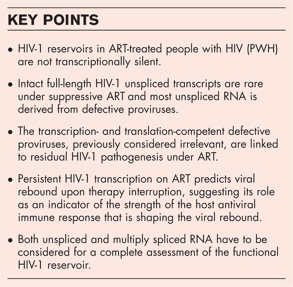
 Box 1:
 no caption available
CELL TYPESResearch efforts over the past 30 years have sought to understand the complexity of the HIV reservoir in order to facilitate the development of interventions that could eradicate the HIV-infected cells (a cure) or enable HIV seropositive individuals to maintain suppressed viremia in the absence of ART (a functional cure) [20,21]. In this context, the qualitative and quantitative assessment of the HIV reservoir in ART treated individuals has been conducted by various research groups employing diverse assays to measure the presence of viral nucleic acids (such as total and integrated DNA, or various forms of RNA) through PCR-based assays either alone or with proviral sequencing, viral protein (p24) detection and/or with functional assessments measuring viral competence through culture-based viral outgrowth assays. In this regard, pioneering studies demonstrated the presence of HIV-infected cells (HIV DNA+ and/or RNA+), albeit at different frequencies, within various blood CD4+ T cell subsets [9–12,22–27]. However, it is important to note that while PCR-based assays are highly standardized, robust, and require fewer cells as compared with culture-based methods, they fail to distinguish between defective and replication competent viruses [28].
Alternative approaches, such as the use of culture-based quantitative viral outgrowth assays (Q-VOAs) to assess the frequencies of HIV-infected cells harboring inducible replication-competent proviruses in treated individuals demonstrated a notable enrichment within blood CD4+ T cells expressing CXCR3 [24,29] and CD32a [30]. Similarly, enrichment was observed within lymph node (LN) CD4+ T cells expressing PD-1 [23], that is, LN Tfh cells (Fig. 1) and CD32a [31] in the same individuals during ART, although at significantly higher frequencies within lymph nodes. Notably, Q-VOAs are highly robust and could be instrumental for measuring reservoir size in cure-oriented clinical trials. However, Q-VOAs rely on large cell inputs of highly purified cell populations in the limiting-dilution format, the necessity for specific stimulations to induce viral production, and potentially underestimate the viral reservoir size due to the assay's sensitivity (reviewed in Eriksson et al.[28]). Furthermore, elegant studies have demonstrated that while around 10% of HIV-infected cells may harbor intact proviruses in treated individuals, only a small fraction (<5%) is induced to produce replication-competent viruses in vitro under routine VOA laboratory settings [32].
 FIGURE 1:
 Schematic representation of potential HIV tissue reservoirs in the body: The figure represents the complexity of the HIV reservoir in terms of its cellular composition, broad anatomical distribution and transcriptional status during ART. Major HIV reservoirs depicted include: blood, lymphoid tissues, i.e. spleen, lymph nodes, gut-associated lymphoid tissues (GALT), bone marrow, lungs, genitourinary systems and central nervous system (CNS). Proposed cellular reservoirs within blood and tissues are depicted. Cell types are color-coded; HIV-infected cells are depicted harboring integrated HIV DNA; transcriptionally active HIV-infected cells are shown with dashed red lines. ‘cTfh’ refers to circulating T follicular helper cells; ‘GI’ refers to gastrointestinal tract. ‘M’ refers to male genitourinary system and ‘F’ refers to female genital tract.
Given these technical limitations, recent developments focused on alternative experimental strategies targeted at the combined evaluation of both the phenotype of HIV-infected cells and cellular transcriptome at single cell level, as seen in the PheP-seq assay [33]. Notably, these new advancements provided an unprecedented high-resolution analysis of proviral landscape of individual HIV-infected cells during ART [33]. This analyses revealed the presence of intact viruses within both tissue-resident memory CD4+ T cells - expressing CD127 and CD69+ T cell population and within circulating resting CD4+ T cells (central memory and effector memory T cells) in lymph node tissues of treated individuals [33], suggesting the capacity of these cells to disseminate infection to other lymphoid organs. Of note, these analyses did not provide evidence supporting the presence of a single phenotypic marker capable of effectively distinguishing virally infected cells from uninfected cells. Therefore, future studies probing the efficacy of these approaches to better identify and define the viral reservoir characteristics would be crucial.
HIV DNA has also been readily detected within local macrophages resident in several mucosal tissues of ART treated individuals such as the gastro intestinal tract [34–36], penile urethra [37], testes [38] and vaginal tissues [39,40] or in tissue-resident macrophages such as Kupffer cells in liver [41], alveolar macrophages in lungs [42] and within microglial cells and perivascular macrophages in the brain [43] by RNA and DNAscope in situ approaches. However, viral RNA has been demonstrated predominantly within vaginal [39] and urethral macrophages [44] of ART suppressed individuals. Moreover, cells harboring inducible replication competent HIV has been shown to be specifically enriched within urethral macrophages isolated from ART suppressed individuals undergoing elective gender reassignment surgery [44] as compared to CD3+ penile urethral cells of the same individuals, suggesting the major contribution of these cells to the total reservoir in these tissues (Fig. 1). Interestingly, recent evidences indicated that tissue macrophages may persist long-term through self-renewing capacity [45], were relatively resistant to viral cytopathic effects [46] and to cytotoxic T lymphocytes (CTL) killing [47], and could contain inducible replication competent viruses, highlighting their contribution as relevant tissue reservoirs [37].
The data regarding dendritic cells (DCs) as potential cellular reservoirs are rare, mainly due to the low frequencies and scarce availability of tissues, and therefore the high reliance on experiments performed either on blood or on in-vitro-derived cells. Notably, within lymph nodes, two distinct populations of myeloid DCs can be identified on the basis of their original tissue location, that is, ‘resident DCs’ and ‘migratory DCs’. In particular, LN resident DCs differentiate in, and spend their entire lives within LN tissues. On the other hand, migratory DCs can migrate from peripheral tissues (e.g., from the genital mucosa to the inguinal draining LNs) bearing antigens. In this context, a recent study evaluated the presence of HIV-infected LN-derived myeloid DCs during HIV infection and under suppressive ART. The study demonstrated that despite detectable levels of antiviral restriction factors within LN DC subpopulations, a large number of individual proviral DNA sequences isolated from LN DCs were genome intact. Moreover, HIV-infected LN DCs harboring inducible and infectious replication-competent HIV could be detected despite years of suppressive therapy, highlighting their potential role as a yet underestimated cellular source of HIV within LN tissues (Fig. 1) [48▪▪]. Moreover, CD1a+ vaginal epithelial DCs harboring integrated HIV DNA could be detected in female genital tract of virologically suppressed women, suggesting that tissue DCs could also serve as potential cellular reservoirs in vivo[49]. However, the potential mechanisms associated with the detection of HIV-infected DCs despite ART remain to be elucidated with possibilities including replenishment through the proliferation of precursor DCs.
HIV infection of hematopoietic precursor or progenitor cells capable of differentiating into distinct cell lineages have also been described [50] (Fig. 1) (discussed in Herd et al.[51]). However, the data regarding the potential susceptibility of hematopoietic precursor or progenitor cells isolated directly ex vivo from bone marrow tissue to HIV infection has long been debated mainly due to conflicting results obtained in part because of insufficient samples, insufficient cell purities, different experimental approaches, the use of differentiation and growth factors, HIV vectors used, infection strategy, and HIV readouts used in different studies [51]. Notably, a recent study detected identical genome intact sequences in hematopoietic progenitor cells isolated directly ex vivo from ART treated individuals using near-full genome proviral sequencing, and suggested their survival to be linked with cellular proliferation [52]. Moreover, clonal proviral sequences obtained from progenitor cells and their daughter cells showed a large homology to virion-sequences obtained from residual plasma viremia, suggesting the contribution of this compartment to residual plasma viremia during ART [52]. Although few, these studies support the possibility that bone marrow compartment may serve as a relevant and a less explored tissue reservoir in ART treated HIV-infected individuals.
ANATOMIC SITESThe majority of studies addressing the HIV reservoir have been performed in peripheral blood CD4+ T cells from treated individuals (Fig. 1) [6,9,22,28,32,53,54]. Although these studies have yielded key insights into the HIV reservoir, circulating CD4+ T cells represent <2% of total body CD4+ T cells at a given instance [55], with the majority of them located within secondary lymphoid organs. In this context, there is a growing recognition of the significance of tissue microenvironments in studying the viral reservoir and assessing cure interventions. Indeed, HIV-infected cell fate (survival, proliferation, elimination, migration and renewal-capacity) within different tissues might be influenced by multiple parameters which may be specific to tissues and distinct from blood [7,17,19,56,57]. In particular, different tissues may harbor specific spatial organization, cellular and cytokine milieu [58]; variation in viral expression levels [23,48▪▪,59,60]; anatomical compartments harboring specific immune effector mechanisms to clear infected cells expressing viral RNA and/or protein [61–63] and finally differential ART drug penetration [59,64].
The characterization of tissue reservoirs has been performed using multiple approaches including either through virological and/or phenotypic evaluation of HIV-infected cells using flow/mass cytometry [23,24,31,48▪▪,65], imaging platforms [7,60,66] and/or through alternative approaches after direct isolation ex vivo under various scenarios, from SIV-infected macaques [60] or HIV-infected individuals on ART [23,24,48▪▪,65], SIV-infected macaques [66] or HIV-infected individuals after analytical treatment interruption [7], and more recently, in autopsy samples from HIV-infected individuals [67▪,68]. Notably, these studies have demonstrated that HIV DNA and/or RNA are detected within multiple tissues, consistent with the infection of multiple tissue-resident and circulating cell types. However, lymphoid tissues (lymph nodes, GALT and spleen) are the predominant sites of HIV infection and persistence of cells with replication competent virus.
Lymph nodes (LNs) represent distinct compartments containing phenotypically and functionally specialized cell subsets as compared to blood. LNs are dynamic and highly structured tissues, consisting of strategically prepositioned LN resident cells within micro-anatomical niches and recirculating cells. The differential location of LN cell subsets within the micro-anatomical niches is associated with distinct cell phenotypes and molecular and functional signatures [19] and different ART drug penetration [59]. In this context, classical in situ hybridization-based approaches in ART SIV NHP models [60], and more recently, the profiling of phenotype of HIV-infected cells and cellular transcriptome at single cell level have indicated that while HIV DNA harboring cells can be detected within both LN extra-follicular and follicular areas [33], transcriptionally active SIV/HIV RNA+ cells are mainly detected within B cell follicles in LNs. Interestingly, SIV/HIV RNA+ was associated with two distinct cell types including [23,69–71]: follicular dendritic cells (FDCs) that are not infected but can bind and retain intact HIV virions within a nondegradative cycling compartment for prolonged periods of time and transmit infectious particles to CD4+ T cells [70,71] or HIV/SIV infected T follicular helper cells (Tfh) (Fig. 1). Notably, SIV/HIV infected Tfh cells were enriched in multiple virological setting including in HIV viremic controllers [72], SIV-infected elite controller macaques [69], HIV viremic individuals and ART-treated aviremic HIV-infected individuals [23,31]. The enrichment of HIV-infected Tfh cells potentially involves nonmutually exclusive phenomena that are not yet fully identified, including: heterogeneous ART drug penetration (particularly protease inhibitors) into distinct areas of LN tissues with limited combined exposure to all infected cells, potentially allowing for environments where low-level, intermittent viral replication can occur [59]; likelihood of maintenance of HIV-infected cells containing replication-competent viruses through both long-term survival and clonal expansion [53,73–76], without necessarily inducing viral expression; relatively low accessibility of GCs to HIV/SIV-specific cytotoxic CD8+ T cells, thereby allowing viral reservoirs that reside in these microenvironments to escape CTL elimination [73,74,76–78], and enrichment of regulatory T cells and DCs expressing immunomodulatory molecules cytokines such as TGF-β and interleukin (IL)-10 and plasmacytoid DCs secreting type I interferon that contribute to promote viral latency in the LN paracortex and relative exclusion from germinal center areas (Fig. 1) [19].
Additional cell types, such as tissue resident macrophages in other sites (such as microglial cells in brain), astrocytes, hematopoietic progenitor cells, among others, in which HIV DNA or RNA has been detected however inducible replication-competent HIV has only been recovered in limited scenarios and with extreme difficulty (Fig. 1) [52,79,80]. In this context, it is important to note that because of limited access to human tissue, there is sparse sampling of many tissue types (i.e., spleen) that may also harbor CD4+ T cells with replication-competent virus [67▪] and addition technical complexities associated with sampling of tissues and the assessment of replication competence by Q-VOAs, have limited the sensitivity of assessments performed to date in various tissues. Therefore, understanding the composition and frequency of the replication-competent reservoirs across different anatomic sites remains a critical issue for future cure-oriented interventional studies.
SPECTRUM OF TRANSCRIPTIONAL ACTIVITY AND REPLICATION COMPETENCEHIV-infected cells in blood were frequently considered as ‘transcriptionally silent’, primarily due to the absence of detectable HIV RNA in most instances, as assessed by PCR methodologies. However, recently, breakthrough assays were developed that either allowed the profiling of individual proviral chromosomal integration site and transcriptional activity with increased sensitivity (PRIPseq) [81▪▪], evaluation of transcriptional activity in individuals under temporary ART initiated during primary HIV infection by distinct cell-associated HIV RNA-based measurements [82], or evaluation of transcriptional activity directly ex vivo in CD4+ T cells from fully suppressed individuals at single-cell level by RNAflow-FISH [83▪▪]. These studies have independently supported the detection of a broad spectrum of transcriptional activity directly ex vivo under these conditions. Furthermore, they have also highlighted the power of assessing quantitative and qualitative nature of transcriptional activity directly ex vivo as a tool to predict features of HIV-specific immune responses in vivo and viral rebound post-ART cessation [82,83▪▪].
Transcriptional activity within infected CD4+ T cells at a specific point is suggested to be associated with several factors including the presence or not of transcriptional blocks arising at various stages during transcription – such as in initiation, elongation and in multiple splicing in resting CD4+ T cells, due to both host-related and virus-related factors [84]. Based on these observations, it is conceivable that diverse mechanisms influencing the levels of transcriptional activity may exist within distinct cell types, tissues, and anatomical compartments as compared to blood (Fig. 1).
Multiple mechanisms have been proposed for the persistence of transcriptionally active HIV-infected CD4+ T cells under long-term during ART. Amongst these, proviral integration within nongenic locations has been strongly associated with significantly weaker viral transcriptional activity, likely because of nonpermissive features of the chromatin at or near the chromosomal integration site as opposed to those integrations within chromosomal locations surrounded by activating epigenetic chromatin signals in their immediate chromatin proximity (observed for both intact and defective proviruses) [85]. The authors highlighted an intriguing aspect, emphasizing the probability of detecting an expanded clone that contained both transcriptionally silent and active proviruses [81▪▪] in suppressed individuals. This suggests the possibility that the transcriptionally active cells may survive over-time by outcompeting host-related immune selection forces despite ART or through intrinsic features of survival and reduced susceptibility to host-mediated elimination [81▪▪,85].
Indeed, the transcriptional status of a cell may change over-time, impacting the fate of an HIV-infected cell and therefore the frequency of total cells harboring transcription at a given time. Cellular factors such as activation (spontaneous or antigen-driven) and/or migration through tissues may induce a threshold of transcription adequate for virus-mediated cytolysis or immune recognition, and therefore promote the elimination of infected cells. On the contrary, homeostatic or antigen-driven clonal expansion of cells harboring or not transcriptional activity may in-turn increase the frequency of HIV-infected cells that may or may not at a later stage become transcriptionally active. Taken together, it appears that the dynamic and evolving nature of transcriptionally active proviruses under the influence of host-mediated selection pressure necessitates longitudinal evaluation.
COMPARTMENTALIZATIONThe various immune environments, immunological selection forces and ART drug penetration capacities within specific tissues (such as within LNs, GALT, and CNS) may foster viral compartmentalization as well as evolution of viral sequences, allowing for new cell types to be infected. In this regard, to better understand the relationship between circulating proviruses and those present in tissues, studies have focused on the assessment of phylogenetic relationship between individual proviral sequences isolated from the tissues and from blood CD4+ T cells [8,24,65,67▪]. Notably, these studies have indicated the possibility of three states of virus in tissue when compared to the blood: equilibrated (where virus in the blood and tissue are very similar), compartmentalized (where blood and tissue viral populations are distinct, indicating separately evolving populations in these compartments), and clonal amplification (where a single variant is greatly expanded within a compartment). In this context, while proviral compartmentalization has been observed in some studies (liver [86], testes [87], female genital tract [88], central nervous system (CNS) [89]), many others did not observe viral compartmentalization (gut [90], LNs [24,65], CNS [91]). The contrasting results obtained might be explained at least in part by the compartments compared the type of samples compared, that is, cells isolated from ART treated individuals or biopsies [91] and/or the techniques used, that is, bulk versus single genome proviral sequencing of a part of viral genome (HIV env[68,92] versus other regions [93,94]) or near full-length sequencing [67▪,91]. Importantly, an elegant study performed an extensive characterization of genetic compartmentalization of proviral sequences present in multiple tissues obtained from autopsies of HIV-infected individuals using near full-length proviral genome sequencing [67▪]. Notably, compartmentalization analyses restricted to distinct proviruses per tissue, revealed no evidence for compartmentalization [67▪]; on the other hand, several analyzed tissue reservoirs (secondary lymphoid organs, lungs, liver and genital tract) harbored large clones (both defective and intact proviruses) that were often shared between distinct tissues (such as lymph nodes, GALT, spleen, liver, lungs; Fig. 1), suggesting a re-circulation of HIV-infected cells between lymphoid and effector tissues even after prolonged ART [67▪].
CONCLUSIONMore than 30 years of research demonstrated that the HIV reservoir is heavily complex in nature and involves multiple cell types located in multiple tissues. In addition, the HIV reservoir is probably heavily dynamic in terms of renewal potential, cell migratory potential but also in terms of transcriptional activity. In this context, the recent development of single-cell and single-viral genome approaches, will probably bring valuable insights into tissue reservoirs.
AcknowledgementsWe would like to thank Aaron Weddle for his assistance in figure preparation.
Financial support and sponsorshipThis work was supported by Swiss National Science Foundation Grant 320030_200912 and by Freedom Forever to M. Perreau and by an educational grant from FONDATION MACHAON to R. Banga.
Conflicts of interestThere are no conflicts of interests.
REFERENCES AND RECOMMENDED READINGPapers of particular interest, published within the annual period of review, have been highlighted as:
▪ of special interest
▪▪ of outstanding interest
REFERENCES 1. Palella FJ Jr, Delaney KM, Moorman AC, et al. HIV Outpatient Study Investigators. Declining morbidity and mortality among patients with advanced human immunodeficiency virus infection. N Engl J Med 1998; 338:853–860. 2. G Williams B, Lima V. Gouws EJCHr. Modelling the impact of antiretroviral therapy on the epidemic of HIV 2011; 9:367–382. 3. Whitney JB, Hill AL, Sanisetty S, et al. Rapid seeding of the viral reservoir prior to SIV viraemia in rhesus monkeys. Nature 2014; 512:74–77. 4. Chun T-W, Carruth L, Finzi D, et al. Quantification of latent tissue reservoirs and total body viral load in HIV-1 infection 1997; 387:183–188. 5. Finzi D, Blankson J, Siliciano JD, et al. Flexner CJNm. Latent infection of CD4+ T cells provides a mechanism for lifelong persistence of HIV-1, even in patients on effective combination therapy. Nat Med 1999; 5:512–517. 6. Siliciano JD, Kajdas J, Finzi D, et al. Long-term follow-up studies confirm the stability of the latent reservoir for HIV-1 in resting CD4+ T cells. Nat Med 2003; 9:727–728. 7. Rothenberger MK, Keele BF, Wietgrefe SW, et al. Large number of rebounding/founder HIV variants emerge from multifocal infection in lymphatic tissues after treatment interruption. Proc Natl Acad Sci USA 2015; 112:E1126–E1134. 8. De Scheerder MA, Vrancken B, Dellicour S, et al. HIV rebound is predominantly fueled by genetically identical viral expansions from diverse reservoirs. Cell Host Microbe 2019; 26:347–358. e7. 9. Chomont N, El-Far M, Ancuta P, et al. Brenchley JMJNm. HIV reservoir size and persistence are driven by T cell survival and homeostatic proliferation. Nat Med 2009; 15:893–900. 10. Gosselin A, Salinas TRW, Planas D, et al. HIV persists in CCR6+ CD4+ T cells from colon and blood during antiretroviral therapy. AIDS 2017; 31:35–48. 11. Khoury G, Anderson JL, Fromentin R, et al. Persistence of integrated HIV DNA in CXCR3+CCR6+memory CD4+ T cells in HIV-infected individuals on antiretroviral therapy. AIDS 2016; 30:1511–1520. 12. Siliciano JD, Siliciano RF. Enhanced culture assay for detection and quantitation of latently infected, resting CD4+ T-cells carrying replication-competent virus in HIV-1-infected individuals. Human retrovirus protocols. Springer; 2005:3–15. 13. Laird GM, Bullen CK, Rosenbloom DI, et al. Ex vivo analysis identifies effective HIV-1 latency-reversing drug combinations. J Clin Invest 2015; 125:1901–1912. 14. Siliciano RF. A reservoir for HIV in patients on combination antiretroviral therapy. Hopkins HIV Rep 1998; 10: 1, 5–6, 11. 15. Chun T-W, Justement JS, Moir S, et al. Decay of the HIV reservoir in patients receiving antiretroviral therapy for extended periods: implications for eradication of virus. J Infect Dis 2007; 195:1762–1764. 16. Tedla N, Dwyer J, Truskett P, et al. Phenotypic and functional characterization of lymphocytes derived from normal and HIV-1-infected human lymph nodes. Clin Exp Immunol 1999; 117:92–99. 17. Schacker T. The role of secondary lymphatic tissue in immune deficiency of HIV infection. AIDS 2008; 22: (Suppl 3): S13–S18. 18. Chun TW, Finzi D, Margolick J, et al. In vivo fate of HIV-1-infected T cells: quantitative analysis of the transition to stable latency. Nat Med 1995; 1:1284–1290. 19. Banga R, Munoz O, Perreau M. HIV persistence in lymph nodes. Curr Opin HIV AIDS 2021; 16:209–214. 20. Deeks SG, Archin N, Cannon P, et al. Research priorities for an HIV cure: International AIDS Society Global Scientific Strategy 2021. Nat Med 2021; 27:2085–2098. 21. Deeks SG, Lewin SR, Ross AL, et al. International AIDS Society global scientific strategy: towards an HIV cure 2016. Nat Med 2016; 22:839–850. 22. Fromentin R, Bakeman W, Lawani MB, et al. CD4+ T cells expressing PD-1, TIGIT and LAG-3 contribute to HIV persistence during ART. PLoS Pathog 2016; 12:e1005761. 23. Banga R, Procopio FA, Noto A, et al. PD-1(+) and follicular helper T cells are responsible for persistent HIV-1 transcription in treated aviremic individuals. Nat Med 2016; 22:754–761. 24. Banga R, Procopio FA, Ruggiero A, et al. Perreau MJFii. Blood CXCR3+ CD4 T cells are enriched in inducible replication competent HIV in aviremic antiretroviral therapy-treated individuals. Front Immunol 2018; 9:144. 25. Buzon MJ, Sun H, Li C, et al. HIV-1 persistence in CD4+ T cells with stem cell–like properties. Nat Med 2014; 20:139–142. 26. Anderson JL, Khoury G, Fromentin R, et al. Human immunodeficiency virus (HIV)-infected CCR6+ rectal CD4+ T cells and HIV persistence on antiretroviral therapy. J Infect Dis 2020; 221:744–755. 27. Serra-Peinado C, Grau-Expósito J, Luque-Ballesteros L, et al. Ribera EJNc. Expression of CD20 after viral reactivation renders HIV-reservoir cells susceptible to rituximab. Nat Commun 2019; 10:3705. 28. Eriksson S, Graf EH, Dahl V, et al. Comparative analysis of measures of viral reservoirs in HIV-1 eradication studies. PLoS Pathog 2013; 9:e1003174. 29. Lee GQ, Orlova-Fink N, Einkauf K, et al. Ouyang ZJTJoci. Clonal expansion of genome-intact HIV-1 in functionally polarized Th1 CD4+ T cells. J Clin Invest 2017; 127:2689–2696. 30. Descours B, Petitjean G, López-Zaragoza J-L, et al. CD32a is a marker of a CD4 T-cell HIV reservoir harbouring replication-competent proviruses. Nature 2017; 543:564–567. 31. Noto A, Procopio FA, Banga R, et al. CD32(+) and PD-1(+) lymph node CD4 T cells support persistent HIV-1 transcription in treated aviremic individuals. J Virol 2018; 92:e00901–18. 32. Ho Y-C, Shan L, Hosmane NN, et al. Replication-competent noninduced proviruses in the latent reservoir increase barrier to HIV-1 cure. Cell 2013; 155:540–551. 33. Sun W, Gao C, Hartana CA, et al. Phenotypic signatures of immune selection in HIV-1 reservoir cells. Nature 2023; 614:309–317. 34. Telwatte S, Lee S, Somsouk M, et al. Gut and blood differ in constitutive blocks to HIV transcription, suggesting tissue-specific differences in the mechanisms that govern HIV latency. PLoS Pathog 2018; 14:e1007357. 35. Yukl SA, Shergill AK, Ho T, et al. The distribution of HIV DNA and RNA in cell subsets differs in gut and blood of HIV-positive patients on ART: implications for viral persistence. J Infect Dis 2013; 208:1212–1220. 36. Zalar A, Figueroa MI, Ruibal-Ares B, et al. Macrophage HIV-1 infection in duodenal tissue of patients on long term HAART. Antiviral Res 2010; 87:269–271. 37. Ganor Y, Real F, Sennepin A, et al. HIV-1 reservoirs in urethral macrophages of patients under suppressive antiretroviral therapy. Nat Microbiol 2019; 4:633–644. 38. Roulet V, Satie AP, Ruffault A, et al. Susceptibility of human testis to human immunodeficiency virus-1 infection in situ and in vitro. Am J Pathol 2006; 169:2094–2103. 39. Shen R, Richter HE, Clements RH, et al. Macrophages in vaginal but not intestinal mucosa are monocyte-like and permissive to human immunodeficiency virus type 1 infection. J Virol 2009; 83:3258–3267. 40. Shen R, Richter HE. Smith PDJAjori. Early HIV-1 target cells in human vaginal and ectocervical mucosa. Am J Reprod Immunol 2011; 65:261–267. 41. Kandathil AJ, Sugawara S, Goyal A, et al. No recovery of replication-competent HIV-1 from human liver macrophages. J Clin Invest 2018; 128:4501–4509. 42. Cribbs SK, Lennox J, Caliendo AM, et al. Healthy HIV-1-infected individuals on highly active antiretroviral therapy harbor HIV-1 in their alveolar macrophages. AIDS Res Hum Retroviruses 2015; 31:64–70. 43. Ko A, Kang G, Hattler JB, et al. Macrophages but not astrocytes harbor HIV DNA in the brains of HIV-1-infected aviremic individuals on suppressive antiretroviral therapy. J Neuroimmune Pharmacol 2019; 14:110–119. 44. Ganor Y, Real F, Sennepin A, et al. Marion SJNm. HIV-1 reservoirs in urethral macrophages of patients under suppressive antiretroviral therapy. Nat Microbiol 2019; 4:633–644. 45. Hashimoto D, Chow A, Noizat C, et al. Tissue-resident macrophages self-maintain locally throughout adult life with minimal contribution from circulating monocytes. Immunity 2013; 38:792–804. 46. Castellano P, Prevedel L. Eugenin EAJSr. HIV-infected macrophages and microglia that survive acute infection become viral reservoirs by a mechanism involving. BIM 2017; 7:1–16. 47. Clayton KL, Collins DR, Lengieza J, et al. Resistance of HIV-infected macrophages to CD8(+) T lymphocyte-mediated killing drives activation of the immune system. Nat Immunol 2018; 19:475–486. 48▪▪. Banga R, Procopio FA, Lana E, et al. Lymph node dendritic cells harbor inducible replication-competent HIV despite years of suppressive ART. Cell Host Microbe 2023; 31:1714–1371. e9. 49. Pena-Cruz V, Agosto LM, Akiyama H, et al. Sagar MJTJoci. HIV-1 replicates and persists in vaginal epithelial dendritic cells. J Clin Invest 2018; 128:3439–3444. 50. Renelt S, Schult-Dietrich P, Baldauf H-M, et al. HIV-1 infection of long-lived hematopoietic precursors in vitro and in vivo. Cells 2022; 11:2968. 51. Herd CL, Mellet J, Mashingaidze T, et al. Consequences of HIV infection in the bone marrow niche. Front Immunol 2023; 14:1163012. 52. Zaikos TD, Terry VH, Kettinger NTS, et al. Hematopoietic stem and progenitor cells are a distinct HIV reservoir that contributes to persistent viremia in suppressed patients. Cell Rep 2018; 25:3759–3773. e9. 53. Hosmane NN, Kwon KJ, Bruner KM, et al. Proliferation of latently infected CD4(+) T cells carrying replication-competent HIV-1: Potential role in latent reservoir dynamics. J Exp Med 2017; 214:959–972. 54. Simonetti FR, Zhang H, Soroosh GP, et al. Antigen-driven clonal selection shapes the persistence of HIV-1-infected CD4+ T cells in vivo. J Clin Invest 2021; 131:145254. 55. Ganusov VV, De Boer RJ. Do most lymphocytes in humans really reside in the gut? Trends Immunol 2007; 28:514–518. 56. Lorenzo-Redondo R, Fryer HR, Bedford T, et al. Persistent HIV-1 replication maintains the tissue reservoir during therapy. Nature 2016; 530:51–56. 57. Yukl SA, Boritz E, Busch M, et al. Challenges in detecting HIV persistence during potentially curative interventions: a study of the Berlin patient. PLoS Pathog 2013; 9:e1003347. 58. O’Neill NA, Eppler HB, Jewell CM, Bromberg JS. Harnessing the lymph node microenvironment. Curr Opin Organ Transplant 2018; 23:73–82. 59. Fletcher CV, Staskus K, Wietgrefe SW, et al. Schmidt TEJPotNAoS. Persistent HIV-1 replication is associated with lower antiretroviral drug concentrations in lymphatic tissues. Proc Natl Acad Sci USA 2014; 111:2307–2312. 60. Estes JD, Kityo C, Ssali F, et al. Beilman GJJNm. Defining total-body AIDS-virus burden with implications for curative strategies. Nat Med 2017; 23:1271–1276. 61. Connick E, Folkvord JM, Lind KT, et al. Kim HOJTJoI. Compartmentalization of simian immunodeficiency virus replication within secondary lymphoid tissues of rhesus macaques is linked to disease stage and inversely related to localization of virus-specific CTL. J Immunol 2014; 193:5613–5625. 62. Connick E, Mattila T, Folkvord JM, et al. White CJTJoI. CTL fail to accumulate at sites of HIV-1 replication in lymphoid tissue. J Immunol 2007; 178:6975–6983. 63. Reuter MA, Del Rio Estrada PM, Buggert M, et al. HIV-specific CD8(+) T cells exhibit reduced and differentially regulated cytolytic activity in lymphoid tissue. Cell Rep 2017; 21:3458–3470. 64. Cory TJ, Schacker TW, Stevenson M. Fletcher CVJCoiH, AIDS. Overcoming pharmacologic sanctuaries. AIDS 2013; 8:190–185. 65. Kuo H-H, Banga R, Lee GQ, et al. Pantaleo GJTJoID. Blood and lymph node dissemination of clonal genome-intact HIV-1 DNA sequences during suppressive antiretroviral therapy. J Infect Dis 2020; 222:655–660. 66. Solis-Leal A, Boby N, Mallick S, et al. Lymphoid tissues contribute to plasma viral clonotypes early after antiretroviral therapy interruption in SIV-infected rhesus macaques. Sci Transl Med 2023; 15:eadi9867. 67▪. Dufour C, Ruiz MJ, Pagliuzza A, et al. Near full-length HIV sequencing in multiple tissues collected postmortem reveals shared clonal expansions across distinct reservoirs during ART. Cell Rep 2023; 42:113053. 68. Chaillon A, Gianella S, Dellicour S, et al. HIV persists throughout deep tissues with repopulation from multiple anatomical sources. J Clin Invest 2020; 130:1699–1712. 69. Fukazawa Y, Lum R, Okoye AA, et al. Lucero CJNm. B cell follicle sanctuary permits persistent productive simian immunodeficiency virus infection in elite controllers. Nat Med 2015; 21:132–139. 70. Busman-Sahay K, Starke CE, Nekorchuk MD, Estes JD. Eliminating HIV reservoirs for a cure: the issue is in the tissue. Curr Opin HIV AIDS 2021; 16:200–208. 71. Heesters BA, Lindqvist M, Vagefi PA, et al. Follicular dendritic cells retain infectious HIV in cycling endosomes. PLoS Pathog 2015; 11:e1005285. 72. Perreau M, Savoye A-L, De Crignis E, et al. Pantaleo GJJoEM. Follicular helper T cells serve as the major CD4 T cell compartment for HIV-1 infection, replication, and production. J Exp Med 2013; 210:143–156. 73. Vibholm LK, Lorenzi JCC, Pai JA, et al. Characterization of intact proviruses in blood and lymph node from HIV-infected individuals undergoing analytical treatment interruption. J Virol 2019; 93:e01920-18. 74. Simonetti FR, Sobolewski MD, Fyne E, et al. Clonally expanded CD4+ T cells can produce infectious HIV-1 in vivo. Proc Natl Acad Sci USA 2016; 113:1883–8. 75. Coffin JM, Wells DW, Zerbato JM, et al. Clones of infected cells arise early in HIV-infected individuals. JCI Insight 2019; 4:e128432. 76. McManus WR, Bale MJ, Spindler J, et al. HIV-1 in lymph nodes is maintained by cellular proliferation during antiretroviral therapy. J Clin Invest 2019; 129:4629–4642. 77. Kline C, Ndjomou J, Franks T, et al. Persistence of viral reservoirs in multiple tissues after antiretroviral therapy suppression in a macaque RT-SHIV model. PLoS One 2013; 8:e84275. 78. Kearney MF, Wiegand A, Shao W, et al. Ongoing HIV replication during ART reconsidered. Open Forum Infect Dis 2017; 4:ofx173. 79. Tang Y, Chaillon A, Gianella S, et al. Brain microglia serve as a persistent HIV reservoir despite durable antiretroviral therapy. J Clin Invest 2023; 133:e167417. 80. Lutgen V, Narasipura SD, Barbian HJ, et al. HIV infects astrocytes in vivo and egresses from the brain to the periphery. PLoS Pathog 2020; 16:e1008381. 81▪▪. Einkauf KB, Osborn MR, Gao C, et al. Parallel analysis of transcription, integration, and sequence of single HIV-1 proviruses. Cell 2022; 185:266–282. e15. 82. Pasternak AO, Grijsen ML, Wit FW, et al. Cell-associated HIV-1 RNA predicts viral rebound and disease progression after discontinuation of temporary early ART. JCI Insight 2020; 5:134196. 83▪▪. Dubé M, Tastet O, Dufour C, et al. Spontaneous HIV expression during suppressive ART is associated with the magnitude and function of HIV-specific CD4(+) and CD8(+) T cells. Cell Host Microbe 2023; 31:1507–1522. e5. 84. Yukl SA, Kaiser P, Kim P, et al. HIV latency in isolated patient CD4
留言 (0)