
記住我
The entry of HIV-1 into latency is responsible for the virus's persistence despite antiretroviral treatment. The formation of the latent HIV-1 reservoir occurs within 10 days of infection and conceals infected cells from antiviral immune surveillance since they cannot produce viral antigens [1]. HIV-1 primarily targets effector CD4+ T cells, which provide an intracellular environment conducive to completing the retroviral replicative cycle due to their activated state [2]. Although many productively infected cells are eliminated through cytopathic effects [3,4], a small surviving subset transitions to become quiescent central memory cells (TCM) [5,6]. Multiple cellular events during this transition induce a state of viral transcriptional silencing at the molecular level. First, key transcription initiation factors, especially Nuclear factor of activated T-cells (NFAT) and nuclear factor (NF)-κB are sequestered in the cytoplasm. Second, Positive transcription elongation factor (P-TEFb), the cellular cofactor for the HIV transactivator protein Tat, is disassembled and removed from the nucleus (refer to the review by Mbonye et al.[7]). These blocks to transcription are reinforced by the formation of epigenetic silencing marks, including transcriptionally suppressive histone methylation [8], histone deacetylation [9], DNA methylation [10▪▪], and reduced levels of 3D chromosomal interactions [11].
Despite these multiple overlapping molecular restrictions, the latently infected cells can efficiently reactivate upon encountering an antigenic challenge or inflammatory stimulus, leading to the release of the virus. When this occurs during the interruption of antiretroviral therapy (ART), there is a systemic viral rebound. Even in the presence of effective ART, reservoir persistence is a dynamic process, that is shaped by multiple overlapping mechanisms, including viral spread from reactivated cells in sanctuary sites with low penetration of ARTs [12] and antigen-driven [13] or homeostatic proliferation [14,15] of the latently infected memory T-cell population.
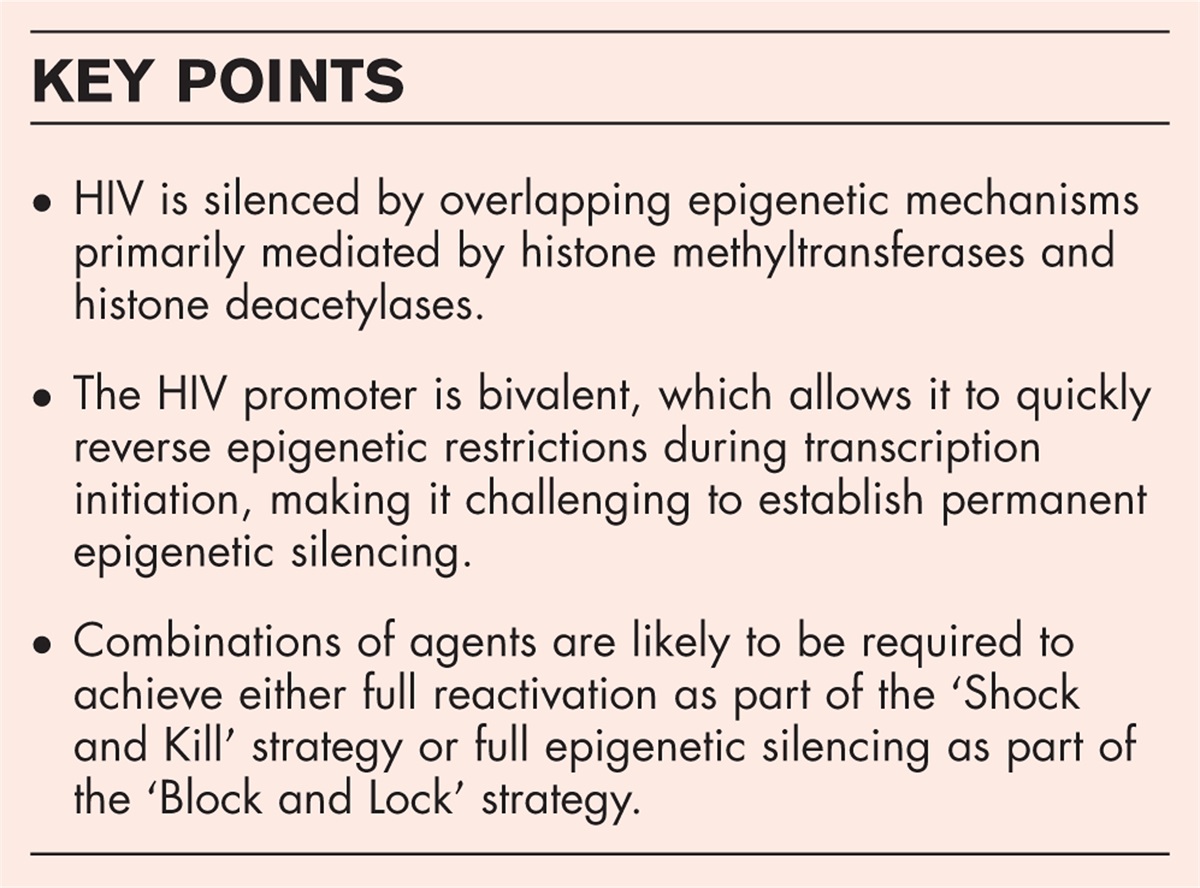
 Box 1:
 no caption available
EPIGENETIC CONTROL OF HIV-1 LATENCYEpigenetic modifications of chromatin and DNA result in heritable changes in gene function that occur without altering the underlying DNA sequence. These modifications, including DNA methylation, histone modification, and noncoding RNA regulation, are pivotal in orchestrating transcriptional regulation.
The impact of epigenetics on HIV-1 latency has been extensively studied since the first description of histone modifications in 2008 and DNA methylation in 2009 [16,17]. Histone modifications, including histone methylation and acetylation, can alter chromatin accessibility and impact the transcriptional activity of the integrated provirus. DNA methylation at the viral promoter regions, particularly CpG islands, can hinder transcription factor binding, inhibiting viral gene expression. However, a challenge for the field is that regulation of HIV-1 latency is a multifaceted process, and research has revealed there is extensive epigenetic heterogeneity of the HIV-1 latent reservoir in T cells [18▪▪,19].
Studies using chromatin immunoprecipitation and inhibitors of epigenetic silencing complexes have shown that the major epigenetic restriction of the HIV provirus is due to histone methylation (Fig. 1). Histone 3 lysine 27 (H3K27) and histone 3 lysine 9 (H3K9) methylation is required to maintain HIV-1 latency in both primary and transformed T cells [8], transformed microglial cells [20], and human primary macrophage [21]. H3K27 methylation is mediated by the Polycomb repressive complex (PRC2) with its main catalytic subunit, EZH2 [22], while H3K9 methylation is driven by multiple proteins such as G9a, SUV39H1/2, SETDB1/2, and GLP [23] (Fig. 1 and Table 1). Inhibitors targeting these methyltransferases have been shown as promising latency-reversing agents (LRAs) for HIV-1 reactivation [8].
 FIGURE 1:
 Exchange of epigenetic modulators and histone 3 modifications between latent and activated HIV-1 proviruses. Top: Latent HIV-1 chromatin serves as a bivalent promoter, which simultaneously displays both activating H3K4me3 and inhibiting H3K27me3 marks, safeguarding HIV-1 DNA from hypermethylation by DNMTs. Consequently, latent HIV-1 DNA exhibits minimal DNA methylation. H3K27me3 formation is mediated by the PRC2 complex, while H3K9 methylation, induced by EHMT2 (G9a), is also enriched in latent HIV-1 chromatin. The presence of HDACs leads to histone deacetylation and induced chromatin compaction. The combined effects of PRC2, G9a, and HDACs potently block HIV-1 transcription, driving HIV-1 into latency. Bottom: Upon induction with a potent stimulus, such as T-cell receptor stimulation, the recruitment of H3K27 demethylase UTX from the MLL2/3-UTX complex results in the demethylation of H3K27 and enhanced methylation of H3K4. This process leads to the relaxation of HIV-1 chromatin. Additionally, the displacement of HDACs from latent HIV-1 further boosts HIV-1 transcription. The bivalency of the promoter and the recruitment of TETs blocks DNA methylation at HIV-1 chromatin after reactivation. Depletion of UTX or inhibition of its demethylase activity displaces TETs from HIV-1 chromatin and helps recruit DNMT3a, inducing hypermethylation of HIV-1 DNA. However, this process can be reversed upon the removal of the UTX inhibitor. Sites of DNA methylation are indicated by the filled circles on the CpG islands.
Table 1 - Components of the major epigenetic silencing complexes that target HIV Protein complex Subcomponents Function Polycomb repressive complex 2 (PRC2) EZH2 (catalytic subunit) - Methylation of histone 3 lysine 27 [8,22] EED SUZ12 RbAp46/48 AEBP2 JARID2 PCL CoREST complex CoREST - Deacetylation of histone 3 lysine 9/14/18The histone methyltransferases are counteracted by specific histone demethylases, including UTX or JMJD3 [24] for the PRC2 methyltransferase group or LSD1/2 for H3K9 methyltransferases [25]. A recent study from our group revealed that latent HIV-1 chromatin behaves analogously to cellular bivalent genes and is characterized by simultaneously carrying the activating H3K4me3 and inhibiting H3K27me3 marks [10▪▪,26]. We also identified the H3K27 demethylase UTX as a crucial activator for the transcriptional reactivation of latent HIV-1 in primary T cells and memory T cells isolated from people with HIV (PWH) since it is required to resolve the bivalency at the 5’LTR [10▪▪].
To permanently block latent HIV-1 reactivation we used GSK-J4, a small inhibitor of UTX, to enhance H3K27 methylation of HIV-1 chromatin. Although GSK-J4 successfully suppressed HIV-1 reactivation in ex-vivo cultured primary T cells isolated from HIV-1 patients, the suppression required the continuous administration of the drug due to the reversibility of H3K27 methylation [10▪▪]. Considering the significant role of H3K9 methyltransferases in HIV-1 latency within T cells and other cell types, it will be important to investigate whether inhibitors targeting H3K9 demethylases are more effective silencers as part of a ‘Block and Lock’ strategy.
The histone deacetylases (HDACs) also play significant roles in epigenetically controlling HIV-1 latency [27]. Unfortunately, clinical studies on the use of HDAC inhibitors to reverse latency have been disappointing, with only very limited viral reactivation reported [28,29]. In an interesting new development, Peterson et al. used combinations of HDAC3-directed PROTAC and CRISPR-Cas9 knockout experiments. They showed that while either HDAC1/2 or HDAC3 targeting can prevent latency, all three enzymes must be targeted to achieve latency reversal [30▪▪]. Latency prevention is associated with increased H3K9ac at the proviral LTR promoter region and decreased H3K9me3, emphasizing an unexpected role of H3K9 modifications in the establishment of latency.
CRISPR-Cas9 technology has also been exploited to perform genome-wide screens aimed at identifying novel epigenetic factors associated with the regulation of HIV-1 latency (Table 2). Although many of the ‘hits’ arising from screens in transformed cells are factors that enhance cellular survival, using this approach, using a custom CRISPR screening strategy (Latency HIV-CRISPR), Hsieh et al. uncovered additional novel epigenetic factors potentially involved in controlling HIV latency in the J-Lat cell model [31▪▪]. The work identified multiple hits from both the nucleosome acetyltransferase of H4 histone acetylation (NuA4 HAT) complex and the related SNF2-Related CBP activator protein (SRCAP) complex, which share many subunits. They pinpointed ING3, a component of the NuA4 HAT complex, as a promising therapeutic target when used in combination with AZD5582, a SMAC mimetic and noncanonical NF-κB pathway activator for HIV-1 reactivation. In a similar screen, Pedersen et al. identified SLTM, FASB, and FASB2-members of the FASB family of chromatin regulators as chromatin and transcriptional silencers of HIV-1 [32▪▪]. Depleting SLTM led to a relaxation of chromatin structure. Moreover, in ex-vivo HIV-1 infected CD4+ T cells, the absence of SLTM enhanced cytopathic effects arising from HIV-1 induction [32▪▪]. In addition to these studies, Ding et al. reported a novel function of Schlafen-5 (SLFN5) as a silencer of HIV-1 transcription. Mechanistically, SLFN5 binds to the RBBP7 protein, a subunit of the PRC2 and G9a complexes, and helps recruit these complexes to latent HIV-1 [33▪].
Table 2 - List of novel epigenetic factors identified by recent studies Complex Function Reference NuA4 HAT complex Histone acetyltransferaseThe chromatin landscape near the integration sites on HIV-1 latency can also play a role in restricting HIV reactivation. Studies of rare people with HIV (PWH) with nonsuppressible HIV-1 viremia (NSV) have shown that the integration sites of the intact producer provirus that are responsible for generating sequencing-matching plasma viruses in the circulation show significant enrichment of activating H3K36 methylation marks [34]. These findings align with a recent study showing that nucleosomes containing H3K36me3 are preferentially targeted by the HIV-1 preintegration complex [35]. H3K36 methylation is also found in microglial cells [36].
Thus, epigenetic regulation mediated by histone modification plays a crucial role in both the establishment and maintenance of HIV-1 latency. Many of these mechanisms are shared between different cell types that are permissive to HIV infection [20,21,36].
EPIGENETIC CONTROL OF UNINTEGRATED HIV-1Like its integrated counterpart, unintegrated episomal HIV DNA is also epigenetically silenced [37,38▪▪]. Silenced episomal HIV DNA is uniquely characterized by an enrichment of H3K9me3 and an absence of activating H3K4me3 or H3Ac [39,40]. Iwarn et al. utilized a genome-wide CRISPR-Cas9 knockout screen to identify the direct involvement of the SMC5/6 complex in this process. The recruitment of SMC5/6 to the viral episomal chromatin resulted in the sumoylation of chromatin histones, subsequently leading to the recruitment of H3K9 methylation and the suppression of transcription [38▪▪]. This similarity might simplify the development of therapeutic approaches using epigenetic inhibitors.
DNA METHYLATION OF LATENT HIV-1DNA methylation occurring at CpG islands (CGIs) found in gene promoters directly impedes transcription initiation and results in transcriptional inhibition [41]. The presence of two CGIs surrounding the transcriptional initiation site in the HIV-1 promoter makes it susceptible to DNA methylation. Some studies have documented significantly elevated levels of DNA methylation at CGIs in transformed cell models of HIV-1 latency, akin to observations in certain human endogenous retroviruses (HERVs) [42▪,43]. However, investigations on ex-vivo primary T cells obtained from well suppressed PWH have consistently identified little or no DNA methylation of the latent HIV-1 [10▪▪,44].
A recent study conducted by Verdikt et al.[42▪] defined a novel role of UHRF1 (ubiquitin like with PHD and ring finger domains 1) in the epigenetic silencing of HIV-1. UHRF1, which is a critical regulator of retrotransposons [45], targets latent HIV-1 5’LTR through its direct binding to methylated DNA and its ability to ubiquitinate several lysine residues of the histone H3 [46,47]. As a result, this initiates the recruitment of DNA methyltransferase DNMT1 and the silencing of H3K9 methyltransferase G9a to the HIV-1 5’LTR.
Motivated by epigenetic studies demonstrating significant DNA hypermethylation in human endogenous retrovirus (HERV) [48], we investigated whether inducing DNA hypermethylation could lead to permanent HIV silencing. We hypothesized that the sustained low levels of DNA methylation seen in latent HIV-1 are due to its bivalent chromatin structure [10▪▪] since bivalency prevents de novo DNA methylation of cellular gene promoters [49,50]. We therefore investigated whether DNA methylation of latent HIV-1 could be enhanced either by depleting UTX expression or utilizing GSK-J4, an H3K27 demethylase inhibitor, for the long-term suppression of latent HIV-1. Our findings indicated that such an approach resulted in enhanced recruitment of DNMT3a, a DNA methyltransferase [51], and elevated DNA hypermethylation of the provirus. Although this effectively blocked HIV-1 reactivation, the effect was reversible upon inhibitor withdrawal [10▪▪]. Our results also suggested that DNA methylation of latent HIV-1 is a dynamic process controlled by both DNA methylases and TET proteins, a group of proteins responsible for the demethylation of DNA in humans [52]. Given the opposing actions of DNMT enzymes and TET proteins within cells, achieving persistent hypermethylation of HIV-1 DNA will likely require inhibition of TET activity in addition to enhancement of DNMT recruitment to the HIV LTR.
ROLES OF HIV-1 ANTISENSE TRANSCRIPTS AND NON-CODING RNAThe recruitment of epigenetic silencing complexes to the HIV LTR is likely to be mediated by RNA scaffolds. Research on long noncoding RNAs (lncRNAs) has identified them as essential components in the control of chromatin architecture, emphasizing their significant roles as decoys or recruiters for various epigenetic silencing complexes, including PRC2 and G9a, both directly implicated in HIV-1 latency [53–55]. Studies of the involvement of long noncoding RNAs in HIV latency remain underdeveloped. In one of the few studies addressing these mechanisms, Qu et al.[56▪▪] have reported an activating role of the MALAT1 lncRNA in HIV-1 transcription. Since MALAT1 interacts with PRC2, it can effectively sequester it from latent HIV-1 chromatin and, in turn, indirectly activate HIV-1 transcription [56▪▪]. Consequently, there is a notable gap in our understanding of the role of lncRNAs in the complex process of controlling HIV-1 latency, indicating the necessity for further research in this field.
The involvement of HIV-1 antisense transcripts and cellular noncoding RNAs in HIV-1 latency has been documented in numerous studies [56▪▪,57,58]. Viral antisense transcripts can be synthesized from a negative-sense promoter (NSP) located at the 3’LTR of HIV-1. Li et al.[59▪] reported that the NSP of HIV-1 shares features with the viral promoter at the 5’LTR, including being occupied by nucleosome histone proteins H3 and H4 and enriched with silencing histone methyltransferases EZH2 and G9a. The concurrent induction of antisense transcripts may limit the efficacy of latency-reversing agents in 'shock and kill’ cure strategies, thereby hindering their impact on sense transcription.
EPIGENETIC APPROACHES FOR ACHIEVING AN HIV CUREThe new insights into HIV epigenetic regulation described above have stimulated a renewed interest in exploiting these mechanisms to achieve HIV Cure. Two strategies have emerged: First, drugs targeting the epigenetic silencing complexes involved in controlling HIV-1 latency can be used as latency-reversing agents (LRAs) to deliberately reactivate the virus for targeting by the immune system ('Shock and Kill’). Although various epigenetic drugs can effectively reverse latency ex vivo[60], most clinical studies have focused on HDAC or BAF inhibitors [61▪▪] and have been generally disappointing. There remains a notable gap in the field, with limited studies exploring small inhibitors targeting histone methyltransferases, which play a crucial role in controlling HIV-1 or other inhibitor classes.
The ‘Block and Lock’ approach aims to achieve prolonged viral control in people with HIV (PWH) without relying on antiretroviral therapy (ART). This strategy involves silencing viral expression and permanently deactivating the latent provirus, mirroring the natural course of endogenous retroviruses over millions of years. So far, the only successful example of the ‘Block and Lock’ strategy is the use of didehydro-cortistatin A (dCA), a specific inhibitor for the Tat protein [62,63], which can achieve long-term suppression of HIV-1 reactivation through the establishment of epigenetic silencing [63]. Considering the high turnover rate of DNA methylation in HIV-1, as described in our study [10▪▪], it seems likely that the reported effects of dCA may not be solely attributed to a prolonged duration of epigenetic silencing but instead due to the selection of the least reactive proviruses during the months of exposure of cells to this drug in the ex vivo studies.
A promising approach for the silencing of HIV that avoids the limitations of drugs involves editing the epigenome of latent HIV-1 using CRISPR-Cas9 technology [64]. However, despite its potential, challenges to the successful implementation of this approach include improving target specificity, preventing undesirable genomic mutations, and, perhaps most importantly, achieving efficient in vivo delivery of the CRISPRs to the rare latently infected cell population [64,65]. Overcoming each of these hurdles will be essential to unlock the full potential of CRISPR-Cas9 technology in the targeted modification of the HIV-1 epigenome.
CONCLUSIONRecent studies have explored novel epigenetic factors involved in the transcriptional regulation of HIV-1 latency, with a particular emphasis on T cells. These investigations have significantly expanded our comprehensive understanding of the intricate molecular mechanisms governing HIV-1 latency. However, the primary focus has predominantly been on exploring the roles of DNA methylation, histone modification, and chromatin modulators, with limited attention directed toward the emerging field of lncRNAs in controlling HIV-1 latency. Furthermore, there is a pressing need to extend the scope of studies beyond T cells, investigating the epigenetic control of HIV-1 latency in other cell types that serve as additional reservoirs for latent HIV-1. Despite these advancements, achieving efficient reactivation or long-term, irreversible epigenetic silencing of HIV-1 remains a persistent challenge.
AcknowledgementsWe thank members of the Karn laboratory for helpful discussions about the mechanisms of HIV latency.
Financial support and sponsorshipWork from the Karn laboratory of HIV epigenetics has been supported by NIH grants R01 AI148083, R01 DA049481, R61 AI169629, R01 MH134316, the CRISPR for Cure Martin Delaney Collaboratory (UM1 AI164568), and the Rustbelt Center for AIDS Research (P30 AI036219).
Conflicts of interestThere are no conflicts of interest.
REFERENCES AND RECOMMENDED READINGPapers of particular interest, published within the annual period of review, have been highlighted as:
▪ of special interest
▪▪ of outstanding interest
REFERENCES 1. Chun TW, Engel D, Berrey MM, et al. Early establishment of a pool of latently infected, resting CD4(+) T cells during primary HIV-1 infection. Proc Natl Acad Sci USA 1998; 95:8869–8873. 2. Nishimura Y, Brown CR, Mattapallil JJ, et al. Resting naive CD4+ T cells are massively infected and eliminated by X4-tropic simian-human immunodeficiency viruses in macaques. Proc Natl Acad Sci USA 2005; 102:8000–8005. 3. Hazenberg MD, Hamann D, Schuitemaker H, Miedema F. T cell depletion in HIV-1 infection: how CD4+ T cells go out of stock. Nat Immunol 2000; 1:285–289. 4. Cooper A, Garcia M, Petrovas C, et al. HIV-1 causes CD4 cell death through DNA-dependent protein kinase during viral integration. Nature 2013; 498:376–379. 5. Chun TW, Finzi D, Margolick J, et al. In vivo fate of HIV-1-infected T cells: quantitative analysis of the transition to stable latency. Nat Med 1995; 1:1284–1290. 6. Chun TW, Carruth L, Finzi D, et al. Quantification of latent tissue reservoirs and total body viral load in HIV-1 infection. Nature 1997; 387:183–188. 7. Mbonye U, Kizito F, Karn J. New insights into transcription elongation control of HIV-1 latency and rebound. Trends Immunol 2023; 44:60–71. 8. Nguyen K, Das B, Dobrowolski C, Karn J. Multiple histone lysine methyltransferases are required for the establishment and maintenance of HIV-1 latency. MBio 2017; 8:e00133-17. 9. Ran XH, Zhu JW, Ni RZ, et al. TRIM5alpha recruits HDAC1 to p50 and Sp1 and promotes H3K9 deacetylation at the HIV-1 LTR. Nat Commun 2023; 14:3343. 10▪▪. Nguyen K, Dobrowolski C, Shukla M, et al. Inhibition of the H3K27 demethylase UTX enhances the epigenetic silencing of HIV proviruses and induces HIV-1 DNA hypermethylation but fails to permanently block HIV reactivation. PLoS Pathog 2021; 17:e1010014. 11. Einkauf KB, Osborn MR, Gao C, et al. Parallel analysis of transcription, integration, and sequence of single HIV-1 proviruses. Cell 2022; 185:266–282. e15. 12. Lorenzo-Redondo R, Fryer HR, Bedford T, et al. Persistent HIV-1 replication maintains the tissue reservoir during therapy. Nature 2016; 530:51–56. 13. Massanella M, Fromentin R, Chomont N. Residual inflammation and viral reservoirs: alliance against an HIV cure. Curr Opin HIV AIDS 2016; 11:234–241. 14. Chomont N, El-Far M, Ancuta P, et al. HIV reservoir size and persistence are driven by T cell survival and homeostatic proliferation. Nat Med 2009; 15:893–900. 15. Liu R, Simonetti FR, Ho YC. The forces driving clonal expansion of the HIV-1 latent reservoir. Virol J 2020; 17:4. 16. Pearson R, Kim YK, Hokello J, et al. Epigenetic silencing of human immunodeficiency virus (HIV) transcription by formation of restrictive chromatin structures at the viral long terminal repeat drives the progressive entry of HIV into latency. J Virol 2008; 82:12291–12303. 17. Kauder SE, Bosque A, Lindqvist A, et al. Epigenetic regulation of HIV-1 latency by cytosine methylation. PLoS Pathog 2009; 5:e1000495. 18▪▪. Wu VH, Nordin JML, Nguyen S, et al. Profound phenotypic and epigenetic heterogeneity of the HIV-1-infected CD4(+) T cell reservoir. Nat Immunol 2023; 24:359–370. 19. Verdikt R, Hernalsteens O, Van Lint C. Epigenetic mechanisms of HIV-1 persistence. Vaccines (Basel) 2021; 9:514. 20. Ye F, Alvarez-Carbonell D, Nguyen K, et al. Recruitment of the CoREST transcription repressor complexes by nerve growth factor IB-like receptor (Nurr1/NR4A2) mediates silencing of HIV in microglial cells. PLoS Pathog 2022; 18:e1010110. 21. Lu F, Zankharia U, Vladimirova O, et al. Epigenetic landscape of HIV-1 infection in primary human macrophage. J Virol 2022; 96:e0016222. 22. Margueron R, Reinberg D. The Polycomb complex PRC2 and its mark in life. Nature 2011; 469:343–349. 23. Levinsky AJ, McEdwards G, Sethna N, Currie MA. Targets of histone H3 lysine 9 methyltransferases. Front Cell Dev Biol 2022; 10:1026406. 24. Agger K, Cloos PA, Christensen J, et al. UTX and JMJD3 are histone H3K27 demethylases involved in HOX gene regulation and development. Nature 2007; 449:731–734. 25. Jeon HY, Hussain A, Qi J. Role of H3K9 demethylases in DNA double-strand break repair. J Cancer Biol 2020; 1:10–15. 26. Bernstein BE, Mikkelsen TS, Xie X, et al. A bivalent chromatin structure marks key developmental genes in embryonic stem cells. Cell 2006; 125:315–326. 27. Rausch JW, Parvez S, Pathak S, et al. HIV expression in infected T cell clones. Viruses 2024; 16: 28. Kroon E, Ananworanich J, Pagliuzza A, et al. A randomized trial of vorinostat with treatment interruption after initiating antiretroviral therapy during acute HIV-1 infection. J Virus Erad 2020; 6:100004. 29. Gay CL, James KS, Tuyishime M, et al. Stable latent HIV infection and low-level viremia despite treatment with the broadly neutralizing antibody VRC07-523LS and the latency reversal agent vorinostat. J Infect Dis 2022; 225:856–861. 30▪▪. Peterson JJ, Lewis CA, Burgos SD, et al. A histone deacetylase network regulates epigenetic reprogramming and viral silencing in HIV-infected cells. Cell Chem Biol 2023; 30:1617–1633. e9. 31▪▪. Hsieh E, Janssens DH, Paddison PJ, et al. A modular CRISPR screen identifies individual and combination pathways contributing to HIV-1 latency. PLoS Pathog 2023; 19:e1011101. 32▪▪. Pedersen SF, Collora JA, Kim RN, et al. Inhibition of a chromatin and transcription modulator, SLTM, increases HIV-1 reactivation identified by a CRISPR inhibition screen. J Virol 2022; 96:e0057722. 33▪. Ding J, Wang S, Wang Z, et al. Schlafen 5 suppresses human immunodeficiency virus type 1 transcription by commandeering cellular epigenetic machinery. Nucleic Acids Res 2022; 50:6137–6153. 34. Mohammadi A, Etemad B, Zhang X, et al. Viral and host mediators of nonsuppressible HIV-1 viremia. Nat Med 2023; 29:3212–3223. 35. Sapp N, Burge N, Cox K, et al. HIV-1 preintegration complex preferentially integrates the viral DNA into nucleosomes containing trimethylated histone 3-lysine 36 modification and flanking linker DNA. J Virol 2022; 96:e0101122. 36. Rheinberger M, Costa AL, Kampmann M, et al. Genomic profiling of HIV-1 integration in microglia cells links viral integration to the topologically associated domains. Cell Rep 2023; 42:112110. 37. Irwan ID, Karnowski HL, Bogerd HP, et al. Reversal of epigenetic silencing allows robust HIV-1 replication in the absence of integrase function. mBio 2020; 11:e01038-20. 38▪▪. Irwan ID, Bogerd HP, Cullen BR. Epigenetic silencing by the SMC5/6 complex mediates HIV-1 latency. Nat Microbiol 2022; 7:2101–2113. 39. Geis FK, Goff SP. Unintegrated HIV-1 DNAs are loaded with core and linker histones and transcriptionally silenced. Proc Natl Acad Sci USA 2019; 116:23735–23742. 40. Wang GZ, Wang Y, Goff SP. Histones are rapidly loaded onto unintegrated retroviral DNAs soon after nuclear entry. Cell Host Microbe 2016; 20:798–809. 41. Mattei AL, Bailly N, Meissner A. DNA methylation: a historical perspective. Trends Genet 2022; 38:676–707. 42▪. Verdikt R, Bendoumou M, Bouchat S, et al. Novel role of UHRF1 in the epigenetic repression of the latent HIV-1. EBioMedicine 2022; 79:103985. 43. Zhang M, Zheng S, Liang JQ. Transcriptional and reverse transcriptional regulation of host genes by human endogenous retroviruses in cancers. Front Microbiol 2022; 13:946296. 44. Boltz VF, Ceriani C, Rausch JW, et al. CpG methylation profiles of HIV-1 pro-viral DNA in individuals on ART. Viruses 2021; 13:799. 45. Dong J, Wang X, Cao C, et al. UHRF1 suppresses retrotransposons and cooperates with PRMT5 and PIWI proteins in male germ cells. Nat Commun 2019; 10:4705. 46. Liang T, Zhang Q, Wu Z, et al. UHRF1 suppresses HIV-1 transcription and promotes HIV-1 latency by competing with p-TEFb for ubiquitination-proteasomal degradation of Tat. mBio 2021; 12:e0162521. 47. DaRosa PA, Harrison JS, Zelter A, et al. A bifunctional role for the UHRF1 UBL domain in the control of hemi-methylated DNA-dependent histone ubiquitylation. Mol Cell 2018; 72:753–765. e6. 48. Mao J, Zhang Q, Cong YS. Human endogenous retroviruses in development and disease. Comput Struct Biotechnol J 2021; 19:5978–5986. 49. Kumar D, Cinghu S, Oldfield AJ, et al. Decoding the function of bivalent chromatin in development and cancer. Genome Res 2021; 31:2170–2184. 50. Macrae TA, Fothergill-Robinson J, Ramalho-Santos M. Regulation, functions and transmission of bivalent chromatin during mammalian development. Nat Rev Mol Cell Biol 2023; 24:6–26. 51. Zhang J, Yang C, Wu C, et al. DNA methyltransferases in cancer: biology, paradox, aberrations, and targeted therapy. Cancers (Basel) 2020; 12:E2123. 52. Zhang X, Zhang Y, Wang C, Wang X. TET (Ten-eleven translocation) family proteins: structure, biological functions and applications. Signal Transduct Target Ther 2023; 8:297. 53. Long Y, Hwang T, Gooding AR, et al. RNA is essential for PRC2 chromatin occupancy and function in human pluripotent stem cells. Nat Genet 2020; 52:931–938. 54. Rosenberg M, Blum R, Kesner B, et al. Motif-driven interactions between RNA and PRC2 are rheostats that regulate transcription elongation. Nat Struct Mol Biol 2021; 28:103–117. 55. Mattick JS, Amaral PP, Carninci P, et al. Long noncoding RNAs: definitions, functions, challenges and recommendations. Nat Rev Mol Cell Biol 2023; 24:430–447. 56▪▪. Qu D, Sun WW, Li L, et al. Long noncoding RNA MALAT1 releases epigenetic silencing of HIV-1 replication by displacing the polycomb repressive complex 2 from binding to the LTR promoter. Nucleic Acids Res 2019; 47:3013–3027. 57. Savoret J, Chazal N, Moles JP, et al. A pilot study of the humoral response against the antisense protein (ASP) in HIV-1-infected patients. Front Microbiol 2020; 11:20. 58. Zapata JC, Campilongo F, Barclay RA, et al. The human immunodeficiency virus 1 ASP RNA promotes viral latency by recruiting the Polycomb repressor complex 2 and promoting nucleosome assembly. Virology 2017; 506:34–44. 59▪. Li R, Caico I, Xu Z, et al. Epigenetic regulation of HIV-1 sense and antisense transcription in response to latency-reversing agents. Noncoding RNA 2023; 9:5. 60. Khan S, Iqbal M, Tariq M, et al. Epigenetic regulation of HIV-1 latency: focus on polycomb group (PcG) proteins. Clin Epigenetics 2018; 10:14. 61▪▪. Hashemi P, Sadowski I. Diversity of small molecule HIV-1 latency reversing agents identified in low- and high-throughput small molecule screens. Med Res Rev 2020; 40:881–908. 62. Kessing CF, Nixon CC, Li C, et al. In vivo suppression of HIV rebound by didehydro-cortistatin A, a ‘Block-and-Lock’ strategy for HIV-1 treatment. Cell Rep 2017; 21:600–611. 63. Li C, Mori L, Valente ST. The Block-and-Lock strategy for human immunodeficiency virus cure: lessons learned from didehydro-cortistatin A. J Infect Dis 2021; 223:46–53. 64. Ueda J, Yamazaki T, Funakoshi H. Toward the development of epigenome editing-based therapeutics: potentials and challenges. Int J Mol Sci 2023; 24:4778. 65. Lyons DE, Kumar P, Roan NR, et al. HIV-1 remission: accelerating the path to permanent HIV-1 silencing. Viruses 2023; 15:2171. 66. Maksour S, Ooi L, Dottori M. More than a corepressor: the role of CoREST proteins in neurodevelopment. eNeuro 2020; 7:ENEURO.0337-19.2020. 67. Leighton G, Williams DC Jr. The methyl-CpG-binding domain 2 and 3 proteins and formation of the nucleosome remodeling and deacetylase complex. J Mol Biol 2020; 432:1624–1639.
留言 (0)