
記住我
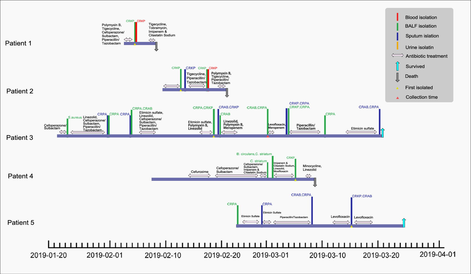
Patients with KPC-NDM-CR-KP infections were admitted to Shandong Provincial Hospital’s ICU from January to April 2019. Most had severe pneumonia, and one had postoperative pulmonary infection. Patients developed symptoms like pulmonary edema, pleural effusion, sputum, and shortness of breath, requiring mechanical ventilation. The strains were isolated within 3–42 days of admission. Patients 1 and 2 died due to severe pneumonia, sepsis, and multi-organ failure, while patient 4 discontinued treatment after septic shock. The other three improved with antibiotic therapy (Table 1, Fig. 1). All strains were highly resistant to β-lactam antibiotics, including carbapenems, but susceptible to tigecycline. JNKPN26 isolated from patient 3 was resistant to polymyxin (MIC > 64 mg/L) (Table 2).
Table 1 Clinical characteristics of patients with KPC-2-NDM-1-CR-hvKPFig. 1
Epidemiology of Klebsiella pneumoniae outbreak cases. Colored text and bars represent the source from which the bacterial species were isolated, whereas colored triangles represent the time at which the bacterial species were isolated. CRKP: carbapenem-resistant Klebsiella pneumoniae; CRPA: carbapenem-resistant Pseudomonas aeruginosa; CRAB: carbapenem-resistant Acinetobacter baumannii; S. aureus: Staphylococcus aureus; E. coli: Escherichia coli; B. circulans: Bacillus circulans; C. striatum: Corynebacterium striatum
Table 2 Microbiological characteristics of clinical isolatedPhylogenetic analysis of these KPs and eleven other CRKPs from the hospital showed that five KPC-NDM-CR-KPs and NDM-CR-KP JNKPN30 formed a separate clade (Subclade Ib of Clade I) (Fig. 2). Six strains had identical PFGE profiles, indicating the same clone. This clade appeared to evolve between 2018 and 2019, possibly due to acquiring a blaNDM-positive plasmid. Genetic analysis of JNKPN23, JNKPN26 and JNKPN30 revealed a chromosome (~ 5470 kb) and three plasmids per isolate, revealing nearly 99.7% coverage more than 200× in depth: a virulence plasmid (~ 220 kb), a KPC-positive plasmid (~ 120 kb), and an NDM-positive plasmid (~ 140 kb), as confirmed by S1-PFGE. High similarity was observed among the KPC-encoding IncFII/IncR plasmid, which co-harboring blaTEM-1, blaCTX-M-65 and blaSHV-12 resistance genes, with more than 99% identity. The IncC plasmid carried blaNDM and several AMR genes including blaCMY-6, sul1, emrE, aadA16, dfrA27, and arr-3, and exhibited a high similarity to others, with 100% coverage and 100% identity. JNKPN30 lacked blaKPC gene, but an IncFII/IncR-type plasmid was identified within it. WGS showed the IncFII/IncR plasmid lost a 25-kb fragment (including ΔISKpn6-blaKPC-2-ISKpn27 and IS26-blaSHV-12-TnAs1 resistance units, as well as ΔTn21) (Fig. 3). Notably, mgrB deletion was observed in the colistin-resistant strain JNKPN26 (Additional file 1: Fig. S1).
Fig. 2
Hospital outbreak evolution analysis of CO-NDM-KPC-CRKP. The simulated outbreak evolution analysis of CRKP strains isolated in one health set. A phylogenetic tree was constructed using the sample separation time to infer evolutionary relationships among clinical isolates (BioProject PRJNA792451). The features of CO-NDM-KPC-CRKP associated with our hospital outbreak are highlighted in bold
Fig. 3
Comparative analysis of hybrid plasmids. Circular maps and alignments of pJ-JNKPN26-2_HNK (GenBank accession no. OR041627) and pJ-JNKPN26-4_HNK (GenBank accession no. OR041629) plasmids, showing their structural features and comparative analysis with other blaKPC and blaNDM harboring plasmids sequenced in this study
All six strains harbored a 200–220-kb pLVPK-like virulence plasmid, identified as IncFIB/IncHI1B, carrying canonical virulence factors such as the regulator of the mucoid phenotype (rmpA and rmpA2), salmochelin (iroBCDN), aerobactin (iucABCD and iutA), and siderophore salmochelin (iroBCDE and iroN). The plasmid lacked antibiotic resistance genes and was devoid of T4SS, making it unable to undergo conjugative transfer. The G. mellonella virulence assay revealed that KPC-NDM-CR-KP JNKPN26 exhibited significantly higher virulence than ATCC13883 but showed similar virulence to the positive control NTUH-K2044. Furthermore, serum bactericidal assays indicated that KPC-NDM-CR-KP strains exhibited serum resistance, with a survival rate of approximately 78% after 60 min of incubation with pooled human serum. These phenotypic findings confirmed that KPC-NDM-CR-KP strains were hypervirulent (Fig. 4).
Fig. 4
Analysis of the virulence and serum resistance of Klebsiella pneumoniae strains. A Survival rates of Galleria mellonella larvae injected with Klebsiella pneumoniae strains. Survival data were plotted using the Kaplan–Meier method, and the groups were compared using the log-rank test. Hypervirulent K. pneumoniae NTUH-K2044 and classic K. pneumoniae ATCC13883 strains were used as lower-virulence comparators. B Evaluation of the serum resistance of ST11 Klebsiella pneumoniae strains. Data are presented as mean (SD). The reference K. pneumoniae strain ATCC 13883 and the hypervirulent strain NTUH-K2044 were used as controls for the susceptible (ATCC 13883) and resistant (NTUH-K2044) grades, respectively. Significant differences (two-tailed unpaired t-test) in viable counts (3 h) of NTUH-K2044 and ATCC 13883 cells are shown in the table. *Unpaired t-test was not used because identical viable counts were recorded. C Virulence gene matrix of Klebsiella pneumoniae strains. The presence and absence of virulence genes are represented by dark blue and light blue boxes, respectively. Red triangles indicate virulence genes located in the plasmids. NTUH-K2044 and ATCC13883 are publicly available K. pneumoniae strains
Mobilization of the pJNKPN26-KPC plasmid with the help of the conjugative pJNKPN26-NDM plasmidConjugation assays demonstrated that the NDM-1-positive plasmid, pJNKPN26-NDM, could efficiently self-transfer to E. coli J53 with a high frequency of 3.56 × 10⁻³. In contrast, KPC-positive plasmid had a much lower transfer frequency of 1.8 × 10⁻⁹. Interestingly, all the KPC-positive transconjugants selected from plates containing EDTA, also carried the NDM bearing IncC plasmids (Table 3).
Table 3 Microbiological characteristics of transconjugantsThe IncC plasmid harbored a complete conjugative transfer-related module, including an oriT region, a relaxase of the MobH family, a type IV coupling protein (T4CP), and a tra gene cluster that coded for type IV T4SS. In contrast, relaxase and T4CP were absent in the pJNKPN26-KPC plasmid, suggesting that the KPC plasmid transfer relied on assistance from the IncC plasmid.
The plasmid state of transconjugants coharboring NDM and KPC was assessed with S1-PFGE. In addition to NDM and KPC located on separate plasmids, similar to the clinical strain (~ 140 kb and ~ 120 kb), two extra S1 patterns were detected in the conjugates. Briefly, the transconjugants co-harboring NDM and KPC displayed three different S1-PFGE profiles: (i) J-JNKPN26-1, which carried two independent plasmids (approximately 140 kb and 120 kb); (ii) J-JNKPN26-2 and J-JNKPN26-3, which contained a novel large plasmid (approximately 270 kb) and two smaller plasmids (approximately 140 kb and 120 kb, respectively); and (iii) J-JNKPN26-4, which possessed a novel large plasmid (approximately 270 kb) that was smaller than the novel plasmids found in J-JNKPN26-2 and J-JNKPN26-3 (Fig. 5, Additional file 2: Fig. S2).
Fig. 5
Mobilization of pJNKPN26-NDM and pJNKPN26-KPC. A XbaI and S1-PFGE of K. pneumoniae JNKPN26, as well as their corresponding transconjugants. Differently colored triangles denote different plasmids: virulence plasmid pJNKPN26-Vir (blue triangles), IncFII/IncR plasmid pJNKPN-KPC (yellow triangles), IncC plasmid pJNKPN-NDM (green triangles), and novel hybrid plasmid (orange triangles). B Schematic representation of a round of conjugation assays and the state of hybrid plasmid after passage. Rounded rectangles of the same color represent the same strains. Blue, red, and yellow circles denote pJNKPN26-NDM or its derivatives, pJNKPN26-KPC or its derivatives, and pJNKPN26-Vir, respectively
In pattern (i), pJ-JNKPN26-1_NDM and pJ-JNKPN26-1_KPC were almost identical with NDM and KPC harboring the plasmids in the clinical strain; nevertheless, inverted repeats (IRs) at the 5′-end repeat of TnAs1 were only present within dnaQ in pJ-JNKPN26-1_NDM. Thus, the recombinant junction mediated by TnAs1 was supposed to occur (Fig. 6).
Fig. 6
Genetic structures of the conjugative hybrid resistance plasmid fusion regions. A Alignment of hybrid resistance plasmids pJ-JNKPN26-1-NDM, pJ-JNKPN26-2_HNK, pJ-JNKPN26-3_HNK, and pJ-JNKPN26-4_HNK with parental plasmids from JNKPN26. Black, yellow, red, cyan-blue, and other colored arrows indicate other function protein, conjugation transfer, resistance gene, IS insertion site, and mobile element protein, respectively. The evidence for the occurrence of the fusion event in transconjugant J-JNKPN261-1 was the emergence of TnAs1 on pJ-JNKPN26-1-NDM. C The resistance genes are indicated by rectangles. A target site and subsequent 8-bp duplications are indicated by a vertical flag. The relative frequencies of IS26- and TnAs1-mediated reactions are indicated by blue and green arrows, respectively. Proposed mechanisms of plasmid fusion. i pJ-JNKPN26-1_NDM and pJNKPN26-2_HNK: TnAs1 attacked the dnaQ gene of pJNKPN26-NDM, leading to the formation of fusion plasmids through a replicative transposition mechanism. ii pJ-JNKPN26-3_HNK: TnAs1 interrupted the uvrD gene of pJNKPN26-NDM, leading to the reverse insertion of pJNKPN26-KPC into pJ-JNKPN26-1-NDM forming plasmid pJ-JNKPN26-3_HNK. iii pJ-JNKPN26-4_HNK: formation of cointegration was mediated by IS26 interrupting ISEcp1
In pattern (ii), three plasmids existed in the transconjugant J-JNKPN26-2—namely, pJ-JNKPN26-2_HNK (270,446 bp, GenBank accession number: OR041627), pJNKPN26-2_NDM (144,158 bp), and pJ-JNKPN26-2_KPC (126,207 bp). The transconjugant J-JNKPN26-3 harbored three plasmids—namely, pJ-JNKPN26-3_HNK (270,443 bp, GenBank accession number: OR041628), pJ-JNKPN26-3_NDM (144,239 bp) and pJ-JNKPN26-3_KPC (126,207 bp).
In pattern (iii), one large plasmid was identified in the transconjugant J-JNKPN26-4 with a size of 267,313 bp (Fig. 3). Comparative genetic analysis revealed that pJ-JNKPN26-4_HNK (GenBank accession number: OR041629) was a fusion plasmid formed from pJ-JNKPN26-KPC and pJNKPN26-NDM, mediated by the insertion sequence IS26. The two IS26 sequences around the infusion site of ISEcp1 shared a 14-bp terminal IR sequence and 12-bp direct target repeats, suggesting that the recombinant junction was supposed to occur (Fig. 6).
Further genomic sequencing suggested that pJNKPN26-KPC underwent complete integration into pJNKPN26-NDM within the dnaQ, uvrD, and ISEcp1 genes in all three patterns. Additionally, genetic comparative analysis showed that TnAs1 in the KPC-2 positive plasmid mediated the formation of pJ-JNKPN26-2_HNK and pJ-JNKPN26-3_HNK hybrid plasmids. The insertion sites in pJ-JNKPN26-2_HNK and pJ-JNKPN26-3_HNK were dnaQ and uvrD, respectively (Fig. 6). PCR targeting the region spanning the integration site of the fusion plasmid pJ-JNKPN26-2_HNK confirmed the hybrid state and fusion site. The TnAs1-mediated fusion event in pJ-JNKPN26-KPC targeting the dnaQ or uvrD gene of pJNKPN26-NDM was reversible.
Stability and mobilization of hybrid plasmidsThe stability of the hybrid plasmids pJ-JNKPN26-2_HNK, pJ-JNKPN26-3_HNK, and pJ-JNKPN26-4_HNK during passage was verified by conducting PCR on the fusion fragments flanking the susceptible recombination sites of the hybrid plasmids. The TnAs1-mediated heterozygous plasmids (pJ-JNKPN26-2_HNK and pJ-JNKPN26-3_HNK) showed poor stability in the heterozygous state on day 7 (stability of 54% and 57%, respectively). In contrast, the IS26-mediated fusion plasmid (pJ-JNKPN26-4-HNK) remained stable (stability of 100%) during passage for 7 days in an antibiotic-free environment, indicating that the plasmid had relatively high stability (Fig. 5). However, the KPC-2 and NDM-1 resistance genes could be detected by PCR from all transconjugate colonies after passage on day 7.
A second round of conjugation assay was performed to evaluate the transfer capability of hybrid plasmids. The results confirmed that all hybrid plasmids could be successfully transferred from the J53 transconjugant to the E. coli C600 recipient strain. The conjugative transfer efficiencies ranged from 4 × 10⁻⁵ to 8.69 × 10⁻⁶ and were lower than those of the exclusive IncC plasmid.
留言 (0)