Cell lines and cell culture
MCF10A, MCF7, T47D, BT474, MDA-MB-453, BT549, MDA-MB-468, HCC1806, MDA-MB-231, and human embryonic kidney HEK-293T cell lines were purchased from the Chinese National Infrastructure of Cell Line Resource (Beijing, China). All human cell lines were authenticated using STR DNA fingerprinting at Shanghai Biowing Applied Biotechnology Co., Ltd. (Shanghai, China). MCF7, T47D, BT474, MDA-MB-453, BT549, MDA-MB-468, HCC1806, MDA-MB-231, and HEK-293T were maintained in DMEM supplemented with 10%FBS (Gibco). MCF10A cells were cultured in DMEM/F12 (Invitrogen) supplemented with 20 ng/ml epidermal growth factor, 5% horse serum, 0.5 µg/ml hydrocortisone, 10 µg/ml insulin, and 100 ng/ml cholera toxin. All cell lines were routinely verified to be mycoplasma negative using a mycoplasma detection kit (Sigma-Aldrich).
Fluorescence-activated cell sorting (FACS)
The ALDH+ cell staining was performed by using an ALDEFLUOR assay kit according to the manufacturer’s guidelines (STEMCELL Technologies). Briefly, cells were suspended in ALDEFLUOR assay buffer containing an ALDH substrate, BODIPY-aminoacetaldehyde (BAAA), and incubated for 30 min at 37 °C. Cells treated with diethylaminobenzaldehyde, an ALDH inhibitor, were used as the control. ALDH+ cells were sorted by flow cytometer (Beckman Coulter, CytoFLEX). Propidium iodide (PI) staining was used to identify and gate out dead and late apoptotic cells.
Mammosphere formation and extreme limiting dilution assay (ELDA)
The single-cell suspension was obtained by trypsinization and sieving through a 40-mm sieve. Single cells were plated in ultralow attachment six-well plates (Corning, 3471) at a density of 1 × 10³ cells/well and cultured in DMEM/F12 medium plus 20 ng/ml epidermal growth factor (EGF), 20 ng/ml basic fibroblast growth factor (bFGF), 20 µl/ml B27, and 4 mg/ml heparin for 10 days. Then, mammospheres were photographed under an inverted microscope (10× objective, Olympus). For ELDA, cells were seeded into 96-well ultralow attachment plates with sphere medium at densities of 5, 10, 20, 50, and 100 cells/well (12 wells per cell density). After 10 days, each well was examined for the formation of tumor spheres. Stem cell frequency was calculated using extreme limiting dilution analysis (http://bioinf.wehi.edu.au/software/elda/).
Cell invasion and migration analysis
Cells (5 × 10⁴) suspended in medium without FBS were plated on the upper chamber membranes (8 μm pore size, Corning) coated with or without Matrigel (BD Biosciences). The inserts were incubated in a medium supplemented with 10% FBS for 16 h. To evaluate the invasion and migration ability, non-invasive or non-migrative cells were removed. Invasive and migrative cells were fixed with methanol, stained with crystal violet, and counted under a light microscope.
RNA pull-down assay
LncRNA was transcribed and amplified in vitro according to the manufacturer’s instructions by using MEGAscript (Ambion, life technologies). Cell nuclear lysates were incubated with biotinylated lncRNA and streptavidin beads for RNA pull-down incubation. In brief, biotinylated RNA was refolded in NEB enzyme buffer with RNase-out (Invitrogen, USA) and then incubated with cell nuclear lysates and streptavidin beads for RNA pull-down incubation. For the pull-down incubations, MDA-MB-231 BCSC lysates were pre-cleared with streptavidin beads and then incubated with biotinylated RNA for 4 h at 4 °C. Beads were collected and washed with RNA binding buffer. RNA-associated proteins were eluted and resolved by SDS/PAGE followed by silver staining (Bio-Rad, USA). Bands were excised and subjected to LC-MS/MS sequencing and data analysis at Jiyun Biotech. Inc (Shanghai, China).
RNA immunoprecipitation
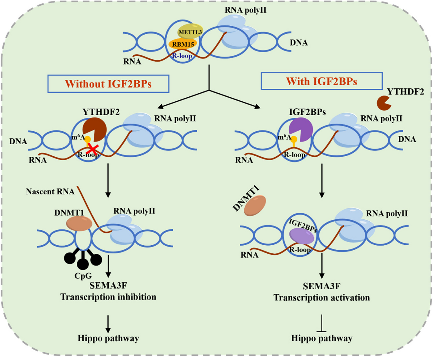
RNA immunoprecipitation (RIP) experiments were performed using the Megna RIP RNA-binding Protein Immunoprecipitation Kit (Millipore). The anti-YTHDF2 antibody (#71,283, Cell Signaling Technology) was used. The co-precipitated RNAs were detected by qRT-PCR. The gene-specific primers used for detecting LINC00115 are listed in Supplementary Table S2.
Plasmid construction
LINC00115, SETDB1, PLK3, and HIF1α cDNAs were purchased from GeneChem (Shanghai, China). They were sequenced and subcloned into the pcDNA3.3 or pLVX-Puro vector (Clontech). PLK3 or SETDB1-truncated constructs were generated by PCR using PLK3 or SETDB1-pcDNA3.3 as templates, and then they were inserted into pcDNA3.3. Point mutations were generated using a site-directed mutagenesis kit (Invitrogen) following the manufacturer’s protocol. GST-HIF1α were inserted into a pGEX-4T-1 vector for expression in E. coli.
shRNA-knockdown, sgRNA-knockout, antisense oligonucleotide (ASO), and transfection assays
LINC00115, SETDB1, HIF1α, and YTHDF2 shRNAs were purchased from Genechem (Shanghai, China). LINC00115-ASO purchased from RiboBio (Guangzhou, China). ALKBH5 shRNAs were gifted by Professor Xudong Wu as previously described [25]. Single-guide RNA (sgRNA) sequences of SETDB1 were designed using the online tool from the MIT online tool (http://crispr.mit.edu). Targeted DNAs and packaging plasmids were transfected into the HEK293T cells using the Hieff Trans Liposomal Transfection Reagent (40802ES08, YEASEN) following the manufacturer’s instruction. The supernatants were collected and filtered at 72 h after transfection. Targeted Cells were infected with 5 µg/ml polybrene (Sigma-Aldrich). Infected cells were selected with puromycin after infection. Multiple monoclonal cultures were screened for sgRNA by Western blotting and RT-PCR analyses.
Immunoprecipitation (IP) and Western blotting (WB) assays
IP and WB assays were performed as previously described [26]. In brief, to detect protein interactions, cells were lysed in IP buffer. These supernatants were immunoprecipitated with the indicated antibodies, slowly shaken on a rotating shaker at 4 °C overnight, and then incubated with Pierce™ Protein A/G Magnetic beads (Invitrogen) at room temperature for 1 h. Then, the binding proteins were eluted and boiled in 1× SDS loading buffer to prepare the samples for immunoblot analysis.
Antibodies and reagents
Antibodies against SETDB1 (1:1000 for WB, 1:200 for IHC and Co-IP, #2996), PLK3 (1:1000 for WB, 1:200 for Co-IP, #4896), GAPDH (1:1000 for WB, #5174), HIF1α (1:1000 for WB, 1:200 for IHC, #48,085), LDHA (1:1000 for WB, #3582), MDR1 (1:1000 for WB, #12,683), Mono-Methyl Lysine (1:1000 for WB, #14,679), Di-Methyl Lysine (1:1000 for WB, #14,117), Tri-Methyl Lysine (1:1000 for WB, #14,680), methyl-Histone H3 (Lys9) (1:1000 for WB, #13,969), ALKBH5 (1:1000 for WB, 1:100 for RIP, #80,283), YTHDF2 (1:1000 for WB, 1:100 for RIP, #71,283), N6-Methyladenosine (m6A) (3 µg for Me-RIP, #56,593), HA (1:1000 for WB, 1: 200 for Co-IP, #3724), His (1:1000 for WB, 1:200 for Co-IP, #12,698) were purchased from CST (Danvers, MA); an antibody against FLAG (1:1000 for WB, 1:50 for Co-IP, F3165) was from Sigma-Aldrich (US); an antibody against Phospho-Ser/Thr (1:1000 for WB, ab17464) was purchased from Abcam (UK). The secondary antibodies, anti-rabbit IgG, HRP-linked antibody (1:5000, #7074), and anti-mouse IgG, HRP-linked antibody (1:5000, #7076) were purchased from CST (Danvers, MA). PLK3-K106 and -K200 mono-methylation rabbit polyclonal antibodies were raised by Proteintech (US) using the peptide E-K(me)-ILNEIELH and LGNFFITENMEL-K(me) (1:1000 for WB, 1: 50 for IHC). Paclitaxel, CHX, and TDD-IN were from Med Chem Express. MG-132 was from Selleck Chemicals.
LncRNAs microarray and data analysis
For microarray analysis, RNA purity and integrity were analyzed by Agilent Bioanalyzer 2100 (Agilent). Qualified total RNA was further purified by RNeasy mini kit (QIAGEN) and RNase-free DNase set (QIAGEN). Sample labeling, microarray hybridization, and washing were conducted in accordance with the manufacturer’s standard protocols. Total RNA was transcribed to double-stranded cDNA, and then cRNA was synthesized. Subsequently, 2nd cycle cDNA was synthesized from cRNA. Following fragmentation and biotin labeling, the 2nd cycle cDNA was hybridized onto microarrays. After washing and staining, the arrays were scanned with an Affymetrix Scanner 3000 (Affymetrix). Affymetrix GeneChip Command Console (version 4.0, Affymetrix) software was used to extract the raw data. The Expression Console (version 1.3.1, Affymetrix) software provided RMA normalization for both gene- and exon-level analysis. Additionally, genespring software (version 14.9; Agilent Technologies) was used to complete the fundamental analysis. Differentially expressed lncRNAs were subsequently identified via fold change and p-values calculated by t-tests. The thresholds for up- and downregulated genes were fold-change > 2 and P ≤ 0.05.
RNA-Seq analysis
RNA-Seq and differentially expressed gene analysis were performed as previously described [26]. In brief, total RNAs were extracted and purified according to the RNeasy Plus kit (Qiagen, 74,104). Libraries were prepared using NEB Next® Ultra TM RNA Library Prep Kit for Illumina® (NEB, Beverly, MA, USA) following the manufacturer’s recommendations. The products were sequenced on the HiSeq3000 platform (Illumina, San Diego, CA, USA). The raw FASTQ files were trimmed for adapter sequences using quart. Then HISAT2 version 2.0.5 software was used to map to the hg19 reference genome with default [27] settings. Genes with an adjusted q value < 0.05 and FC > 2 were assigned as differentially expressed.
RNA extraction and quantitative real-time PCR
Total RNA was isolated from lung tissue and cells using TRIzol reagent (Invitrogen, USA) according to the manufacturer’s instruction. Total RNA from each sample was reverse transcribed to cDNA using the PrimeScript RT kit (Takara, Japan). Quantitative real-time PCR (qPCR) was performed on a Roche Light Cycler 480 System (Roche, Switzerland) using the SYBR Premix Ex Taq II RT-PCR Kit (Takara, Japan) to determine the transcript levels of the target genes. Supplementary Table S2 list the primer sequences. GAPDH was used as the internal reference gene for lung tissues and cells, and the relative expression of the lncRNAs was calculated by the − 2△△Ct method [28].
MeRIP-qPCR
Total RNAs were extracted with RNAiso Plus (TaKaRa). For meRIP, the procedure was described previously. In brief, purified mRNAs (5 µg) were digested by DNase I (M0303, NEB) and then fragmented into around 200 nt fragments by incubation at 95 °C for 25 s in RNA Fragmentation Reagents (Ambion, AM8740), followed by standard ethanol precipitation and collection. Anti-m6A antibody (10 µg antibody for 5 µg mRNAs; Synaptic Systems) was incubated with 40 µl Protein A beads (Sigma, P9424) in IPP buffer (150 mM NaCl, 0.1% NP-40, 10 mM Tris-HCl, pH 7.4) for 2 h at room temperature. The fragmented mRNAs (5 µg) were incubated with the prepared antibody-bead mixture for 4 h at 4 °C. By washing three times, bound RNA was eluted from the beads with 0.5 mg/ml N6-methyladenosine (BERRY & ASSOCIATES, P3732) in IPP buffer. The eluted RNA was extracted by Enol:Chloroform:Isoamylol (pH < 5.0, Solarbio life science, P1025) and then generated to cDNA using 5 x All-In-One RT MasterMix (ABM, G490). The enrichment of m6A was quantified by qPCR. The sequences of qPCR primers are listed in Supplementary Table S2.
RNA fluorescence in situ hybridization (FISH)
For in situ detection of LINC00115 in breast cancer tissues, FITC-labeled LINC00115 probes, U6 probes, and 18S probes were designed and synthesized by PinpoRNA (China). Cell or tissue FISH assay was performed with a Fluorescence in situ Hybridization Kit (PinpoRNA, China) according to the manufacturer’s instructions. Confocal laser scanning microscopy (Leica, Germany) was used to observe the images.
Drug sensitivity assays to paclitaxel and TDD-IN
Cells were seeded onto 96-well plates at an initial density of 5 × 10³ cells per well and treated with different doses of paclitaxel and TDD-IN. Sensitivity was assayed using CellTiter-Glo (Promega), which measures cellular ATP levels as a surrogate for cell number and growth, according to the manufacturer’s protocol. The solution was diluted 1 part CellTiter-Glo to 2 parts PBS before a 1:1 addition to the volume on the plate. Luminescence was measured using a PerkinElmer Envision.
Mouse experiments
All animal experiments were conducted under the Institutional Animal Care and Use Committee (IACUC)-approved protocols at Ren Ji Hospital, Shanghai Jiao Tong University School of Medicine in accordance with NIH and institutional guidelines. For the immunodeficiency mouse model, MDA-MB-231 PTX-resistant cells (1 × 10⁵) or their derivatives were suspended in 100 µl of PBS and then injected into the tail vein of nude mice (6 mice for each group). For in vivo drug treatment, 7 days after inoculation, tumor-bearing mice were treated randomly with or without TDD-IN (50 mg/kg), paclitaxel (10 mg/kg), and LINC00115-ASO (10 mg/kg) intraperitoneally every day 21 days after injection, luciferin was injected and the primary/metastatic tumors were detected by BLI with the IVIS 100 (Caliper Life Sciences, Hopkinton, MA, USA). Metastasis in the lungs was detected by bioluminescence imaging (BLI). Animals were treated with isoflurane and then sacrificed with CO2 when showing a symptom of cachexia or multiple organ failure. All lobes of the lung were harvested and the number of macroscopic metastatic nodules on the surface of the lung was counted.
Histology and immunohistochemical staining
Hematoxylin-eosin (H&E) and immunohistochemistry (IHC) stainings were performed on 4-µm formalin-fixed paraffin-embedded tissue sections. The mice’s lung sections were stained with H&E and scanned using a Scanscope XT digital slide scanner (Aperio Technologies). Digital images of lung sections were used to analyze the metastatic burden. IHC for patients’ tissues was performed using anti-SETDB1 (1:100), anti-PLK3K106me1 (1:50), anti-PLK3K200me1 (1:50), and anti-HIF1α (1:100) antibodies. The staining extent score was on a scale of 0–3, corresponding to the percentage of immunoreactive tumor cells (0-10%, 11-25%, 26-75%, and 76-100%, respectively) and the staining intensity (negative, score = 0; weak, score = 1; strong, score = 2; very strong, score = 3). A score ranging from 0 to 3 was calculated by multiplying the staining extent score with the intensity score, resulting in a low (0–1) level or a high (2–3) level value for each specimen. The stained tissues were scored by three individuals blinded to the clinical parameters.
Human tissue samples
All paraffin-embedded sections of clinical TNBC metastatic lymph nodes were collected at Ren Ji Hospital, Shanghai Jiao Tong University School of Medicine in accordance with a protocol approved by Shanghai Jiao Tong University Institutional Clinical Care and Use Committee of Renji Hospital (Shanghai, China). These patients received chemotherapy, and were with locally advanced or metastatic TNBC progressed after standard chemotherapy including taxol. Informed consent was obtained from all patients. These specimens were examined and diagnosed by independent pathologists. All the research was performed according to the provisions of the Declaration of Helsinki of 1975.
Statistical analyses
The significance of the data between experimental groups was determined by one-way analysis of variance (ANOVA) with Newman-Keuls post-test or unpaired two-tailed Student’s t-test. Pearson Chi-square test or Fisher exact test (two-sided) was used to evaluate IHC score levels between different clinicopathological variable groups. Survival analysis was calculated using the log-rank test and the Kaplan-Meier method. Statistically analyzed data are expressed as the mean ± S.D. or mean ± S.E.M., as indicated. P < 0.05 was considered statistically significant. All statistical analyses were performed with GraphPad Prism version 8.3 (GraphPad Software Inc., San Diego, CA, USA).
Data availability
LncRNAs microarray and RNA-Seq data reported in this study have been deposited with the Gene Expression Omnibus under the accession GEO ID: GSE245145. The data supporting the finding of this study are available within the article and its Supplementary Information files or available from the corresponding author on reasonable request.


留言 (0)