In the present study, we compared p53 IHC expression with the mutation status of TP53 as determined by NGS in patients with CRC. In the 92 samples analyzed, TP53 mutations were detected in 72 cases (62.0%), with 53 (73.6%) variants harboring a missense variation and 19 (26.4%) variants having non-sense or frameshift mutations, including 10 stop codon variants. There were five frameshift variants, two in-frame deletions, one in-frame insertion, and one splice donor variant. The frequency of TP53 mutations examined in the present study was consistent with previous studies [19,20,21,22,23]. Therefore, we believe that the present study provides reliable data on TP53 mutations. Based on a previous study, the cut-off value for p53 IHC expression reflecting missense mutations was 80%, and the cut-off value for nonsense/frameshift mutations was 0% in the present study [15]. In addition, expression intensity and distribution were important for p53 IHC expression pattern classification. However, there may be serious limitations to this cut-off value, given that it is unclear how < 80% p53 IHC expression should be classified. For example, a case with 70% p53 IHC expression, with a diffuse and strong staining pattern, is difficult to assign into any of the four-tier classifications. However, there is no such case in the present study. Further consideration may be required for this cut-off value.
Previous studies have reported on using IHC staining for p53 as a tool to assess TP53 mutation status [12,13,14, 23]. However, after the introduction of NGS, sequencing of the TP53 gene in cancer cells has increasingly been used [9, 14]. Previous studies have shown the correlation between p53 IHC expression patterns and TP53 variants status detected by NGS [9, 14]. Köbel et al. adopted a three-tier classification, consisting of wild-type, overexpression, and complete absence [14]. According to this report, the p53 IHC expression patterns showed excellent concordance with the TP53 variant status [14]. The sensitivity of IHC for detecting gain-of-function variations, loss-of-function variations, and the wild-type expression of p53 was 100, 76, and 100%, respectively [13]. In addition, the specificity of IHC for detecting gain-of-function variations, loss-of-function variations, and wild-type expression of p53 was 95, 100, and 96%, respectively [13]. Recent studies have shown that p53 IHC expression patterns can be categorized as wild-type patterns or aberrant-type patterns, and this classification was found to be significantly correlated with TP53 NGS classification in CRC [9, 13]. In gastric cancer, IHC p53 expression patterns were significantly correlated with TP53 variations detected by NGS [24]. In the present study, we adopted a four-tier classification that categorizes samples into wild-type, overexpression, null-type, and cytoplasmic p53 patterns. We found that this classification could predict TP53 mutation patterns with high sensitivity and specificity. Our data suggest that p53 IHC patterns have a good predictive value for TP53 mutations.
Weakly-stained wild-type cases have frequently been misclassified, resulting in false positive mutation predictions [14]. Weakly-stained assays performed without intrinsic controls cannot reliably distinguish null-type from wild-type phenotype [14]. Therefore, we propose the use of intrinsic controls as an internal reference for IHC scoring. Despite a common consensus that p53 IHC cannot detect p53 wild-type protein due to its rapid degradation, the DO7 antibody used in this study detects p53 expression in normal cells, including stromal fibroblasts and lymphocytes, when used with recently improved polymer-based IHC detection systems [25]. It is possible that p53-positive intraepithelial lymphocytes in a complete absence case could be falsely assessed as p53 wild-type tumor cells [24]. Use of improved polymer-based IHC detection systems is needed to differentiate wild-type from null-type p53 expression [25]. Our data strongly support the contention that further assay comparison and training in interpretation are required for p53 IHC to be used as a diagnostic and predictive test.
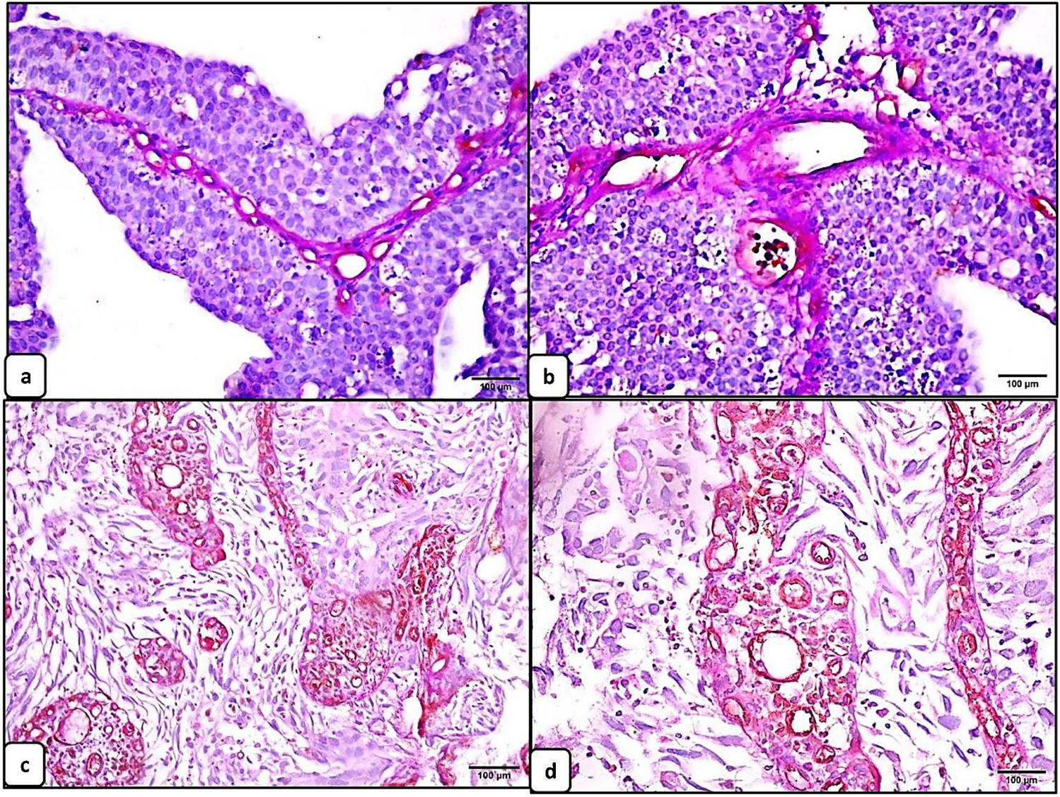
It should be noted that the p53 cytoplasmic pattern was not a single pattern but presented as combinations of cytoplasmic and nuclear staining in a previous study [26]. The cytoplasmic staining pattern shows a spectrum of cytoplasmic stain intensity from weak to strong and from heterogeneous to uniform [26]. Importantly, if a range of nuclear expression patterns of varying intensity involving a few cells to < 80% of tumor cells was observed, such cases were classified into the p53 cytoplasmic pattern. Moreover, a whole slide section from which TMA cores were taken showed a variable amount of nuclear staining with the cytoplasmic staining in this study, supported by previous studies [14, 15]. Focal nuclear staining in tumors with p53 cytoplasmic patterns may be challenging to detect with TMAs rather than by whole slide sections [26]. This may explain some of the differences between previous studies and our data [26]. Finally, determining the prognostic value of the cytoplasmic pattern is very important, and further studies will be needed to evaluate its clinical significance.
There are some limitations to our study. First, the sample size may be too small to identify a correlation between p53 IHC patterns and TP53 mutation status. However, given that the present sample size was higher than the minimum sample size we determined in our sample size calculations, we believe that this sample size is sufficient to investigate the association between p53 IHC patterns and TP53 mutation status. Second, we did not examine the association of p53 IHC patterns with patient prognosis, but aim to do so in a subsequent study. In addition, we excluded CRC samples with microsatellite instability (MSI). Therefore, the present cohort is not representative for evaluating molecular alterations of CRC with such a phenotype. Finally, the p53 IHC pattern and the TP53 mutation status were not obtained from the same site in the present study. Therefore, the p53 IHC pattern may not reflect the TP53 mutation status. However, both sampling sites were obtained from adjacent areas. Moreover, we used isolated tumor gland samples to increase the data accuracy [18]. We suggest that the p53 IHC pattern reflects the TP53 mutation status in the present study.
In conclusion, to our knowledge, this study is the first to examine the correlation of the proposed p53 IHC patterns with TP53 mutation status in CRC. We revealed that the interpretation of p53 IHC patterns is highly reliable and reproducible, and can serve as an excellent surrogate approach for assigning p53 IHC classes. In addition, we showed a high agreement supported by optimal laboratory protocols with adequate controls. Experience, training, and proper p53 IHC staining protocols will be required for routine diagnostic pathology. Nevertheless, the combination of p53 IHC and sequencing should be helpful in considering the p53 functional status for clinical applications.


留言 (0)