UFCM imaging of breast normal and tumoral tissue
UFCM is a new method allowing the intraoperative examination of fresh tissue with limited processing. UFCM images provide a large field-of-view of surgical specimens (up to 20 cm2) allowing the simultaneous analysis of the main tumor and the surrounding breast tissue. The largest UFCM data collection from breast surgical specimens to date has been collected in the present study. None of the postoperative assessments showed artifacts that were attributed to UFCM imaging, indicating that UCFM is not tissue-traumatic as reported by Elfgen et al. [12].
The UFCM image allows the evaluation of the global architecture of the tissue at low magnification while higher magnifications (up to digital 40 ×) allow to explore and identify morphological features of the tumors. With an axial resolution of about 30 µm, UFCM images are richer than 3–4 µm histological sections in terms of cell density.
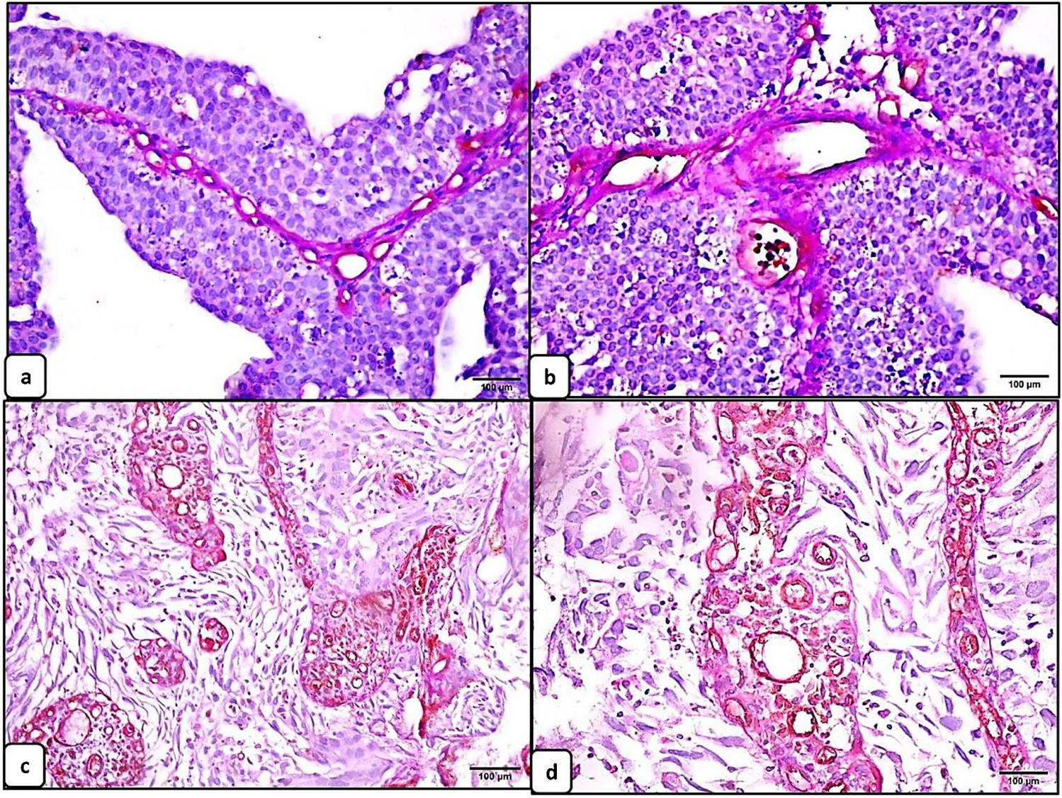
The different components of the normal tissue are usually recognized with confidence: adipocytes, fibroblasts, lobules, ducts, lymphocytes, nerves, and vessels. The adipose tissue is well preserved on fresh tissue with the UFCM technique and do not constitute an issue as observed in frozen sections. The 30-µm axial resolution provides a three-dimensional observation of vessels, ducts, and cysts which is confusing at the beginning and requires adequate training.
UFCM is providing lower level of magnification than conventional optical microscopes increasing the contribution of tissue architecture assessment in the diagnosis. The non-organoid organization of epithelial proliferations, the high density of cells, and stellar organization are typical of IC at low magnification. For IC-NST, at higher magnification, the architecture (cords or tubules), the size of the nuclei (compared to the size of the lymphocyte), and the stroma confirm the diagnosis.
The Indian files, the abundant stroma and the small size of tumor cells are characteristic of ILC visualized at high magnification. There are two difficulties for ILC: (1) the differential diagnosis with inflammation, especially in the scar of the preoperative biopsy. The size of the nuclei of ILC cells can be similar to activated lymphocytes; in these cases, the architecture of the lesion and the stromal organization may be of help for the diagnosis; (2) the differential diagnosis with IC-NST could also be difficult when ILC is not composed of dyscohesive cells and is presenting an architecture resembling to IC-NST, as previously shown [17]. These issues for the diagnosis of ILC can also be encountered in conventional histology, and this is usually addressed with E-cadherin immunohistochemical staining.
For DCIS, intraductal proliferation in dilated ducts could be easily recognized. The architecture of the proliferation is different compared to HES because of the 30-µm axial resolution of the microscope. The cribriform and the micropapillary pattern can be visualized but necessitate some training to be identified. High-grade DCIS has large nuclei, identifiable nucleoli, and lymphocytic infiltration around ducts. Low-grade DCIS may be difficult to differentiate from LCIS or atypical ductal hyperplasia. Microcalcifications and necrosis could not be seen on UFCM images. This phenomenon was likely due to the absence of fluorescence dye both in calcifications that lack nucleic acid for binding and in necrotic areas in which nucleic acids are highly degraded.
In the HIBISCUSS study, we selected patients with a pre-operative diagnosis of the most frequent breast carcinomas: IC-NST, DCIS, and ILC. The analysis of all lesions (tumoral or not) present in the UFCM images has been performed. However, some rarer IC subtypes and benign lesions, as well as tumor after neoadjuvant chemotherapy, are missing in the current atlas, and further studies addressing these topics are underway.
UFCM image acquisition
The pseudo-color mode used in the UFCM images replaces the standard grayscale in CM. It resembles to histological sections that are more familiar to pathologists, and this facilitated the interpretation of the images [18, 19]. With an approximate axial resolution of 30 µm, UFCM images provide a light three-dimension visualization of the tissue surface, especially for ductal and lobular structures. UFCM also permitted to visualize the network of small normal vessels in three dimensions.
In most cases, the core tumor mass was not imaged on the UFCM images and presented as an empty hole of various dimensions surrounded by tumor cells. This was caused by the lack of contact between the center of tumor and the imaging interface during UFCM acquisition, likely due to the scirrhous fibrous stroma in the center of IC. To limit this artifact, we used a surgical instrument to gently flatten out the specimen along the imaging window. This artifact may be considered an indirect marker of IC with abundant fibrous stroma.
Among the artifacts seen on UFCM images, the main problem was the stitching effect due to specimen movement during acquisition. This problem was seen only after image generation, necessitating repeated image acquisition of the surgical specimen. It was caused by “slow spreading” of the surgical specimen until its stabilization. A newer version of the UFCM device with a prediction tool of moving artifacts has been recently released to support users in reducing the occurrence of this specific artifact.
UFCM clinical implementation
In 2019, the committee of the College of American Pathologists focused for the first time on ex vivo microscopy applications, studying the potential advantages of CM over conventional histopathology [20]. UFCM recently joined the clinical market offering a larger field-of-view (20 cm2) and faster acquisition (less than 1 min per image) compared to other CM commercially available [14]. The histological interpretation of the UFCM images is close to the HES semiology and the implementation of the technique for intraoperative examination of specimens, especially margins, could be a streamlined process. However, an UFCM training is recommended for pathologists to make a histological diagnosis on UFCM images with confidence. Some issues related to both image interpretation and tissue handling should be regarded to avoid misdiagnosis. Recently, two clinical studies, with 50 and 40 patients respectively, reported margins assessment of lumpectomies with UFCM [15, 16]. The interpretation was devoted to marginally trained surgeons and a pathologist experienced in CM. They noticed a sensitivity from 30.7 to 33.3% and a specificity from 60.3 to 85.1% for surgeons and sensitivity from 43.8 to 53.8% and a specificity from 84.1 to 85.2% for the pathologist. Togawa et al. underlined that the low performance in their study was mainly due to artifacts caused by the use of electric surgical knives with high intensity and to a delay between surgery and image acquisition that caused excessive surface tissue drying. These issues could seriously impair image quality compromising further interpretation. In contrast, Sandor et al. used cold blades and scissors and had no heavy delays between surgical excision and UFCM imaging to produce good image quality and concluded that intraoperative reading of UFCM images by surgeon and pathologist could lead to a potential reduction of 30% and 58.3% of re-operation, respectively [15]. Additionally, the retrospective analysis conducted by an expert pathologist on UFCM images in Sandor et al. showed that as much as 75% of the re-operations could have been prevented with additional training on UFCM images. This finding underscores the need for adequate training material, and the present atlas as well as the learning program for pathologists and surgeons already developed in HIBISCUSS project [17] are believed to address this need. Finally, the creation of this large UFCM images database could be used to develop a deep learning model for image interpretation, which would constitute a supporting tool for decision-making of the clinician [21]. Due to the large field of view and false-coloring display of images, the atlas is currently specific to the Histolog Scanner but it could be used for other devices that would be developed in the future with the same approach.


留言 (0)