
記住我


Immersive virtual reality (VR) represents a major technological advance in mental health care and its application might substantially shape the future of pain management.22 However, although VR is already being used in pain management,11,26,31,37,48 knowledge of its specific impact on pain perception is largely unknown.
Immersive VR creates interactive, computer-generated worlds that replace real sensory perceptions with digitally generated ones.6,18 As participants move their head to look around, the computer updates the displayed images so that participants experience a surrounding 3D-stereo scene that changes dynamically. When what we see matches our movements, the brain concludes that this is our environment. A mental representation of this world is created, which is thereby experienced as real. This virtual manipulation of context in turn interacts with the perception of self and body.33 However, little is known about the interaction of such a virtual manipulation with the perception of pain.
An established tool to investigate the perception of acute pain is quantitative sensory testing.2 In addition to determining pain threshold and pain tolerance, suprathreshold pain measures, such as temporal summation of pain (TSP) and spatial summation of pain (SSP), can be examined. In addition, pain-inhibitory capacities can be tested through conditioned pain modulation (CPM). Such psychophysical tests permit to examine important mechanisms of pain modulation, such as increased activity of ascending nociceptive pathways or decreased activity of descending pain-inhibitory mechanisms.2
For several pain modalities, patients with chronic pain showed significantly lower pain thresholds and pain tolerance levels,45 increased TSP and SSP,34,45 and deficient CPM compared with healthy controls.10,12,27,34,36,45 However, little is known about the influence of an immersive environment on these parameters. Such knowledge could provide valuable information for future therapeutic applications of VR in pain management. For instance, demonstrating that immersive manipulation has a particular influence on certain aspects of endogenous pain modulation would provide a starting point for the differentiated use of this technique in patients with deficits in this domain.
In a previous study with healthy pain-free controls, immersive VR compared with a nonimmersive control condition resulted in increased pain thresholds for heat. Here, the immersion in a pleasant natural scenery was most effective.8 Similar results have been reported on the influence of mental imagery on pain perception. Mental imagery can significantly modulate pain in certain chronic pain syndromes.29 Consistent with this, psychophysiological studies have described a small increase in pain thresholds under imagination.40,46 Although these psychological interventions are increasingly being used in the management of pain, little is known about their influence on the contributing mechanisms that either facilitate or inhibit nociceptive experiences, as assessed by TSP, SSP, and CPM. It is also unclear whether individuals with and without pain respond differently to the influence of an immersive environment. Consequently, it seems worthwhile to compare the effects of immersive VR with those of mental imagery, in addition to controlling for nonimmersive VR.8,46 Recognizing the link between these interventions and pain modulation could be an important first step in developing more targeted interventions. For example, if a link could be established between VR and CPM in patients with pain, this knowledge could be used in the future to specifically activate deficient CPM using VR.
The aim of this study was to investigate (1) whether an immersive VR environment has a specific influence on different psychophysical indicators of pain perception and pain modulation, (2) whether the influence of an immersive VR environment goes beyond the effects of mental imagery or a nonimmersive control condition, and (3) whether specific differences in their response to VR can be identified between individuals with and without chronic pain. We postulated that the VR environment has an inhibitory effect on pain perception parameters and interacts with pain modulation outcomes (hypothesis 1), that this VR effect is stronger than the effect of mental imagery or a nonimmersive control condition (hypothesis 2), and that these effects can be demonstrated in both individuals with and without chronic pain (hypotheses 3).
2. Methods 2.1. Recruitment and characterization of the sampleTwenty-nine individuals with chronic pain (CP) and 31 healthy controls (HC) were recruited by public announcements at the University of Heidelberg and through social media. Individuals were included if they were aged 18 to 65 years and could speak, read, and understand German. Individuals with pain or injury in the intended test area (calf), pregnancy, current abuse of drugs, use of opioids, antipsychotics, or benzodiazepines were excluded. Individuals were eligible for the pain group if they had experienced pain for more than 3 months; for inclusion in the pain-free control group, participants were not allowed to experience current or previous chronic pain conditions. Recruitment and conduct of this study were in accordance with the Declaration of Helsinki, and the protocol was approved by the Ethics Committee of the University of Heidelberg (No.: S-578/2020).
Age, gender, marital status, level of education, employment status, and smoking status were assessed by questionnaires. In addition, participants were instructed to rate their “chronic pain” over the past 4 weeks before the study appointment for pain intensity and pain-related interference with daily life. Pain intensity was defined as how severe a person perceived their pain. It was assessed using a numeric rating scale (NRS), ranging from 0 = “no pain” to 10 = “worst pain imaginable.” Based on the German version of the Chronic Pain Grade questionnaire,25,47 pain-related interference for the domains “interference with usual activity,” “interference with daily activity,” “interference with leisure time activity,” and “interference with ability to work” was assessed individually for each domain using an NRS ranging from 0 = “no interference” to 10 = “worst imaginable interference.”
2.2. Experimental procedureParticipants were informed about the study and gave written consent in a meeting before the experimental session. Herein, individuals were told that this was a feasibility study and that the aim of the study was to test the tolerability and acceptability of VR in individuals with chronic pain. All experiments were conducted in a quiet laboratory at the Heidelberg University Hospital. Throughout all 4 test conditions, participants kept the VR headset on. Participants sat comfortably on sofas while resting their feet on a height-adjustable footrest, so that the leg muscles could relax completely, and there was good freedom of movement for the upper body and arms. Psychophysical outcomes of mechanical pain measures (pressure cuffs) were recorded on the proximal lower legs using pressure cuff algometry. Cuff algometry has proven to be a valid and reliable tool for measuring pain perception and pain-modulating mechanisms,9,15,16,28 and a close relationship between changes in cuff algometry and clinical pain conditions has been demonstrated.21,23,35,43,44
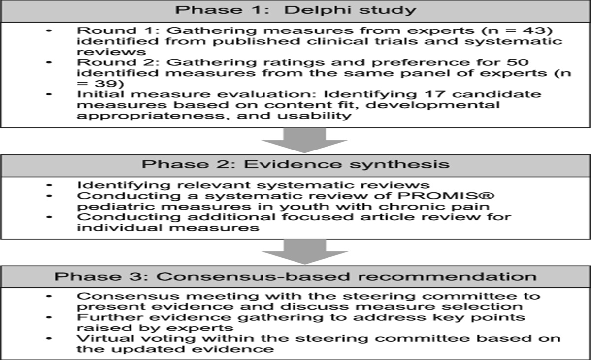
Within each test condition (baseline, imagery, view, and VR), 4 pain paradigms were measured: (1) general deep tissue pain sensitivity on the right leg and on the left leg (PDT: pain detection threshold, PTT: pain tolerance threshold), (2) spatial and (3) temporal summation of pain (SSP and TSP), and (4) CPM. The first test condition was the baseline assessment of individual pain thresholds for cuff pressure to determine the intensity of the test and conditioning stimuli. After the baseline assessment, the other 3 test conditions were performed in a randomized order (Fig. 1).
 Figure 1.:
 Illustration of the experimental setup and procedure. Participants went through 4 conditions, including (1) baseline, (2) immersive VR, (3) 2D control view, and (4) mental imagery. In each condition, participants underwent (1) the pain sensitivity (pain detection threshold and pain tolerance threshold) assessment followed by (2) the assessment of temporal pain summation, (3) spatial pain summation, and (4) the conditioned pain modulation. The first condition after baseline was counterbalanced, and the order of conditions following was randomized after generating 6 sequences to control for time effects.
All test conditions were started by a 5-minute induction period during which participants were exposed to the respective test environment without taking a measurement. The duration of a complete test session (baseline, VR, video, pictures) was usually between 10 and 15 minutes in total, plus the 5-minute induction phase, so that the total duration of all test conditions was usually between 60 and 75 minutes. During each test condition, participants were instructed to remain in their respective test context while simultaneously focusing their attention on their legs and continuously verbally reporting the painfulness of the pressure of each applied test stimulus using an NRS. These verbal pain ratings were documented directly by the assessor using an electronic NRS (eNRS) in parallel with the test application. The NRS uses a 10-cm line with the numbers 0 to 10, with the end point “no pain perceived” marked at the left end of the line and “maximum pain tolerated” marked at the right end of the line. To ensure accurate reporting of pain levels, a standardized training session was conducted before the study. This session introduced participants to the use of NRS and calibrated the pressure cuffs using spatial summation paradigm. Calibration allowed participants to become familiar with the cuffs and adjust their NRS scores accordingly. To prevent the experimenter from influencing the participants' pain ratings, 2 steps were taken. First, participants were informed of the calibration phase, and its results were not used in the study. Second, the experimenter received training not to affect the participants' ratings and to only follow their instructions for eNRS adjustments.
2.3. Experimental intervention 2.3.1. Sequence of test conditionsSubjects were exposed to 4 experimental conditions during 1 single session: (1) a baseline condition (“baseline”) where no intervention took place, (2) an immersive and interactive VR landscape scene (“VR” condition), (3) a mental image of the landscape scene instructed to subjects through a standardized audio file with headphones (“Imagery” condition), and (4) a nonimmersive 2-dimensional version of the landscape scene (“view” condition). All scenarios were selected and constructed in such a way that the intended psychophysical indicators of pain perception and modulation could be well integrated into them using pressure cuff algometry. Before testing, individuals were asked about their expectations of the extent to which they thought that VR was a suitable treatment option for their pain. After testing, subjects were asked how well they engaged with virtual reality. These measures were taken using a 5-point Likert scale ([1] very poor; [2] poor; [3] neutral; [4] good; [5] very good).
2.3.2. Baseline condition (baseline)The baseline test was the first test condition for each participant. The other 3 conditions were subsequently administered in randomized order. Before testing started, subjects were familiarized with pain testing and the electronic NRS. Participants did not wear the VR headset during the baseline condition. They received the VR headset after the baseline testing and kept it on during all subsequent conditions.
2.3.3. Virtual reality test conditionFor the VR test condition, we used a commercially available and ad hoc immersive and interactive (ie, head movements moved the contexts) VR context in which participants were immersed in a landscape scene on a tropical island with a white sand beach (Nature Treks VR, Version 1.26; 2019 Greenergames, United Kingdom). The island is vegetated by palm trees and grasses. One part of the island includes a rocky landscape. The subject can walk completely around the island. The island is surrounded by water through which the subject can swim to 2 neighboring islands (Fig. S1). In this context, subjects were able to freely move around the immersive environment using head movements and a handheld controller. Although in the real world, the individuals sat comfortably in the experimental chair the whole time, with their legs hanging loosely from the experimental chair, they were able to determine the direction in which their virtual self could move by manually controlling the right-hand controller of the VR device. The headset we used was the Oculus Quest (Oculus VR, Menlo Park, California, United States). Before starting the test procedures, all participants were instructed to move freely in the virtual world for 5 minutes. After this immersion induction phase, the measurement protocol was started as in the other 3 conditions. For the measurement of pressure cuff algometry during the immersive VR test condition, subjects were asked to continue moving through the virtual space, while simultaneously focusing their attention on their legs and to rate the sensations induced in their legs by the pressure cuffs through standardized instructions that were the same for all 4 test conditions and were read by the experimenter.
2.3.4. Mental imagery condition (imagery)The imagery test condition was based on a purely mental imagination of the natural scene. The VR headset showed a black screen during the whole condition. Participants were instructed to close their eyes during the imagination journey to better follow the standardized auditory instructions for the imagination journey by means of the headset, but they could leave their eyes open if they felt more comfortable. The headset with the dark screen ensured that all participants were exposed to the same visual stimuli and that this test condition was comparable to the other conditions. Then, the imaginary journey began, and participants were asked to follow the instructions without any manipulation for the first 5 minutes. After this induction phase, the measurement protocol was started as in the other 3 conditions. The specific instructions given through the headphones of the VR headset closely followed the contents of the VR context to create an imaginary world in the subjects, which was as similar as possible to the other 2 test conditions. At the same time, the instructions were designed in such a way that the measurements could be well integrated into the imaginary journey (see supplemental material for more details, available at https://links.lww.com/PAIN/B938). For the measurement of pressure cuff algometry during mental imagery, subjects were asked to direct their attention to and evaluate the sensations in their legs induced by the pressure cuffs by means of a standardized instruction, while simultaneously remaining in their imagery of the nature scene.
2.3.5. Nonimmersive control condition (view)To evaluate the contribution of the VR-based immersive experience, we included a control condition with a nonimmersive, noninteractive 2D version of the VR context. This was a video recording created by a screen recording that contained exactly the same content as the VR test condition and was also played through the VR headset. Therefore, we recorded a representative scene from the VR Nature Treks program as a video stream using a screen recording software (ShareX; frame resolution: 1280 × 720 pixels; frame rate: 30 frames per second), which included all the representative locations and animations of the original 3D-VR application. This recorded virtual reality movie scene was then stored as a file on the dedicated computer where it could be accessed by the experimenter and played as a 2D video on the VR headset, allowing participants to view it as a 2D version without having to be in the game. Before starting the test procedures, all participants were instructed to watch the movie for 5 minutes. After this condition induction phase, the measurement protocol was started as in the other 3 conditions. For the measurement of pressure cuff algometry during the nonimmersive 2D control condition, subjects were asked to focus their attention on and evaluate the sensations induced in their legs by the pressure cuffs through a standardized instruction, while simultaneously continuing to watch the movie sequence of the nature scene presented by means of headset.
2.4. Test paradigms 2.4.1. Setup and sequence of pressure cuff algometryA pressure cuff system (NociTech; Aarlborg, Denmark) was used to examine pain thresholds and pain tolerance levels (PDT and PTT), spatial (SSP) and temporal pain summation (TSP), and CPM.16 Four psychophysical paradigms were run sequentially, including (1) PDT/PTT, (2) SSP, (3) TSP assessment on the dominant leg, and (4) the CPM protocol with PDT/PTT assessment on the dominant leg and conditioning on the nondominant leg. The cuffs were applied slightly below the knees around the most voluminous parts of the gastrocnemius muscle with participants in a reclined sitting position. The cuffs were automatically inflated according to the amount indicated in the protocol. During pressure application, participants were asked to continuously verbally report their pain on a 10-point NRS. Maximum cuff pressure was limited to 100 kPa with 100 seconds as the maximum time of stimulation per setting (as the pressure increased by 1 kPa/s), and participants could immediately deflate the cuffs by reporting that their PTT was reached. Each subject was familiarized with the cuff algometry device during a training phase at the beginning of the session, during which the subject was seated and the cuff was placed around the upper arm. Subjects were trained on how to stop the cuff inflation (see supplemental material for more details, available at https://links.lww.com/PAIN/B938).
2.4.2. Pain threshold and pain toleranceThe PDT and PTT protocols were performed under a steadily increasing pressure (1 kPa/second) separately for both legs up to a possible maximum of 100 kPa. The pressure at which participants reported first perceiving the pressure as painful was defined as the PDT and marked as 1 out of 10 on the electronic numeric rating scale.16,32 After reaching the PDT, subjects started to continuously rate their pain intensity during further cuff inflation. The pressure at which participants indicated that they felt the pain was no longer tolerable was defined as the pressure PTT and marked as 10 out of 10 on the electronic numeric rating scale.16,32 For subjects who reached the maximum pressure of 100 kPa, this pressure was taken as a conservative estimate of the PDT and PTT.19 First, the values on the dominant leg were determined, followed by the values on the nondominant leg. The values on the nondominant leg were only used to determine the intensity of the test stimulus during the CPM paradigm. They were not included in the estimation of the outcome measures.
2.4.3. Spatial summation of painSSP relates to the increase in pain intensity because of an increase in the size of the stimulated body area.28 Here, this was achieved by activating pressure cuffs on the dominant and nondominant legs simultaneously. The spatial summation of pain is the result of the integration of neuronal activity at different levels of the neuroaxis.1 Because pain does not increase linearly with stimulated area, but logarithmically, the SSP effect is also referred to as subadditive. The subadditivity interacts with the descending pain system39 and suggests that the SSP effect is a unique intermixture of facilitatory and inhibitory processes.1,39 In our study, the degree of spatial summation of pain (SSPPDT and SSPPTT) was assessed by the ratio between the cuff PDT (PTT) measured with both cuffs simultaneously divided by the mean PDTs (PTTs) of the 2 single cuffs (left and right legs, respectively).17
2.4.4. Temporal summation of painTemporal pain summation measures the increase in pain perception because of repeated or prolonged stimulation. The TSP response is based on the windup effect of neurons in the dorsal horn of the spinal cord.38 In our study, TSP was assessed once by applying 10 cuff stimuli on the dominant leg. Each stimulus was 1 second in duration and 1 second apart.14 The applied pressure corresponded to the PDT plus 70% of the range between PDT and PTT (see above). Participants were instructed to rate their pain continuously on the NRS. Participants had the chance to rate the stimuli as painful but below the value of 1 to indicate the PDT. We scored these values as NRS of 0.5.
The TSP effect was calculated as the average of the pain ratings for the last 3 stimuli minus the average of the pain ratings during the first 3 stimuli as used in previous studies.16,20,35
2.4.5. Conditioned pain modulationConditioned pain modulation is defined as the inhibition of pain elicited by a painful (test) stimulus by the presentation of another (conditioning) stimulus. Conditioned pain modulation reflects the degree of pain reduction elicited by stimulus interference. Although CPM is mainly used as an inhibitory pain modulation paradigm, the underlying processes involve both complex central facilitatory and inhibitory mechanisms of pain processing; the specific mechanisms involved in this pain characteristic are not yet fully understood.10,49 The CPM protocol in this study included rapidly ramping up pressure on the nondominant leg, which was constant from then on, and simultaneously steadily increasing pressure on the dominant leg to capture the conditioned PDT and PTT. The CPM effect (CPMPDT and CPMPTT) was calculated as the difference between the PDT (and PTT) of the dominant leg during conditioning and the PDT (and PTT) measured at baseline. Inhibitory CPM effects were defined as all CPM effects in which the test stimulus was perceived as less sensitive (=higher PDT and PTT) under conditioning stimulus than before conditioning stimulus (indicated by positive CPM values). Correspondingly, facilitatory CPM effects were defined as all CPM effects in which the test stimulus was perceived more sensitively (=lower PDT and PTT) under conditioning stimulus than before conditioning stimulus (indicated by negative CPM values). The conditioning stimulus was a painful cuff conditioning stimulus of 110 seconds with an intensity of the PDT plus 70% of the range between PDT and PTT applied to the nondominant leg. The test stimulus was started 10 seconds after the onset of the conditioning stimulus.
2.5. Statistical analysesData analysis was performed with R (Version: 4.1.2, R Core Team, 2021) in R studio (Version: 382, RStudio Team, 2021). One person with chronic pain was excluded from the study because she did not rate any stimulus in the 3 intervention conditions resulting in a final sample of 28 individuals with chronic pain and 31 healthy controls. Table 1 contains an overview of the sociodemographic and clinical variables of the sample. Descriptive statistics (mean [M] ± standard deviation [SD]) were calculated for the psychophysical variables and are displayed in Table 2. To examine whether the perceived engaging with VR and therapy expectations had an influence on changes in PDT/PTT compared with the baseline condition, Pearson correlations were calculated between the PDT/PTT differences and these 2 variables.
Table 1 - Sociodemographic characteristics. All With pain Without pain No. of subjects 59 28 31 Age, M ± SD 54.2 ± 13.4 53.5 ± 12.4 54.7 ± 14.4 Female gender, N % 33 (55.9%) 17 (60.7%) 16 (51.6%) Marital status, % Married 33 (55.9%) 16 (57.1%) 17 (54.8%) Steady relationship 14 (23.7%) 6 (21.4%) 8 (25.8%) Education (>10 y in school), N % 39 (67.2%) 16 (57.1%) 23 (76.7%) Working status, N %** Employed (full time) 17 (31.5%) 3 (11.1%) 14 (51.9%) Employed (part time) 15 (27.8%) 9 (33.3%) 6 (22.2%) Employed (sometimes) 3 (5.56%) 2 (7.41%) 1 (3.70%) Unemployed 0 0 0 Student/in training 1 (1.85%) 1 (3.70%) 0 (0.00%) Retired 6 (11.1%) 6 (22.2%) 0 (0.0%) Others 12 (20.3%) 6 (22.2%) 6 (22.2%) Experience with VR (yes/no) N % 8 (13.6%) 2 (7.14%) 6 (19.4%) Smoking, N % 9 (15.8%) 5 (18.5%) 4 (13.3%)There were 5 missing values for working status (4 HC and 1 CP) and 2 missing values for smoking (1 HC and 1 CP).
Significant differences between groups are indicated (*P < 0.05; **P < 0.01; ***P < 0.001).
M, mean; SD, standard deviation; N, number; %, percentage; VR, virtual reality.
P, P value (calculated by independent t test for continuous variables); SD, standard deviation; m, mean; CPM, conditioned pain modulation; SSP, spatial summation of pain; TSP, temporal summation of pain; PDT, pain detection threshold; PTT, pain tolerance threshold; Ba, significant different from baseline; Vr, significant different from VR; Im, significant different from imagery; Vi, significant different from view.
As the SSP, CPM, and TSP effects are not observable in one measurement, we performed a preanalysis for every combination of group and condition to examine whether the expected effects occurred and how strong they were. To test the CPM effect, we compared the PDT and PTT under the conditioned stimulus with the initial PDT/PTT. To test the SSP effect, we compared the PDT and PTT under the spatial run (where the pressure in both cuffs increased simultaneously) with the mean of the PDT/PTT values of the experimental runs where only the dominant or nondominant leg was tested. To test the TSP effect, we compared the mean of the first 3 pain ratings with the mean of the last 3 pain ratings. Comparisons were made using dependent t-tests and effect sizes (Cohen dz). Data and their differences were analyzed for extreme outliers (Q3 + 1.5 × IQR; Q1 − 1.5 × IQR) and for the assumption of normal distribution (Shapiro–Wilk test). For variables violating the assumption of normal distributed differences, additional Wilcoxon tests were performed.
To examine the influence of group (with pain vs without pain), condition (Baseline, VR, View, Imagery), and their interaction on the acute pain perception and modulation, we performed a mixed analysis of variance (ANOVA) for every dependent variable with group as the between-subject factor and condition as the within-subject factor. Missing values as a result of technical errors were excluded from the analyses (n = 4 for CPMPDT and CPMPTT [1 CP, 3HC] and n = 5 for TSP [3 CP, 2 HC]). Data were analyzed for extreme outliers (Q3 + 1.5 × IQR; Q1 − 1.5 × IQR) and the assumptions of normal distribution (Shapiro–Wilk test), homogeneity of variances (Levene test), homogeneity of covariances (box M test), and sphericity (Mauchly test) were tested. Greenhouse–Geisser correction was applied if the assumption of sphericity was violated. Although the F-statistic tends to be robust against violations of the normality assumption41 and (in case of equal sample sizes) also against violations of homoscedasticity,4,24 we additionally employed a robust mixed ANOVA30 for every variable violating the assumptions to ensure validity of our results. For post hoc tests, we followed significant effects with dependent t-tests and Welch t tests. Partial eta-square (ηp2), Cohen d, and Cohen dz and their noncentral confidence intervals5 were calculated as effect sizes. Interpretation of the strength was based on Cohen7: ηp2 = 0.01 and d(z) = 0.2 are indicating a small effect, ηp2 = 0.06 and d(z) = 0.5 are indicating a medium effect, and ηp2 = 0.14 and d(z) = 0.8 are indicating a large effect.7
For variables violating the assumption of normality, additional Wilcoxon tests were performed as post hoc tests. To improve clarity and the ease of comprehension of the tables and figures throughout the article, we decided to report the results of the parametric tests (t test and ANOVA), except for the tests for which the nonparametric results differed from the parametric ones (including preanalysis). This presentation aids the interpretation of the results. Where possible, we have summarized the results of the preanalysis and the post hoc tests to improve readability. The detailed results are presented in Table S2, Table S3 and Table S4. The significance of post hoc tests was controlled using the false discovery rate.3 The R-code for the preanalysis and main analysis is available online (https://github.com/JR-psych/VR-Code).
3. Results 3.1. Recruitment and characteristics of participantsA total of 28 individuals with chronic pain and 31 age-matched and gender-matched individuals without pain (54.2 + 13.4 years; 26 men) were included. Age, gender, and level of education did not significantly differ between the groups (Table 1). Clinical pain diagnoses of participants according to ICD-11 included chronic primary widespread pain (fibromyalgia syndrome: n = 10), chronic musculoskeletal pain (back pain: n = 8, osteoarthritis: n = 4, leg pain: n = 2), chronic headache and orofacial pain (tension type headache n = 2, migraine: n = 2), chronic neuropathic pain (burning feet syndrome: n = 1, diabetic polyneuropathy: n = 1, CRPS: n = 1, nerve injury: n = 2), and chronic visceral pain (gastritis: n = 1, endometriosis: n = 1), with several individuals also suffering from multiple chronic pain conditions. The mean average pain intensity of the clinical pain symptomatology was in the moderate range with 5.2 ± 4.3 on the NRS, and the mean maximum pain intensity was 7.6 ± 1.7 on the NRS. Pain-related interference was 5.2 ± 4.3 for usual activities, 5.1± 2.9 for daily activities, 5.4 ± 2.6 for leisure activities, and 5.9 ± 2.7 for work ability on the NRS. The current intensity of the clinical pain symptomatology of individuals with pain did not differ before and after test conditions (P = 0.189).
3.2. Testing for assumptionsShapiro–Wilk test showed significant results in at least one condition for every dependent variable except SSPPDT, indicating a violation of the normality assumption. Levene test showed significant results in at least one condition for PDT, CPMPDT, CPMPTT, and TSP, and the box M test showed significant results for PDT, SSPPDT, SSPPTT, CPMPDT, CPMPTT, and TSP indicating a violation of the homoscedasticity assumption. Extreme outliers were found for SSPPTT, CPMPDT, CPMPTT, and TSP. There was no reason to assume that the outliers were related to technical errors. As a result, we did not exclude any outliers from the analysis. Instead, we performed a sensitivity analysis showing that our analysis was not affected by extreme outliers except for CPMPDT, which is discussed below. We excluded 13 participants (9 HC and 4 CP) for the TSP analysis because they did not rate any stimulus in any condition as painful.
3.3. Pressure pain threshold and tolerance 3.3.1. Global effectsWe found a significant large main effect for group (pain vs pain free) with individuals with pain showing significantly lower PDT (F[1, 57] = 22.946., P < 0.001, ηp2 = 0.29, 95% CI 0.11, 0.46) and lower PTT (F[1, 57] = 14.143, P < 0.001, ηp2 = 0.20, 95% CI 0.05, 0.37) scores than those without pain, suggesting an overall higher pain sensitivity in these individuals (Fig. 2). For condition (baseline, VR, view, and imagery), we also found a significant effect for PDT (F[2.3, 131.32] = 17.327, P < 0.001) and PTT (F[2.47, 140.79] = 11.05, P < 0.001). Furthermore, we found a significant interaction effect between group and condition for PDT (F[2.3, 131.32] = 5.299, P = 0.004) but not for PTT (F[2.47, 140.79] = 1.313, P = 0.274). For PDT, the maximum pressure of 100 kPa was reached for one individual without pain in the imagery condition and for 2 individuals without pain in the view condition. For PTT, the maximum pressure of 100 kPa was reached for 10 (8 HC and 2 CP) individuals in the baseline condition, 15 (12 HC and 3CP) individuals in the imagery condition, 16 (13 HC and 3CP) individuals in the view condition, and 18 (14HC and 4CP) individuals in the VR condition.
 Figure 2.:
 Pressure-pain threshold and tolerance levels in the 4 conditions in individuals with and without chronic pain. (A) Pressure cuff pain detection thresholds (PDT) and (B) pain tolerance threshold (PTT) for the dominant leg of both groups in each condition. In the box plots, the lower boundary of the box indicates the 25th percentile, and the upper boundary of the box indicates the 75th percentile; the bold black bar represents the median. The black point and it's thin vertical lines inside the box plot represent the mean and its standard errors. Whiskers above and below the box indicate the 10th and 90th percentiles. Bold points outside the box plot represent outliers. Significant differences between groups and conditions are indicated (*P < 0.05; **P < 0.01; ***P < 0.001).
留言 (0)