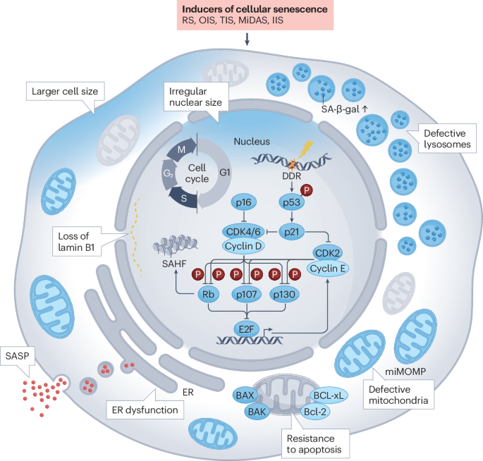
Spatiotemporal control over cell–matrix interactions using dynamic micropatterns
Cells are responsive to the molecular makeup and spatial architecture of the surrounding microenvironment, and dysregulated cell–cell and cell–extracellular matrix (ECM) interactions can contribute to many pathological conditions, including fibrosis and cancer. Owing to the remarkable complexity of most human tissues it is often difficult to pinpoint the molecular mechanisms that regulate these cellular responses in vivo. The effect of the local microenvironment on different cellular processes can be studied by confining cells on microscale substrates with well-defined geometries and biochemical compositions. Ideally, these ‘micropatterns’ should be dynamic and enable investigators to control cell adhesion to previously inaccessible areas at will. This can be achieved using light- or temperature-induced surface modifications or different click chemistries, but such techniques invariably involve specialized equipment or extensive synthesis and offer limited control over the actual matrix components the cells are interacting with.
To investigate the role of ECM composition in the geometric control of cell polarity and migration, we developed a new method for dynamic micropatterning. Biotinylated polyethylene glycol-grafted poly-l-lysine surfaces were patterned using ultraviolet light and different matrix components or monoclonal antibodies were adsorbed to the irradiated regions to make them permissive for cell adhesion. Using the method, we could confine cells onto the micropatterns and later release them to spread and migrate on different streptavidin-conjugated matrix components or biomimetic peptides. The new micropatterned substrates are versatile and easy to fabricate, and the biotin–streptavidin capture chemistry ensures that the transition from confined to unconfined state is fast, robust and fully biocompatible.


留言 (0)