Various microvascular changes in eye signs were observed in SLE-PAH patients, including decreased vessel number (ischaemic areas), increased vessel number (reticulum deformity), changes in vascular morphology (twisting, dilation, microangioma) and vascular venous injury (haemorrhage and wound spots). Decreased VD and MFI were associated with poor cardiopulmonary function. Compared to conventional risk assessment indicators for pulmonary hypertension, WHO functional class (HR 0.12, 95% CI 0.02–0.75, p = 0.024), 6MWD (HR 1.02, 95% CI 1.00–1.03, p = 0.039), NT-proBNP (HR 0.99, p = 0.010), VD (HR 10.11, p = 0.006) and MFI (HR 7.85, p = 0.008) appeared to be more effective in predicting inadequate therapeutic response and poor prognosis of SLE-PAH.
PAH is a severe and progressive pulmonary vascular disease characterized by elevated pulmonary artery pressure and increased pulmonary artery resistance, leading to right ventricular failure and mortality. The REVEAL registry study in the United States revealed that CTD-related PAH accounts for 25.3% of all PAH cases. SLE and systemic sclerosis (SSc) are the most common CTDs associated with PAH [12]. An analysis of the causes of death among SLE patients in China over the past 30 years found that SLE-PAH was the third leading cause of death among SLE patients [13]. The pathogenesis of CTD-PAH is complex, making treatment challenging, as even with novel targeted combination therapies, the three-year mortality rate among moderate- to high-risk patients remains as high as 56% [14].
Assessing the risk of pulmonary arterial hypertension (PAH) is crucial in guiding standardized treatment and reducing mortality rates among PAH patients. Therefore, PAH risk assessment methods were introduced in the 2012 REVEAL study, the 2015 European PAH guidelines, and the 2018 6th World Symposium on Pulmonary Hypertension (WSPH) [10, 15, 16]. Evaluation indicators include clinical manifestations, the level of cardiac function, plasma brain natriuretic peptide (BNP) levels, cardiac echocardiography and haemodynamic levels. However, in these risk stratifications, some risk parameters require high measurement conditions, such as cardiac index (CI) and mixed venous oxygen saturation (SvO2) derived by right heart catheterization, limiting their practicality. Research has been conducted to identify simpler risk assessment indicators for daily clinical practice, such as age, right atrial area, pulmonary arteriole diameter, serum iron levels, red cell distribution width, and blood uric acid levels [17,18,19]. However, the predictive value of some risk assessment indicators used in CTD-PAH remains controversial. Current studies, particularly those involving nailfold video capillaroscopy [20], have revealed the importance of microvascular damage in systemic sclerosis (SSc)-associated PAH and support the hypothesis of systemic microvascular involvement in idiopathic PAH. These findings further suggest that although our research primarily focused on SLE and CTD-PAH, microvascular damage and its related clinical manifestations may also be equally important in other types of PAH.
Vessel density (VD) and microvascular flow index (MFI) are essential parameters in microcirculation [21]. VD refers to the number of blood vessels in a unit area. The level of vascular density reflects the distribution of microvessels and the degree of blood supply. The formation and regulation of vascular density involve multiple mechanisms, including angiogenesis, proinflammatory cytokine release, and vascular constriction and dilation. For instance, angiogenic factors such as VEGF stimulate the formation of new blood vessels. The release of cytokines and chemical mediators may induce vascular constriction or dilation, thereby regulating vascular density. MFI assesses the haemodynamic characteristics of microcirculation by measuring the average flow velocity and density of red blood cells in microvessels. It reflects the blood flow velocity and volume in the microcirculation. Clinically, changes in MFI effectively indicate alterations in blood perfusion. Higher MFI values indicate faster blood flow velocity and larger vascular blood volume [7].
VD and MFI have gained increasing attention as crucial assessments for haemodynamic changes and systemic microcirculation disorders in critically ill patients. Research on microcirculation evaluation in sepsis and critical care monitoring has shown that VD and MFI can determine the fluid resuscitation needs of ICU patients [22, 23]. There is currently a lack of relevant research on VD and MFI for assessing SLE-PAH. A previous cross-sectional study showed that VD and MFI in conjunctival microvasculature were associated with risk levels of mortality in SLE-PAH [7]. Further investigation into the measurement of VD and MFI in SLE-PAH can contribute to evaluating the systemic haemodynamic changes caused by PAH and a better understanding of the pathophysiological mechanisms of SLE-PAH.
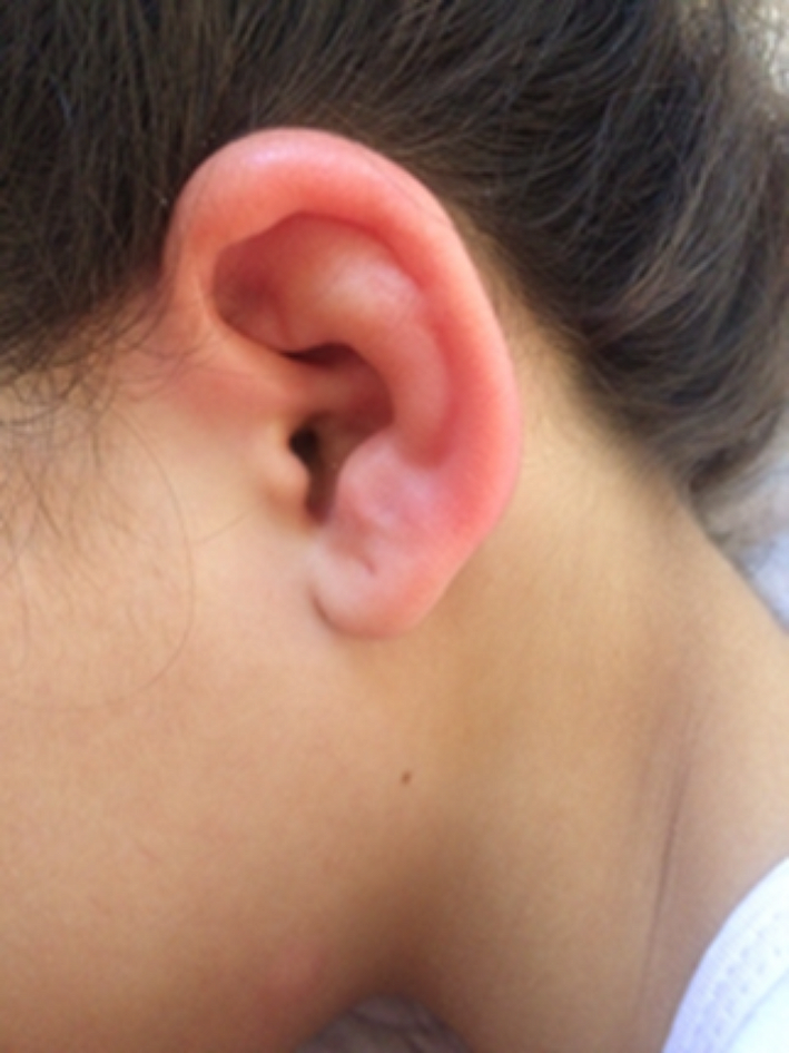
The conjunctival microcirculation, as a crucial window into systemic microcirculation, indirectly reflects the overall state of microcirculation through the observation and measurement of conjunctival vascular density, vessel diameter, blood flow velocity, vascular branching pattern, endothelial wall, vascular reactivity and perfusion status [24]. The normal course of conjunctival microcirculation involves arterioles leading to capillaries and then proceeding through venules. Arterioles have a relatively straight course, while venules exhibit slight curvature. The calibres of both arteries and veins are uniform, with an arterial/venous ratio of approximately 1:2. Capillaries form a branching network. Under normal conditions, blood flow dynamics in arterioles appear as linear flow, while venules exhibit linear or granular flow. Capillaries show granular flow with occasional mild red blood cell aggregation. In comparison with other common methods for sublingual microcirculation and nailfold microcirculation, conjunctival microcirculation offers advantages such as being unaffected by external temperature and having a stronger correlation with visceral blood vessels [21, 25]. However, it also has certain limitations, including difficulties in cooperation during testing, complexity in examination procedures, and greater requirements for equipment and personnel expertise. To solve complex operational problems, eye signs were assessed through a clinical observational study involving a large sample size. Focusing on the distinct manifestations of conjunctival microcirculation changes in patients with a hypercoagulable state, a conjunctival vascular panel was defined, including vessel twisting, dilation, haemorrhages, ischaemic areas, reticulum deformity, microangioma, and wound spots (Fig. 1). This set of distinctive features is referred to as “eye signs.” Compared to conjunctival microcirculation examination, the detection of eye signs is simpler, as they can be observed using a conventional slit lamp or even with the naked eye. Clinical validation of eye signs demonstrates their high accuracy in assessing thrombosis or hypercoagulability, exhibiting a strong correspondence with nailfold capillary microcirculation [26, 27].
In patients with PAH, elevated pulmonary arterial pressure leads to increased pulmonary vascular resistance. This results in decreased blood flow speed and capacity in pulmonary capillaries, causing a reduction in pulmonary blood volume and oxygen supply [28]. Consequently, there is a decline in oxygenation function and impairment of cardiac performance, which are caused by the severe condition of PAH and extremely elevated vascular resistance. Therefore, in cases of PAH occurring in SLE, bulbar conjunctival microvessels can exhibit widespread changes, such as twisting, haemorrhages, dilation, and microangioma. However, more importantly, in this study among patients with SLE-PAH who had a poor prognosis after treatment, microcirculatory VD and MFI were significantly reduced. Thus, clinically, for patients with significant reductions in VD and MFI, intensified treatment is necessary. Additionally, close monitoring of the trends in VD and MFI can provide an important basis for assessing treatment efficacy and predicting prognosis. This is very important for guiding clinical practice in SLE-PAH.
This study was conducted at a single centre and involved a relatively small sample size. Moreover, it included only patients from China, which may limit the generalizability of the findings to other populations. To confirm the clinical significance of vessel density (VD) and the microvascular flow index (MFI) in pulmonary arterial hypertension (PAH), further research with more diverse and larger sample sizes is essential. While additional clinical trials are necessary to refine parameter calculations and achieve standardization, the undeniable convenience and feasibility of conjunctival microcirculation assessment make it a promising method that warrants further clinical promotion and application in various demographic settings.


留言 (0)