Currently, the disruption of epigenetic landscapes is a well-recognized aspect of cancer, including abnormal DNA methylation patterns. DNA methylation and demethylation at the C-5 position occur in a cyclic manner involving many enzymes and proteins, influencing the regulation of gene transcription and expression. DNA methylation can be simply described as the transfer of a methyl group, to the 5′ position of a pyrimidine ring of cytosines in a CpG site. DNA demethylation can occur either passively or actively. Passive DNA demethylation can be described as the “dilution” of methylation levels after rounds of cellular replication due to the absence of methylation of the new DNA strand. Active DNA demethylation occurs via the activity of a group of enzymes called TET (ten-eleven translocation) family (TET1, TET2, and TET3) which realize active DNA demethylation through an iterative stepwise oxidization of 5-mC to 5hmC [15, 16]. Due to their roles in numerous metabolic pathways, 5-mC and 5hmC have both been investigated as potential epigenetic markers. However, unlike 5-mC, the content of 5hmC has revealed better stability and stronger robustness in different tissues. There is a growing body of literature showing the differences in 5hmC expression between cancerous and healthy tissues [17, 18]. A decreased nuclear level of 5hmC in comparison with normal tissue has been reported in some human cancers, such as melanoma, glioma, parathyroid carcinoma, hepatocellular carcinoma, cervical carcinoma, and hematologic malignancies [1, 4, 6, 19, 20]. Aligned with previous studies in other organs, our results in the present series of 318 thyroid tumors show that decreased or loss of expression of 5hmC was associated with malignant tumors when compared with benign tumors. However, the scores of 5hmC expression do not allow us to distinguish low-risk neoplasms from malignant neoplasms, confirming the borderline nature of these lesions.
In our study, information about tumor size was available for 308 cases and was grouped based on the cutoff value of 4 cm as suggested by the AJCC/TNM Staging System 8th edition [21]. We found a significant association between the extension score of 5hmC and tumor size (p value 0.003). Lymphatic invasion presented statistical significance in the evaluation of H-score, whereas vascular invasion represented statistical significance for 5hmC intensity, extension, and H-score. In accordance with these results, Tong et al. [8] compared the expression of 5-mC and 5hmC in 88 PTC, 20 MNG, and adjacent normal tissues based on a 3-tiered H-score evaluation. They found that PTC presented significantly less 5hmC than MNG. Curiously, for 5-mC, no difference was noted. These results support the sensitivity of 5hmC as a distinct epigenetic marker for establishing more stable DNA interactions over 5-mC, as shown in previous studies [15]. Tong et al. [8] also compared the H-score of PTC with and without lymph node metastasis and, in line with our results, found that the group with lymph node metastasis presented a lower score. In our series, loss of 5hmC expression was significantly associated with adverse pathological characteristics, such as minimal/major ETE, invasive/infiltrative capsule status, lymphatic invasion, vascular invasions, bilaterality, multifocality, and biological behavior (Table 6).
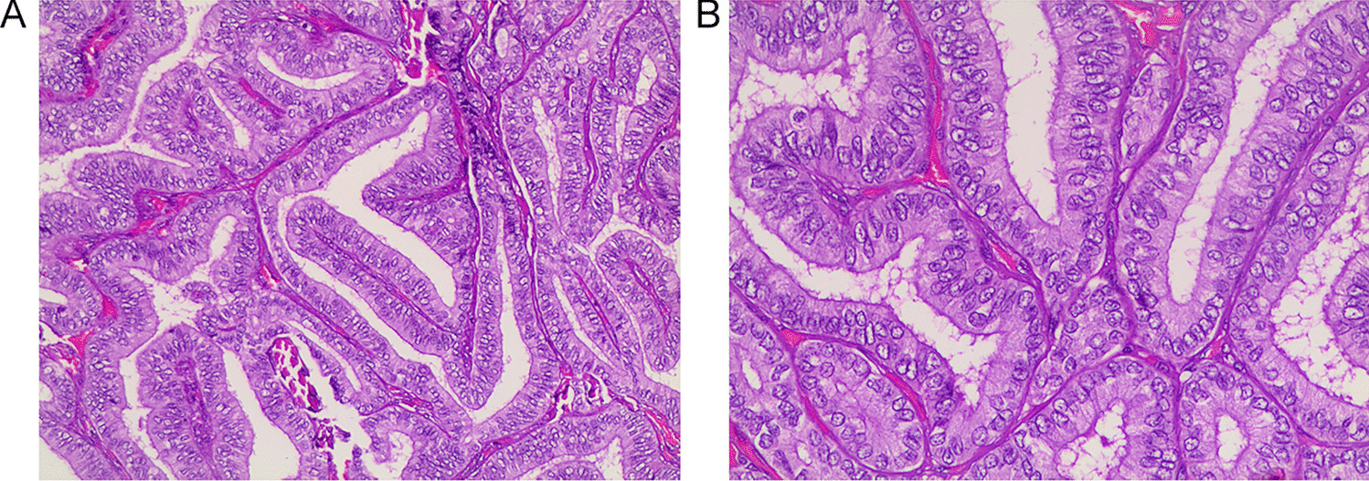
Table 6 5hmC in thyroid neoplasm—a collated summary from discussed studiesOur results also showed a decreased level of 5hmC expression in the group of tumors with invasive/infiltrative growth patterns, with significant differences in intensity (p value < 0.001) and H-score (p value < 0.001). Interestingly, when we compared the invasive (n = 87) versus infiltrative (n = 102) growth patterns, 5hmC expression was similarly distributed across the score evaluations of intensity, extension, and H-score. A study by Seok et al. [10] focused on follicular-patterned thyroid neoplasms and grouped them as group I (NIFTP, 5 cases), group II (encapsulated FVPTC with capsular invasion, 14 cases), group III (infiltrative FVPTC, 10 cases), and group IV (PTC with a predominantly follicular pattern and well-formed papillae (< 1%), 11 cases). All cases were analyzed for 5hmC immunohistochemistry using the H-score, and 34 cases were evaluated for BRAF mutation analysis. 5hmC was highly preserved in groups I, II, and III, whereas group IV cases were noted with moderately reduced nuclear 5hmC [10] (Table 6).
In our cohort, TERT promoter mutations were found in 12 cases out of 183 PTCs, and 9 of them presented low H-scores (p value 0.003). Similar to our results, in 2020, Oishi et al. [11] showed that TERT promoter-mutated PTCs and ATCs have significantly decreased nuclear 5hmC levels in comparison with normal thyroid tissues and in PTCs with the wild-type TERT promoter. Later, the same authors added 63 PTCs with wild-type TERT promoter, ten PTCs with TERT promoter mutations, and four ATCs, to evaluate 5hmC expression by IHC, and no difference was found in the two groups [11]. A recent study of Hysek et al. [12] examined a dataset consisting of 26 FTCs, 2 FT-UMPs, and 1 OCA. However, they were unable to confirm previous observations in papillary thyroid carcinomas regarding the relationship between 5hmC loss and TERT mutations.
Concerning RAS family genes, in the same subgroup of our cohort, RAS and HRAS mutations were associated with the highest H-score, reaffirming the association of these mutations with better prognosis, as has been demonstrated in the literature [14] (Table 6).
The current analysis also verified for the first time an association between a decreased 5hmC H-score and both multifocality (p value 0.007) and bilaterality (p value 0.043). Once again, these results relate the loss of this marker with clinicopathological features associated with poorer prognosis and the need for more aggressive treatment [22, 23].
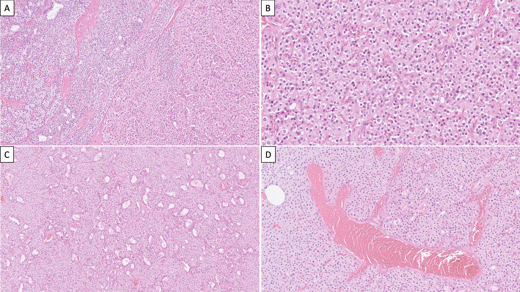
In our results, 5hmC expression was significantly higher when we compared benign tumors with low-risk (p value < 0.001) and malignant thyroid tumors (p value < 0.001), while no differences were observed when comparing low-risk tumors versus malignant tumors. Seok et al. [9] analyzed 5hmC location together with isocitrate dehydrogenase 1 (IDH1) mutations focusing only on an ATC cohort composed of 9 cases that occurred de novo and 15 ATCs that were derived from either PTCs (12 cases) or FTCs (3 cases). The H-score was markedly reduced in ATCs and moderately reduced in PTC and FTC components. In contrast, in the nonneoplastic thyroid, the expression was highly preserved. This study emphasizes the role of 5hmC in the multistep carcinogenesis of thyroid tumors, as has been shown in other human cancers. Additionally, the positioning of low-risk tumors closer to malignant tumors found in our study highlights the current concern with overdiagnosis and overtreatment in thyroid tumors. Since we could not demonstrate clear differences in 5hmC expression between low-risk tumors and minimally invasive follicular pattern carcinomas (IEFV-PTC, FTC, and OCA), it is not possible to use this marker to discriminate these two classes of neoplasms. The lack of differences with widely invasive follicular pattern-carcinoma cases can be attributed to the few cases with wide invasion (2 OCA cases) in our series.
None of the previously mentioned studies specifically addressed thyroid tumors with oncocytic morphology as a substantial dataset, as well as the accepted separate class of oncocytic tumors (OA and OCA). Our data revealed for the first time a link between 5hmC expression and oncocytic morphology.
In our cohort, 33% of the cases presented oncocytic morphology, including well-differentiated tumors with oncocytic morphology (n = 52) and oncocytic tumors (14 OA and 39 OCA), according to the 5th edition of the WHO Classification of Endocrine and Neuroendocrine Tumors. There was a strong statistical association between oncocytic morphology and lower 5hmC intensity, extension, and H-score evaluation (p values < 0.001, < 0.001, and 0.037, respectively). This association remained a trend in the Mann–Whitney test evaluation (p value 0.06), presenting an association between this morphology and lower H-score values. This leads us to hypothesize that perhaps the genetic variations/mutations in mtDNA and the metabolic alteration characteristics of these tumors may play a relevant role in this association [24,25,26,27,28,29]. Most probably, these disruptive mutations in mtDNA, characteristic of oncocytic tumors, can lead to significant changes in the levels of some oncometabolites such as α-KG and succinate, affecting the activity of cellular epigenetic regulatory enzymes. Interestingly, in ongoing work in our group, we found that in thyroid cancer cytoplasmic hybrid (cybrid) cell lines harboring mtDNA mutation significant changes in alpha-KG and succinate levels (data not shown). This has already been observed in pheochromocytomas and other tumor types, with mutations in IDH, FH, and SDH, in which DNA is globally hypermethylated, promoting a metastatic behavior [1, 9, 25, 27]. Also, global hypermethylation has also been described in well-differentiated thyroid neoplasm [30].
Thus, the low 5-hmC staining we found, particularly in oncocytic tumors, leads us to suggest that the low 5-hmC staining in these tumors may be reflecting the pronounced hypermethylation pattern of the tumors. However, further studies are needed to clarify the specific role that 5-hmC may play in thyroid tumorigenesis, particularly in the context of oncocytic tumors.


留言 (0)