Not All Parafibromin Deficiency Relates to Parathyroid Carcinoma: The Role of Morphological Assessment
During gross examination, the weight was 1.1 g. The gland measured 20 × 15 × 7 mm and displayed a brownish cut surface. No macroscopic features indicative of malignancy, such as a grey/white cut surface or unclear relations to the surroundings, were observed. The entire tumor was submitted for microscopic evaluation.
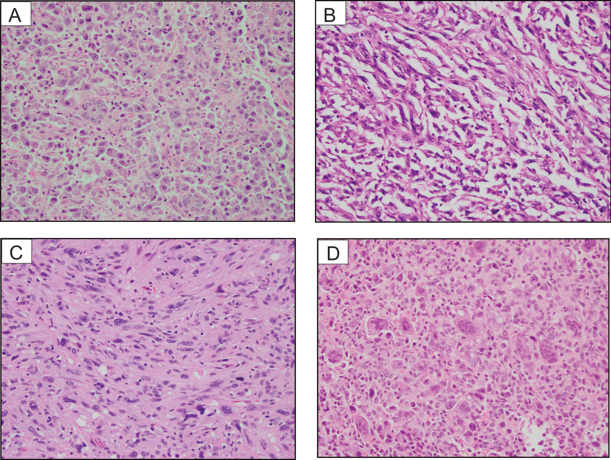
Histology revealed a hypercellular parathyroid gland characterized by chief cells arranged in microacinar patterns and solid areas containing eosinophilic cells (Fig. 1A). The stromal fat content was diminished. Notably, there was a general absence of mitotic activity and tumor necrosis. The tumor was circumscribed, and invasive growth was not observed. No atypical histological features such as trabecular growth, fibrous bands, or prominent nuclear pleomorphism were noted. Upon closer examination, the eosinophilic cells exhibited a distinct pinkish cytoplasm but lacked the characteristic cytoplasmic granularity of oxyphilic cells (Fig. 1B). Additionally, these cells showed perinuclear cytoplasmic clearing (“halo”). Some regions also displayed microcystic features and arborizing vasculature (Fig. 1C, D). These morphological features have been associated with parafibromin-deficient parathyroid tumors [1]. Tumor cells were positive for PTH and APC but negative for galectin-3 and PGP9.5 (Fig. 2A). The Ki-67 index was low (2.4%). Parafibromin exhibited positive staining in cells at the tumor margin and in the majority of chief cells (Fig. 2B). However, the nuclear stain was negative in the eosinophilic cell component, displaying a vague, granular cytosolic stain, interpreted as aberrant (Fig. 2C, D).
The favored diagnosis was a parafibromin-deficient parathyroid adenoma, and the patient was found to harbor a pathogenic CDC73 gene mutation involving the nuclear localization signal of parafibromin.


留言 (0)