
記住我


Rats were weighed weekly during postoperative weeks 0–6 (Fig. 2). No significant differences were observed between the Sham, EGC-0, EGC-L, and EGC-H groups. The EGC-M group showed significantly reduced weight accumulation (p < 0.05) compared to the Sham and EGC-0 groups. Despite the EGC-M rats weighing between 400 and 550 g by week 6, they showed an increasing trend in weight gain over time. After sacrifice, the organs appeared grossly normal, suggesting that Elgucare feeding had no adverse effects on growth.
Fig. 2
Animal weight changes during postoperative weeks 0 to 6. Values are presented as mean ± SD (n = 6). Key: *, a significant difference (p < 0.05) compared to the EGC-0 group; #, a significant difference (p < 0.05) compared to the Sham group in a two-way ANOVA with Tukey’s HSD test
Plantar tinglingThis study used a needle testing method to assess the rat’s response to mechanical stimulation of their plantar surface by applying weight to the plantar surface of the left hind paw and recording the force that elicited the paw withdrawal reflex (Fig. 3). At postoperative week 2, there were no significant differences between groups. At weeks 4 and 6, paw withdrawal occurred at a stimulus of approximately 44 g for rats in the EGC-0 group compared to approximately 58 g for the Sham group. This difference reflects significantly greater pain severity in the EGC-0 group (p < 0.05). At week 4, paw withdrawal thresholds of the EGC-M and EGC-H groups were 54 ± 6 g and 57 ± 5 g, respectively, significantly higher than the EGC-0 group (p < 0.05).
Fig. 3
Plantar tingling test results at postoperative weeks 2 (A), 4 (B), and 6 (C). Values are presented as mean ± SD (n = 6). Key: *, a significant difference (p < 0.05) compared to the EGC-0 group; #, a significant difference (p < 0.05) compared to the Sham group in a one-way ANOVA with Tukey’s HSD test
After feeding for six weeks, all three EGC-fed groups differed significantly from the EGC-0 group (p < 0.05) in the paw withdrawal threshold, which had increased to 63 ± 5 (EGC-L), 58 ± 8 (EGC-M), and 60 ± 6 g (EGC-H). There was no difference between the EGC-fed groups and the Sham group. According to the plantar tingling data, the pain in EGC-fed groups showed substantial relief as feeding duration increased and even approached that of the Sham group after administering Elgucare for six weeks. These findings indicate the effectiveness of Elgucare in alleviating pain.
Gait analysisA fully automated rat gait analysis system was used to analyze the max contact mean intensity, print area, and swing speed of the left hind paw while walking (Fig. 4). At postoperative week 2, there were no significant differences in the maximum contact mean intensities between groups. At week 4, the mean intensity of the EGC-0 group was significantly lower than that of the Sham group (p < 0.05). At week 6, the mean intensities of the EGC-0, EGC-L, EGC-M, and EGC-H groups were all significantly lower than that of the Sham group (p < 0.05).
Fig. 4
Gait analysis results at postoperative weeks 2 (column A), 4 (column B), and 6 (column C). Values are presented as mean ± SD (n = 6). Key: *, a significant difference (p < 0.05) compared to the EGC-0 group; #, a significant difference (p < 0.05) compared to the Sham group in a one-way ANOVA with Tukey’s HSD test
There were no significant differences in the print area across groups at postoperative weeks 2 and 4. At postoperative week 6, the print areas of the EGC-0, EGC-L, and EGC-M groups were significantly lower than that of the Sham group (p < 0.05). The study data showed no improvement in rat maximum contact mean intensity and print area after administering Elgucare.
Swing speed analysis showed that the EGC-0 group differed significantly from the Sham group at postoperative weeks 2, 4, and 6 (all p < 0.05). At postoperative weeks 2 and 4, swing speed was significantly lower for the EGC-M group than for the Sham group (p < 0.05). However, at week 4, the high-dose EGC-H group’s swing speed was 114 ± 16 cm/s, significantly higher than the EGC-0 group’s 74 ± 18 cm/s (p < 0.05), indicating that Elgucare aids improvement in swing speed.
IVD thicknessMicro-computed tomography was used to scan and record images of the vertebral column and measure the IVD thickness (Fig. 5). At week 2, IVDs were significantly narrower in the EGC-0, ECG-M, and EGC-H groups than in the Sham group (p < 0.05). However, the IVD thickness for the EGC-L group was approximately 381 ± 38 μm, significantly higher than the EGC-0 group’s 328 ± 19 μm (p < 0.05) but not significantly different from the Sham group. At week 4, there were no significant differences among groups. At week 6, the IVD thicknesses of the EGC-0, EGC-L, EGC-M, and EGC-H groups were significantly narrower than that of the Sham group. The Sham group’s IVD thickness was approximately 498 ± 53 μm by week 6, thicker than in weeks 2 and 4, which is believed to reflect growth in rat body habitus with time. At postoperative weeks 2, 4, and 6, IVD thicknesses in the EGC-0 group were 328 ± 19, 349 ± 24, and 382 ± 33 μm, respectively, indicating that the IVD thickens with time, a phenomenon attributed to intrinsic repair after damage. However, by week 2, the IVDs of the EGC-L, EGC-M, and EGC-H groups had already reached thicknesses of approximately 350–400 μm, approaching that of the EGC-0 group at week 6.
Fig. 5
IVD thicknesses at postoperative weeks 2 (A), 4 (B), and 6 (C). Values are presented as mean ± SD (n = 6). Key: *, a significant difference (p < 0.05) compared to the EGC-0 group; #, a significant difference (p < 0.05) compared to the Sham group in a one-way ANOVA with Tukey’s HSD test
Histopathological analysisThe rats were sacrificed at postoperative week 6, and their vertebrae were collected for histopathological examination. H&E staining was used to observe histopathological morphology, while safranin O/fast green staining was used to observe the cartilage’s histopathological morphology (Fig. 6). For the EGC-0 group, the H&E stained sections showed many aggregated cells, primarily within cartilage vaults. Further examination using the safranin O stain showed chondrocyte aggregation in the NP. For the EGC-M and EGC-H groups, cell aggregation was also noted in several rats. However, no cell aggregation was observed in the stained sections of the Sham and EGC-L groups, which showed normal IVD histological morphology.
Fig. 6
Histopathological analysis results at postoperative week 6. Arrows indicate sites of cell aggregation. Scale bars = 500 μm
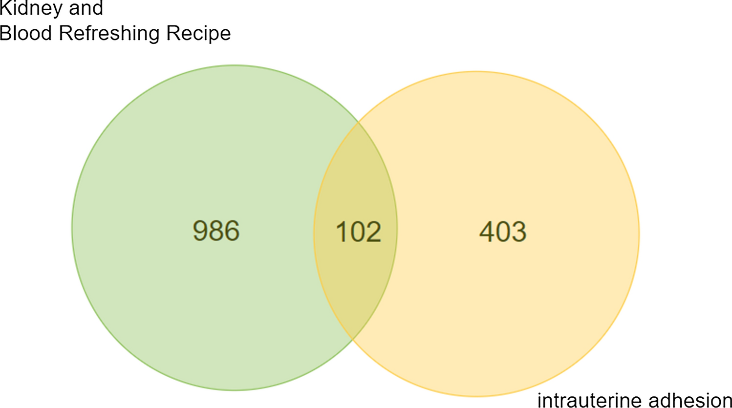
This study destroyed the IVD by needle puncture, and the results showed that histological morphology remained normal in the AF. Therefore, we will elaborate only on the NP findings. H&E and safranin O staining results are shown in Fig. 7; both sets of findings agree after interpretation. All IVDs showed normal histology in the Sham group. Cell aggregation was observed in 67% of the EGC-0 specimens; only 33% showed normal morphology. The proportions of rats with normal histology in the EGC-L, EGC-M, and EGC-H groups were 100%, 50%, and 83%, respectively, indicating that administering Elgucare increased the likelihood that IVD histology returned to normal.
Fig. 7
Proportions of H&E (A) and safranin O/fast green (B) staining interpretations at postoperative week 6
留言 (0)