
記住我


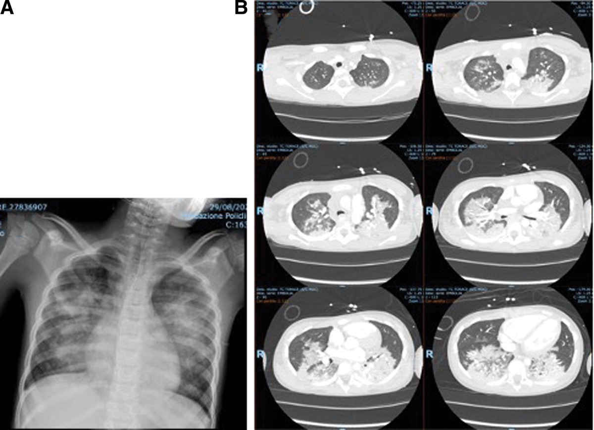
A 20-month-old boy from Nepal with the presumed diagnosis of pulmonary tuberculosis (TB) was referred to our pediatric TB clinic in August 2018 because of ongoing fever, cough, weight loss and lack of radiologic improvement despite 1½ months of first-line antitubercular therapy (ATT) consisting of isoniazid, rifampicin, pyrazinamide, and ethambutol. In July 2018, his chest radiograph at the referring hospital showed mediastinal lymphadenopathy (left > right) with homogenous opacity of the left upper lobe (Figure 1). A repeat chest radiograph in August 2018 was similar. His tuberculin skin test was positive with 14 mm of induration. Xpert MTB/Rif assay performed on gastric lavage was negative. The referring doctor had not done a TB culture. His exposure history was notable for an aunt who had recently been diagnosed with pulmonary TB (PTB) and had been taking ATT for the past 1½ months. Her sputum for acid-fast bacillus had been positive, but TB culture report was not available.
 FIGURE 1.:
 Chest radiograph showing mediastinal lymphadenopathy (left > right) with homogenous opacity of left upper lobe.
On evaluation in August 2018, his weight was 6.8 kg (<3rd SD as per WHO growth chart). Heart rate was 110 beats/min, respiratory rate was 32 breaths/min, blood pressure was 110/80 mm of Hg and oxygen saturation in room air was 100%. Systemic physical examination was normal. Leukocytes were 15,300 cells/mm3 with 23% polymorphs, 74% lymphocytes and 3% eosinophils. Hemoglobin was 10.4 gm/dL, and platelets were 399,000 cells/mm3. ESR was 2 mm at the end of 1 hour. HIV ELISA test was negative. He was continued on the intensive phase of ATT for another month with no improvement, raising suspicion of drug-resistant TB. High-resolution computed tomography of the chest in October 2018 to look for mediastinal nodes, showed collapse/consolidation of the anterior segment of the left upper lobe with surrounding areas of ill-defined infiltration and necrotic areas with foci of air and a well-defined air-filled cystic lesion, measuring 0.9 × 0.7 cm. Necrotic left hilar lymph nodes (1.5 × 1.1 cm) were also seen. No tracheobronchial stenosis or mediastinal mass were seen (Figure 2). Xpert Rif/MTB assay on the bronchoalveolar lavage (BAL) fluid was negative; TB MGIT also did not grow any organisms. He was continued on the same ATT with the exception of pyrazinamide, which was stopped. During follow-up in August 2019, a repeat chest CT (see Figure, Supplemental Digital Content 1 https://links.lww.com/INF/F174) showed a persistent patchy area of consolidation in the apico-posterior and anterior segments of the left upper lobe and an increase in the cystic/necrotic areas abutting the thymus. A CT-guided biopsy of the consolidation was performed leading to the diagnosis.
 FIGURE 2.:
 CT chest in August 2018 showing collapse consolidation of the anterior segment of the left upper lobe with necrotic left hilar lymph nodes.
DENOUEMENTHistopathology showed pulmonary and pleural tissue. Lymph node tissue was not seen. There were increased number of eosinophils, aggregates of neutrophils, foamy histiocytes in the alveolar spaces with fibroblasts, and fibrosis. Focally, cuticle of a parasite and its gonads were noted with adjacent foreign body giant cell response (see Figure, Supplemental Digital Content 2 https://links.lww.com/INF/F175). Necrotizing epithelioid granulomas were not seen. Xpert MTB/Rif on the biopsy sample was negative. TB MGIT culture was also negative. Based on histopathology, the diagnosis of Paragonimiasis was made, ATT was stopped and the child was treated with praziquantel (PZQ) for 3 days. He was subsequently lost to follow-up. On hindsight, there was a history of feeding the child raw freshwater fish. A stool test was not considered, as clinically paragonimiasis was not suspected. Also, it was not possible to get sputum in a 20-month-old child. Biopsy was done mainly to rule out drug-resistant TB.
Paragonimiasis is a zoonotic disease, which is acquired by the intake of raw or undercooked freshwater crabs, crayfish, or raw meat of wild deer or boar. The larval stage of parasite is released after digestion of the crab or crayfish and migrate to the lung causing lung fluke or to other tissues.1,2 Found in the subtropical areas of the Americas, Asia, and Africa, pulmonary paragonimiasis is the most common (76–90% of cases) clinical manifestation whereas pleural effusion and subcutaneous nodules are among the common extrapulmonary forms.2 The clinical and radiological findings of the lungs are remarkably similar to those of pulmonary tuberculosis.1 In a study conducted by Qian et al, the misdiagnosis rate at admission was 45.5%, and the most commonly confused disease was tuberculosis due to their similar symptoms and signs2 as was seen in our patient. Clinically the presentation may be similar. However, history of environmental exposure can provide a clue. By identifying the distinctive golden brown, ellipsoidal, or oval operculated Paragonimus eggs in clinical specimens such as sputum, aspirated fluids, and feces by microscopy, a conclusive diagnosis can be made.3 By using the AMS III concentration technique, Komiya et al revealed that 65.1% of the fecal samples from 189 patients with pulmonary paragonimiasis contained Paragonimus eggs.4 Inflammatory cells, eosinophils, Paragonimus eggs, and Charcot-Leyden crystals can be seen when the exudates are examined. A histological section, on the other hand, will show fibrocollagenous tissue, inflammatory cells, eosinophils, fragments of worms, and deformed eggs, as seen in our case.2 A useful test for differentiating between pulmonary paragonimiasis and pulmonary tuberculosis is the intradermal tuberculin test; however, the tuberculin test may not help to make a diagnosis of TB disease in areas endemic for both paragonimiasis and TB, as it may just suggest latent TB as was seen in our patient.5 Common hematological findings were leukocytosis with relative lymphocytosis, eosinophilia, and generally increased ESR.6 The chest radiographs may show patchy consolidations (62–71%), pleural thickening (28%), cystic or cavitary lesions (11–14%), effusion (9–10%), and nodular lesions (8–13%).6 Computerized tomography (CT) is superior method for visualizing lung lesions compared with a chest radiograph.7
In a study by Henry et al about chest CT findings in Paragonimus infection, 25% patients showed findings like our patient (peripheral nodule surrounded by ground-glass attenuation), 50% had an additional pleural track. So, radiological findings were not more characteristic of a parasitic infection than of TB.8
Two WHO-recommended medications for the treatment of human paragonimiasis are PZQ and triclabendazole.2 PZQ is the first line of treatment for paragonimiasis, according to the American Academy of Paediatrics’ recommendations, with the suggested dose of 150 mg/kg given orally 3 times a day for 3 days.2 The earlier recommended 3-day PZQ course did not show ideal treatment effectivity and 63.6% needed more than one course of PZQ, while triclabendazole in a dose of 10 mg/kg had a better efficacy.2 In highly endemic regions of northeastern India, targeted active case detection and treatment of infected cases along with community education were implemented to diminish the prevalence of paragonimiasis.9 Treatment limitations included instances of underdosing and reinfection due to persistent high-risk cultural dietary practices seen in Vietnam despite more than 15 years of mass screening, treatment, and education.9 Three (3%) of the 96 TB patients who underwent screening for 1 year showed pulmonary paragonimiasis.10 In areas with a dual burden, paragonimiasis should be taken into account in the TB diagnostic algorithms.10
In conclusion, the patient reported here, presented with symptoms suggestive of PTB and had contact with a relative suffering from PTB; he showed no improvement on ATT and was diagnosed to have lung fluke on a CT-guided biopsy. Since the clinical and radiological findings of the lung fluke are remarkably similar to those of pulmonary tuberculosis, thus in areas with a dual burden, paragonimiasis should be taken into account in the TB diagnostic algorithms.10
REFERENCES 1. Maticorena Agramonte VF, Ormeño Julca AJ, Coveñas Coronado CDP, et al. [Pulmonary paragonimiasis. Pediatric case report]. Arch Argent Pediatr. 2019;117:e659–e663. 2. Qian M, Li F, Zhang Y, et al. A retrospective clinical analysis of pediatric paragonimiasis in a Chinese children’s hospital from 2011 to 2019. Sci Rep. 2021;11:2005. 3. Singh TS, Sugiyama H, Rangsiruji A. Paragonimus & paragonimiasis in India. Indian J Med Res. 2012;136:192–204. 4. Komiya Y, Yokogawa M. The recovering of Paragonimus eggs from stools of paragonimiasis patients by AMS III centrifuging technic. Jpn J Med Sci Biol. 1953;6:207–211. 5. Sawada T, Takei K, Sato S, et al. Studies on the immunodiagnosis of paragonimiasis. 3. Intradermal skin tests with fractionated antigens. J Infect Dis. 1968;118:235–239. 6. Singh TS, Mutum SS, Razaque MA. Pulmonary paragonimiasis: clinical features, diagnosis and treatment of 39 cases in Manipur. Trans R Soc Trop Med Hyg. 1986;80:967–971. 7. Vanijanonta S, Bunnag D, Harinasuta T. Radiological findings in pulmonary paragonimiasis heterotremus. Southeast Asian J Trop Med Public Health. 1984;15:122–128. 8. Henry TS, Lane MA, Weil GJ, et al. Chest CT Features of North American Paragonimiasis. Am J Roentgenol. 2012;198:1076–1083. 9. Tidman R, Kanankege KST, Bangert M, et al. Global prevalence of 4 neglected foodborne trematodes targeted for control by WHO: A scoping review to highlight the gaps. PLoS NeglTrop Dis. 2023;17:e0011073. 10. Das M, Doleckova K, Shenoy R, et al. Paragonimiasis in tuberculosis patients in Nagaland, India. Glob Health Action. 2016;9:32387.
留言 (0)