
記住我


Pulmonary manifestations of malaria are not unfrequently observed in children; respiratory distress can occur in up to 40% of cases of severe falciparum malaria, whereas a clinical acute respiratory distress syndrome (ARDS) is more frequent in adults/pregnant women1; it has been described with Plasmodium falciparum as well as with P. vivax and P. ovale, while a severe prognosis can be due mostly to the absence of intensive care facilities. Secondary bacterial pneumonia contributes to morbidity and mortality. Beyond inflammatory mediators, even fluid overload and cardiac failure (due to severe anemia) can contribute to the ARDS pathophysiology.2 We observed 3 clinical cases of pediatric falciparum malarial ARDS, having similar clinical course but quite different imaging and complications. All children recovered from respiratory failure with noninvasive support, beyond specific intravenous antimalarial treatment. General characteristics of the 3 patients, as well as laboratory test results, are reported in Table 1.
TABLE 1. - General Characteristics and Laboratory Test Results of the 3 Children Admitted to PICU with Plasmodium falciparum Malaria-related ARDS Patient #1 Patient #2 Patient #3 General characteristics Age (years)/sex 9/male 12/male 13/male Weight (kg) 35 70 77 Born in endemic area No No Yes Antimalarial chemoprophylaxis Inappropriate None None Length of stay in endemic area (days) 30 7 180 Rural area Yes No No Time intervals (days): Home return–onset of symptoms 5 8 11 Onset of symptoms–hospital admission 5 3 1 Hospital admission–PICU admission 1 5 2 PICU length of stay 6 4 5 Duration of CPAP 4 3 4 Hospital length of stay 17 9 9 Laboratory results Day 1 Day 2 Day 3 Day 1 Day 2 Day 3 Day 1 Day 2 Day 3 Hemoglobin (g/dL) 12.4 11.6 9.1 10.8 11.6 10.4 13.5 12.8 12.7 White blood cell (WBC) count (×109/L) 9.40 11.05 8.81 7.86 10.46 8.01 6.00 7.65 6.62 Platelet count (×109/L) 6 12 14 97 74 181 68 75 77 Parasitemia (% infected erythrocytes) 13 3 <1 1 1 <1 1.2 <1 <1 Creatinine (mg/dL) 1.58 1.53 1.32 0.71 0.66 0.57 0.84 0.68 0.63 Blood urea nitrogen (mg/dL) 48 51 45 9 9 8 9 11 13 Bilirubin (mg/dL) 2.3 2.0 2.3 0.7 0.4 0.4 2.4 1.8 1.0 Alanine transaminase (ALT) (UI/L) 74 71 48 85 104 91 49 43 45 Albumin (g/L) 29 27 25 29 24 23 27 26 28 Procalcitonin (ng/mL) 42.66 33.03 28.91 3.26 2.74 1.98 4.24 3.78 2.16 C-reactive protein (mg/L) 201.0 222.0 129.6 100.5 74.4 105.6 128.1 130.0 104.6 Blood gas analysis: Ph 7.37 7.46 7.41 7.47 7.44 7.50 7.44 7.39 7.38 paO2/FiO2 108 122 232 80 87 210 86 206 378 paCO2 39 44 46 36 39 38 36 37 40 Lactates 2.7 2.0 1.2 0.7 0.9 1.1 1.3 1.1 0.7CPAP, Continuous positive airway pressure; PICU, pediatric intensive care unit.
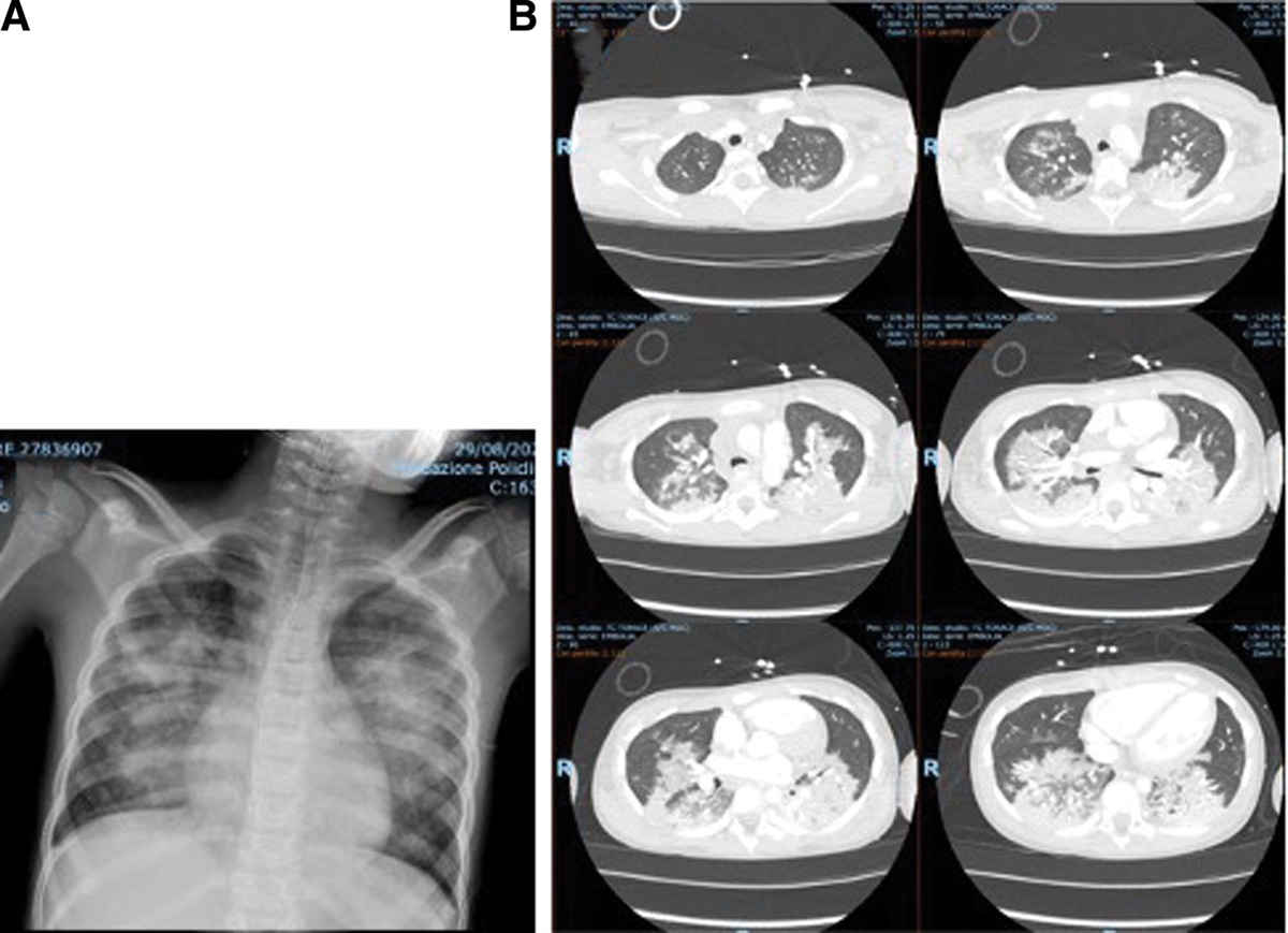
A 9-year-old boy of Nigerian origins was referred to the hospital with a high fever and malaise. He came back from a journey to Nigeria 5 days earlier, after a 30-day stay. No malarial prophylaxis was performed. Apart from polypnea, no other respiratory symptoms were observed (Fig. 1A). An intensive care referral was decided due to severe thrombocytopenia (platelet count 6000/mL) and oliguric acute kidney injury (creatinine 1.58 mg/dL, urine output <0.5 mL/kg/hour). Both C-reactive protein and procalcitonin were increased. Intravenous artemisinin was started. Hemodynamic stabilization and an increase in urine output were obtained, while respiratory failure progressively worsened (Fig. 1B). Packed erythrocytes and albumin transfusion were needed. Meanwhile, severe hypoxia was evidenced by arterial blood gas analysis (P/F ratio 102) and he was placed on helmet-continuous positive airway pressure (CPAP) (FiO2 0.75). Laboratory evaluation demonstrated a hemophagocytic complication (triglycerides 500 mg/dL, ferritin 2900, hemophagocytosis on bone marrow biopsy) requiring dexamethasone and intravenous immunoglobulin administration.
 FIGURE 1.:
 Chest radiograph (1a) and CT scan (1b) of patient 1.
Case 2A 12-year-old boy of Nigerian parents was admitted to the hospital with a high fever and chills. He returned to Italy 8 days before the onset of symptoms, after a 7-day stay in Nigeria. Falciparum malaria was diagnosed, and he was referred to the pediatric intensive care unit (PICU) due to impending respiratory fatigue and hypoxia (Fig. 2A). Chest CT showed multiple diffuse inflammatory infiltrates (Fig. 2B). Artesunate intravenous treatment was started, together with intravenous antibiotics. C-reactive protein and procalcitonin levels were markedly increased. Total-face mask CPAP (15 cmH2O) was needed for 5 days; thereafter, nasal prongs oxygen supplementation could be restored. However, 20 days after PICU discharge, control throat swabs resulted positive for COVID-19 infection. Surprisingly, no respiratory failure relapse occurred.
 FIGURE 2.:
 Chest radiograph (2a) and CT scan (2b) of patient 2.
Case 3A 13-year-old boy of Guinean parents was admitted with a high fever and respiratory distress. He came to Italy 10 days before, after a 6-month stay in Guinea, without any malarial prophylaxis. On admission, he was markedly polypneic (65–70/min) and he was able to utter a few words. His PaO2/FiO2 was 82; helmet-CPAP was started at 12 cmH2O, achieving a progressive PaO2/FiO2 increase (>350 after 24 hours). Chest CT showed predominant right lung involvement, while bilateral basal infiltrates and pleuro-pericardial effusion were present (Fig. 3A, B). Intravenous artemisinin was shifted to oral treatment after reaching parasitemia disappearance. After 3 days, oxygen supplementation with high-flow nasal cannulas was started, having a respiratory rate of 20/min and FiO2 of 0.24.
 FIGURE 3.:
 Chest radiograph (3a) and CT scan (3b) of patient 3.
DISCUSSIONTachypnea is frequently observed in children with malaria; high fever, acidosis/anemia and lung involvement may be equally responsible. Indeed, acute pulmonary edema and ARDS represent the most severe pulmonary manifestations of systemic malaria, sometimes occurring after a few days of treatment2,3 and still carry a poor prognosis in developing countries.4 Eventually, particularly for more virulent falciparum malaria, delayed diagnosis and clearing of the blood forms of the parasite can be life-threatening.
As a result of severe overwhelming inflammation, malaria as well other systemic parasitic infection may rarely induce hemophagocytosis.5,6 No data are available regarding falciparum ARDS in the presence of hemophagocytosis: the role of hyperinflammation in promoting a gravity-dependent and marked lung compaction pattern cannot be excluded. As reported in our cases, the radiological features of ARDS in falciparum malaria can be quite different depending on the lung compaction degree and fluid overload.
In the last decade, interesting insights have been proposed regarding the role of endothelial injury and the C-protein network in malarial ARDS pathophysiology, leading to pulmonary coagulopathy as a potential therapeutic target,7 disclosing the role of dysregulation of EPCR and thrombomodulin in the lung resulting in respiratory distress because of pneumocyte cell injury and apoptosis.
As in cerebral malaria, the binding of EPCR and ICAM-1 has been implicated in iRBC binding to the pulmonary endothelium.8 Thus, modern proteomic and transcriptomic analysis has revealed the critical roles of parasite biomass and perturbations of the APC–EPCR axis leading to severe disease development.
Moreover, recent genetic studies on var gene expression, despite different clinical presentations across different countries, in both children and adults, the same var gene expression profiles are predictive of severe disease.9
Up to now, no specific treatments have been outlined for malarial ARDS, thus respiratory management following nonmalaria ALI/ARDS therapeutic guidelines10 are implemented, beyond early administration of parenteral antimalarial therapy, supportive therapy and management of complications. In milder cases of respiratory involvement, spontaneous ventilation using a face mask with a high-flow gas delivery system can be used. CPAP may be added to improve PaO2 without increasing FiO2; when it reaches more than 0.6 and CPAP of more than 10 cm H2O is needed to achieve PaO2 at least 60 mm Hg, use of noninvasive positive pressure ventilation (NPPV) or tracheal intubation/invasive ventilation should be considered. Both CPAP and NPPV have been rarely described in malarial ARDS,11 although the use of NIV in ARDS with mild to moderate hypoxemia has been reevaluated even recently.12
In conclusion, malaria should be considered as an important cause of curable ARDS even in children, both in endemic areas and in travelers returning in nonendemic areas. Laboratory studies can confirm the diagnosis, even if repeated assessments may be needed to detect parasitemia. Antimalarial treatment can be lifesaving, whereas patients with respiratory failure deserve an early PICU admission for evaluation and correct respiratory support institution.
REFERENCES 1. Taylor WRJ, Hanson J, Turner GDH, et al. Respiratory manifestations of malaria. Chest. 2012;142:492–505. 2. Sarkar PK, Ahluwalia G, Vijayan VK. A talwar critical care aspects of malaria. J Intensive Care Medicine. 2010;25:93–103. 3. Mohana A, Sharma SK, Bollinenic S. Acute lung injury and acute respiratory distress syndrome in malaria. J Vector Borne Dis. 2008;45:179–193. 4. Bruneel F, Tubach F, Corne P, et al.; Severe Imported Malaria in Adults (SIMA) Study Group. Severe imported falciparum malaria: a cohort study in400 critically ill adults. PLoS One. 2010;5:e13236. 5. Muthu V, Dhooria S, Sehgal IS, et al. Malaria-associated secondary haemophagocytic lymphohistiocytosis: report of two cases & a review of literature. Indian J Med Res. 2017;145:399–404. 6. Bhagat M, Kanhere S, Kadakia P, et al. Haemophagocytic lymphohistiocytosis: a cause of unresponsive malaria in a 5-year-old girl. Paediatr Int Child Health. 2015;35:333–336. 7. Maknitikul S, Luplertlop N, Grau GER, et al. Dysregulation of pulmonary endothelial protein C receptor and thrombomodulin in severe falciparum malaria-associated ARDS relevant to hemozoin. PLoS One. 2017;12:e0181674. 8. Avril M, Bernabeu M, Benjamin M, et al. Interaction between endothelial protein C receptor and intercellular adhesion molecule 1 to mediate binding of Plasmodium falciparum-infected erythrocytes to endothelial cells. MBio. 2016;7:e00615–e00616. 9. Duffy F, Bernabeu M, Babar PH, et al. Meta-analysis of Plasmodium falciparum var signatures contributing to severe malaria in African children and Indian adults. MBio. 2019;10:e00217–e00219. 10. Emeriaud G, López-Fernández YM, Iyer NP, et al.; Second Pediatric Acute Lung Injury Consensus Conference (PALICC-2) Group on behalf of the Pediatric Acute Lung Injury and Sepsis Investigators (PALISI) Network. Executive Summary of the Second International Guidelines for the Diagnosis and Management of Pediatric Acute Respiratory Distress Syndrome (PALICC-2). Pediatr Crit Care Med. 2023;24:143–168. 11. Agarwal R, Nath A, Gupta D. Non-invasive ventilation in Plasmodium vivax related ALI/ARDS. Intern Med. 2007;46:2007–2011. 12. Emeriaud G, Pons-Òdena M, Bhalla AK, et al.; Pediatric Acute Respiratory Distress Syndrome Incidence and Epidemiology (PARDIE) Investigators and Pediatric Acute Lung Injury and Sepsis Investigators (PALISI) Network. Noninvasive Ventilation for Pediatric Acute Respiratory Distress Syndrome: Experience From the 2016/2017 Pediatric Acute Respiratory Distress Syndrome Incidence and Epidemiology Prospective Cohort Study. Pediatr Crit Care Med. 2023;24:715–726.
留言 (0)