
記住我


The local ethics committee approved this retrospective study. Written informed consent was obtained from all patients.
Study designFrom 2007 until 2020, in-bore MRI-guided prostate biopsy was performed in 289 consecutive patients with suspected prostate cancer.
According to the MR findings, we obtained 3 to 6 cores per patient. From the resulting 1060 cores, 340 cores from the TZ could be identified. A total of 17 cores had to be excluded because of incomplete documentation.
321 cores in 141 patients remained for evaluation (Fig.1).
Two experienced radiologists reviewed all image sets in in a blinded consensus. One of both radiologists with more than 15 years of experience in prostate imaging performed all biopsies since 2008.
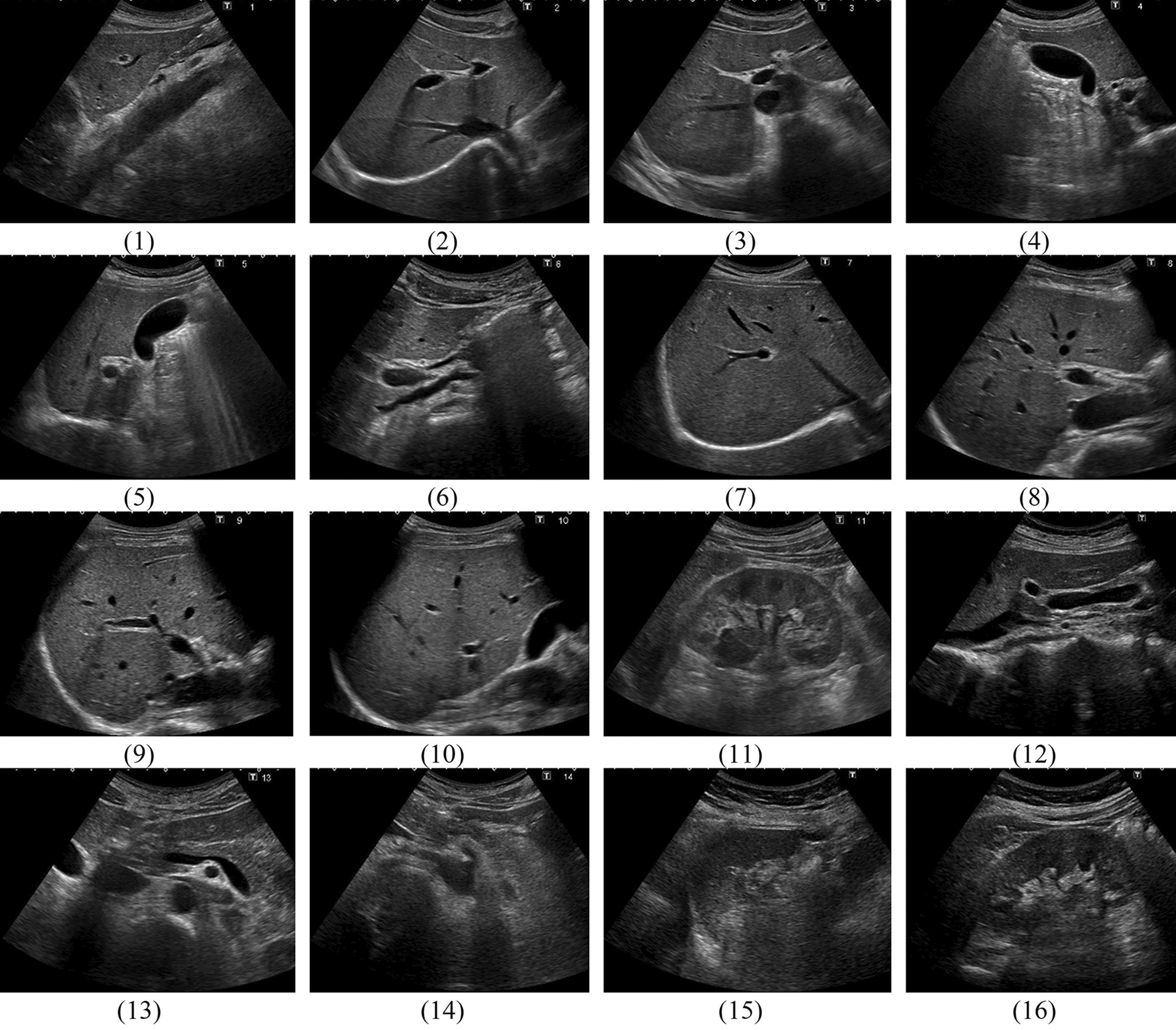
Single early-phase perfusion images were evaluated separately for the presence of a TZ prostate cancer (consensus tumor early perfusion). This was based on a combination of the image criteria for the whole slice. Afterward, the T2w images including the markers of the biopsy trace were presented to describe the perfusion pattern for each biopsy position in detail. The criteria for the perfusion pattern were (1) asymmetry; (2) signal strength compared to the surrounding tissue of the TZ and (3) homogeneity versus internal heterogeneity (Figs. 2 and 3).
Fig. 2
Unspecific heterogeneous perfusion pattern (short arrows in b) of the transition zone in benign prostate hyperplasia, without marked asymmetry and marked high signal intensity; patient of 57 years, PSA 5.1 ng/ml, prostate volume 57 ml, no carcinoma; a T2-weighted imaging axial with marked biopsy traces (long arrows); b T1-weighted early-phase dynamic perfusion; c diffusion-weighted imaging with b = 1400; d apparent diffusion coefficient map
Fig. 3
Typical tumor perfusion pattern (short arrows in b) of the left anterior transition zone, with marked asymmetry and with marked high signal intensity, moderate reduced heterogeneity; patient of 70 years, PSA 39 ng/ml, prostate volume 36 ml, prostate carcinoma G2 Gleason 3+4; a T2-weighted axial imaging with marked biopsy traces (long arrows); b T1-weighted early-phase dynamic perfusion; c diffusion-weighted imaging with b = 1400; d apparent diffusion coefficient map; e: calculated dynamic enhanced integral EI colour map
Furthermore, the calculated dynamic color maps were evaluated for the presence of a TZ prostate cancer to take into account the dynamic character of the perfusion sequence (consensus tumor color map).
Patient demographics and clinical dataPatients with suspicious lesions underwent biopsy after a consensus-based decision by a radiologist and a urologist on an individual base. The main criteria were the level and time course of PSA, together with the image interpretation of mpMRI, which was related to the respective guidelines/PI-RADS version. All imaging and interventional procedures were performed according to current guidelines. Further systematic biopsies before or after the targeted biopsy were not subject of this study. We targeted at least one lesion with the highest suspicion on the left and the right sides of the prostate.
The automatic biopsy gun provides a notch length of 17 mm. Thus, the position of the targets was retrospectively documented in up to 4 subsequent T2w slices of the preceding diagnostic MRI (long arrows in Figs.1a and 2a). All cores were retrospectively reevaluated according to PI-RADSv2.1. For this study, we evaluated exclusively cores of the TZ.
Sample descriptionA sample of 321 biopsy cores from 141 patients, 36 of which were carcinoma positive, i.e., had at least one core with neoplastic alterations, was analyzed. Overall, 77 cores were positive for prostate carcinoma. Tumor grading was G1, G2, and G3 in 14, 42, and 20 cores, and Gleason score was 3 + 3, 4 + 3, 3 + 4, 4 + 4, 5 + 5 in 32, 21, 16, 6, and 1 cores; for one core, no Gleason score was provided. Descriptive characteristics are summarized in Table 1.
Table 1 Descriptive characteristics of the carcinoma positive and negative patient subgroupsMpMRI of the prostate and in-bore MR-guided biopsyMR imaging was performed using a 1.5 Tesla (ESPREE, Siemens Healthineers) in 119 patients with endorectal coil and a 3-Tesla (PIONEER, GE Healthcare) scanner in 22 patients.
The standard protocol fulfilling the requirements of the Consensus Meeting on the Standardization of Prostate MRI and the appropriate version of the prostate imaging included T2w imaging in three planes. DWI was performed using four b-values (0, 100, 800, 1400 in 1.5 T and 0, 100, 800, 1500 in 3 T). The apparent diffusion coefficient was calculated from b = 0, 100, and 800 s/mm2. Volume-interpolated gradient echo sequences with a temporal resolution of 8.3 s were used for DCE in 1.5T. The 3 T scanner used a 3D two-point Dixon (DynamIc SCan Optimization DISCO) with a temporal resolution of 8.3 s.
Gadoteric acid was applied as contrast media in a weight-adapted standard dose (0.1 mmol/kg body weight) with an injection rate of 3 ml s−1. The protocol parameters of the DCE sequences are listed in Table 2.
Table 2 Protocol parameters of the DCE sequencesPost-processing of all DCE datasets was carried out on a GE Advantage Workstation HPZ 800. For a representation of the perfusion kinetics in a fast visual assessment, we created simple colour maps as maximum slope images MSI and enhanced integral EI maps with constant settings.
For the biopsy, the patient was positioned using a biopsy positioning device in the supine position in the 1.5 Tesla scanner (preproduction model by Invivo) or in the prone position in the 3-Tesla scanner (DynaTRIM by Philips).
An adjustable needle guide was inserted transrectally under the control of sagittal and coronal fast imaging. In the correct position, the biopsy was performed using an 18-G MR-compatible fully automatic biopsy gun.
Each biopsy procedure was documented by fast T2w imaging in axial plane, and sagittal or coronal plane with the needle inside the lesion. All biopsy specimens were evaluated in the same histopathologic institute including Gleason grading of prostatic carcinoma.
Data analysisStatistical analysisWe used logistic regression to predict the histological status (cancer vs. no cancer) for each core. As (bivariate) predictors, we used the PI-RADSv2.1 score, its three constituents (V2.1 score T2w, V2.1 score DWI, V2.1 score DCE), lesion size (only available for carcinoma positive patients) as well as the diagnostic consensus variables (positive or negative) based on asymmetry, signal strength, homogeneity, early perfusion imaging, colormap imaging, MSI, and EI, respectively. Using the PI-RADS score and the diagnostic consensus variables as predictors, we subsequently fitted a multiple logistic regression model. From each model’s predictions, we calculated sensitivity, specificity, and receiver operating characteristic (ROC) curves, which graphically represent the different trade-offs between sensitivity and specificity that can be realized by a given predictive model, and their respective areas under the curve (AUC), which vary between 0.5 (chance level predictions) and 1 (perfect predictive accuracy of the model).
留言 (0)