By investigating clinical characteristics and CECT features of 13 S-HCC patients, we found that S-HCCs were frequently seen in elderly male without any clinical symptoms but with HBV infection, elevated AFP and higher Ki-67 level. The CECT manifestations including: large diameter, frequently right lobe of liver involvement, obvious heterogeneity, a dominant enhancement pattern of “slow-in and and slow-out” and hepatic surface retraction were valuable clues to diagnose S-HCC. Moreover, lesions with lobular or wavy contours, ill-defined margins, infiltrative morphology and intrahepatic metastasis contributed to the diagnosis of S-HCC.
As a type of rare malignant tumor, S-HCC is usually characterized by atypical symptoms such as abdominal pain, weight loss, fatigue or discovered in routine medical examination without any symptoms, making diagnosis difficult at an early stage [15]. As our results showed that most patients complained with abdominal pain or tumors were found in asymptomatic physical examination, with a larger average diameter of 66.7mm. Moreover, most patients had HBV infection (53.8%, 7/13), but none of them had HCV infection, indicating that S-HCC was related to hepatitis B, to some extent. Several studies have revealed the similar phenomenon [6, 12, 15, 16]. As Lu et al. found that 19 patients (67.9 %) were positive for serum HBs antigen, and none were positive for HCV antibody in their study [16]. However, a recent meta-analysis demonstrated that there was no significant difference between S-HCC patients and conventional HCC patents in HBV infection [17]. These different results may be due to the small sample size, especially the lack of study in the population with HCV infection. A majority of previous studies found that AFP levels were increased in most S-HCC patients, while both this incidence and the degree of AFP elevation were lower compared to conventional HCC [2, 15, 18]. In our study, 53.8 % of patients had elevated AFP levels, similar to Wang’s and Li’s studies, suggesting that the elevation of AFP is not only an indication of conventional HCC, but also of S-HCC [15, 19].
Different from conventional HCC with a “wash-in and wash-out” enhancement pattern, most S-HCC in our study presented with the “slow-in and and slow-out” enhancement mode in CECT, which were similar to previous studies [7, 12, 13, 20, 21]. Since the major histopathological characteristics of S-HCC were the peripheral viable cancerous tissue accompanied by fibrous stroma, with central necrosis or hemorrhage [4, 7]. Peripheral tumor fibrous stroma may block or delay the flow of contrast agents in CECT examination, resulting in slow peripheral enhancement in the AP, progressive or persistent enhancement toward the tumor center in the PVP and mild descending enhancement in the EP, as our results displayed. In addition, owing to the sarcomatoid components with dominant part of poorly differentiated cells which were growing rapidly, the neovasculature was insufficient for blood and oxygen supplying, leading to central necrosis [7, 22]. As the ischemia or necrosis features in CECT were more frequently seen with a high frequency of 54% (7/13) in our cohort. Several studies reported that S-HCC with various enhancement patterns depended on the tissue component and it’s proportion, such as different proportion of hepatocellular, sarcomatous or necrosis components [5, 13]. The CT manifestations varied when certain histopathological components ranged from focal to prominent, leading to a dilemma of summarizing the typical image features of S-HCC. However, tumors with a “slow-in and and slow-out” enhancement pattern different from HCC should be taken S-HCC into account to some degree.
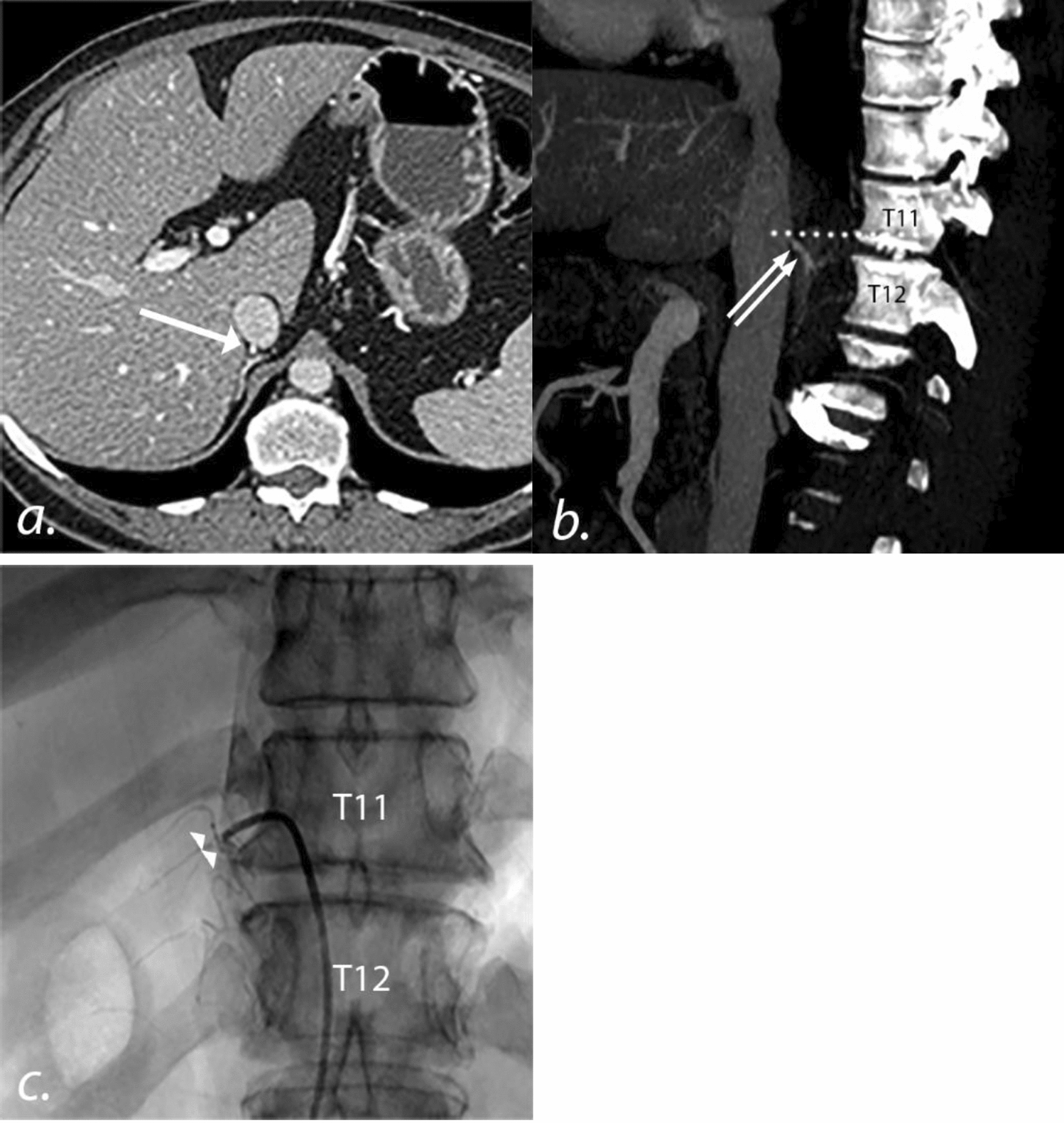
The imaging features of S-HCC were reported to be similar to those of ICC [21, 23]. For example, hepatic surface retraction, a typical feature closely associated with ICC, was seen in 4 patients (31%) in our study. A large amount of fibrous tissue accompanied by aggressive ICC cells would pull or invade adjacent subcapsular area, leading to hepatic surface retraction [24, 25]. We assumed that this similar feature may be attributed to the analogous pathological characteristics of the two malignant tumors, with peripheral viable cancerous tissue and fibrous stroma pulling adjacent liver tissue. Interestingly, there were three lesions located near the liver subcapsular area within the four cases, being easily to be shape of hepatic surface retraction, similar to Shi and his colleagues’ results [13]. In addition, among the four lesions with hepatic surface retraction, three tumors lacked capsules and one had partial capsule, indicating that most of them were prone to invade peripheral regions owing to the lack of limitation of complete capsule. Furthermore, it should be noted that the differential diagnosis should also include liver metastases, liver abscess, scirrhous subtype of HCC and primary hepatic sarcoma, which all could present as “slow-in and and slow-out” enhancement pattern, with larger size, lobulated or wavy contours and infiltrative morphology in some cases [12, 26, 27].
Most researches found that patients with S-HCC had higher extra-hepatic metastases, lymph node metastases and poor prognosis [5, 20, 21, 27]. Distinct genetic patterns of S-HCC compared to conventional HCC may contributed to their aggressive biological behaviors, in spite of the extreme lack of molecular studies about S-HCC. Zhang et al. demonstrated that S-HCC had a high rate of rearrangement and homozygous deletion in CDKN2A gene, a tumor suppressor gene causing a loss of gene funtion [6]. It was reported that genetic and epigenetic aberrations of CDKN2A resulted in enhanced carcinogenesis and poor prognosis in various cancers, such as ovarian cancer, lymphoma, and prostate cancer, etc [28, 29]. Hence, in agreement with previous studies, two patients with adjacent organs invasion (15.4%), two and four patients occurred lymph node metastasis (15.4%) and intrahepatic metastasis (30.7%), respectively, in our study, maybe partially owing to the CDKN2A mutation [5, 15, 17].
There were several limitations in our study should be noted. First, this was a single and small sample study as the very low prevalent rate of S-HCC, and since to it’s retrospective nature, selection bias were inevitable. Second, limited to most patients without MRI examinations in the early years as expensive costs, we didn’t analyze the MRI features of S-HCC. Third, we did not provide the prognosis and survival data of the patients, because most of them were lost in contact, leading to incomplete survival data of the entire study cohort. Finally, due to the absence of a control group in our study as limited patients sample, we were unable to generate statistical data on the difference between S-HCC and other hepatic tumors.
In conclusion, we explored and analyzed the CECT and clinical features of a series of S-HCC patients. S-HCC generally presented with obvious heterogeneity and a CTCT enhancing pattern of “slow-in and and slow-out”. The valuable clues in the diagnosis of S-HCC preoperatively also included: patients with HBV infection, elevated AFP level, large tumor size, irregular and unclear tumor margin, intrahepatic metastasis and hepatic surface retraction. Radiologists pay more attention to the above mentioned CT imaging findings and clinical data in detail might achieve an accurate diagnosis of S-HCC.


留言 (0)