Accuracy of Nodule Volume and Airway Wall Thickness Measurement Using Low-Dose Chest CT on a Photon-Counting Detector CT Scanner
Objectives
A comparison of high-resolution photon-counting detector computed tomography (PCD-CT) versus energy-integrating detector (EID) CT via a phantom study using low-dose chest CT to evaluate nodule volume and airway wall thickness quantification.
Materials and Methods
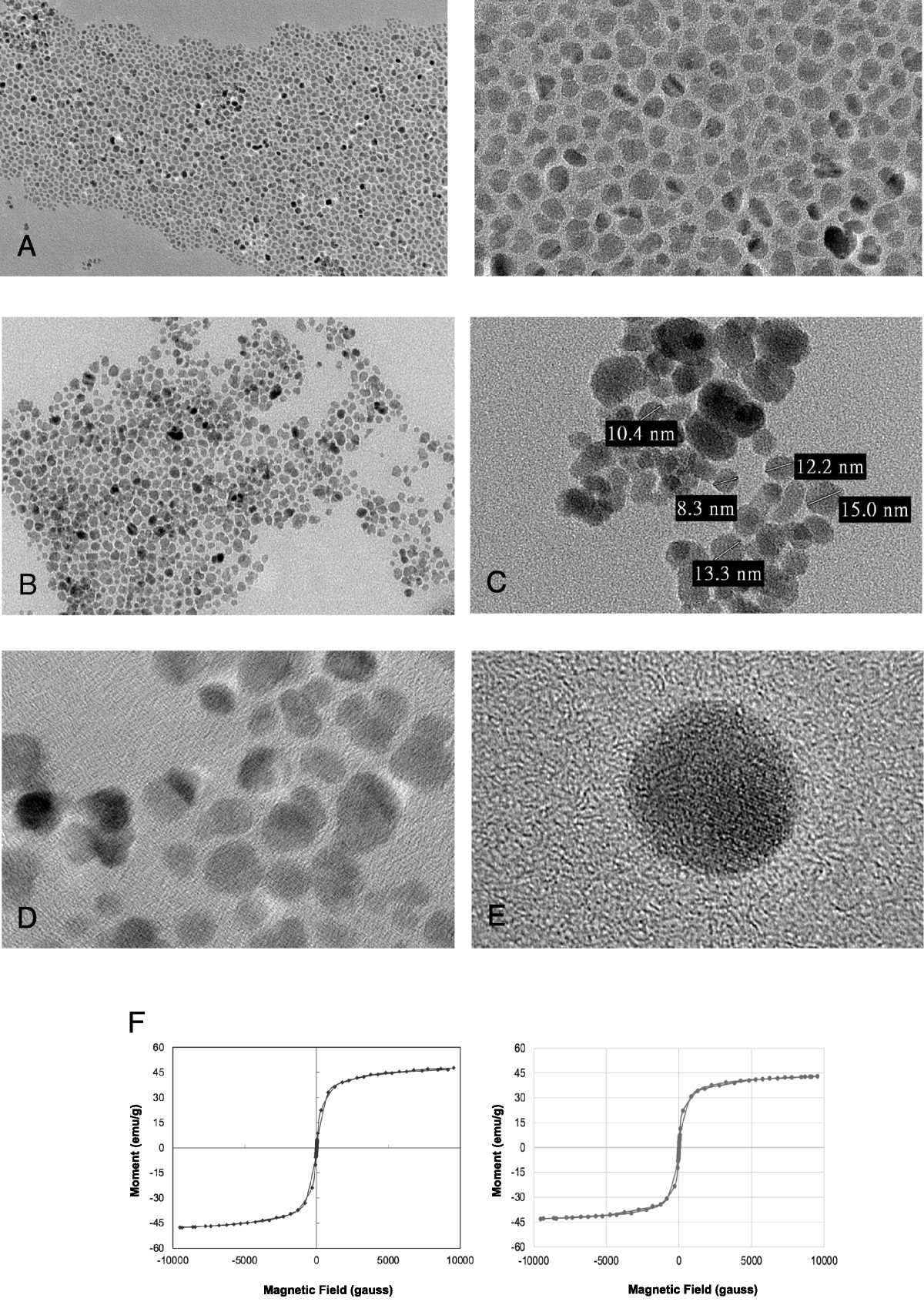
Twelve solid and ground-glass lung nodule phantoms with 3 diameters (5 mm, 8 mm, and 10 mm) and 2 shapes (spherical and star-shaped) and 12 airway tube phantoms (wall thicknesses, 0.27–1.54 mm) were placed in an anthropomorphic chest phantom. The phantom was scanned with EID-CT and PCD-CT at 5 dose levels (CTDIvol = 0.1–0.8 mGy at Sn-100 kV, 7.35 mGy at 120 kV). All images were iteratively reconstructed using matched kernels for EID-CT and medium-sharp kernel (MK) PCD-CT and an ultra-sharp kernel (USK) PCD-CT kernel, and image noise at each dose level was quantified. Nodule volumes were measured using semiautomated segmentation software, and the accuracy was expressed as the percentage error between segmented and reference volumes. Airway wall thicknesses were measured, and the root-mean-square error across all tubes was evaluated.
Results
MK PCD-CT images had the lowest noise. At 0.1 mGy, the mean volume accuracy for the solid and ground-glass nodules was improved in USK PCD-CT (3.1% and 3.3% error) compared with MK PCD-CT (9.9% and 10.2% error) and EID-CT images (11.4% and 9.2% error), respectively. At 0.2 mGy and 0.8 mGy, the wall thickness root-mean-square error values were 0.42 mm and 0.41 mm for EID-CT, 0.54 mm and 0.49 mm for MK PCD-CT, and 0.23 mm and 0.16 mm for USK PCD-CT.
Conclusions
USK PCD-CT provided more accurate lung nodule volume and airway wall thickness quantification at lower radiation dose compared with MK PCD-CT and EID-CT.


留言 (0)