Electrophysiology, Pathology, and Imaging of Pulsed Field Ablation of Scarred and Healthy Ventricles in Swine
BACKGROUND:
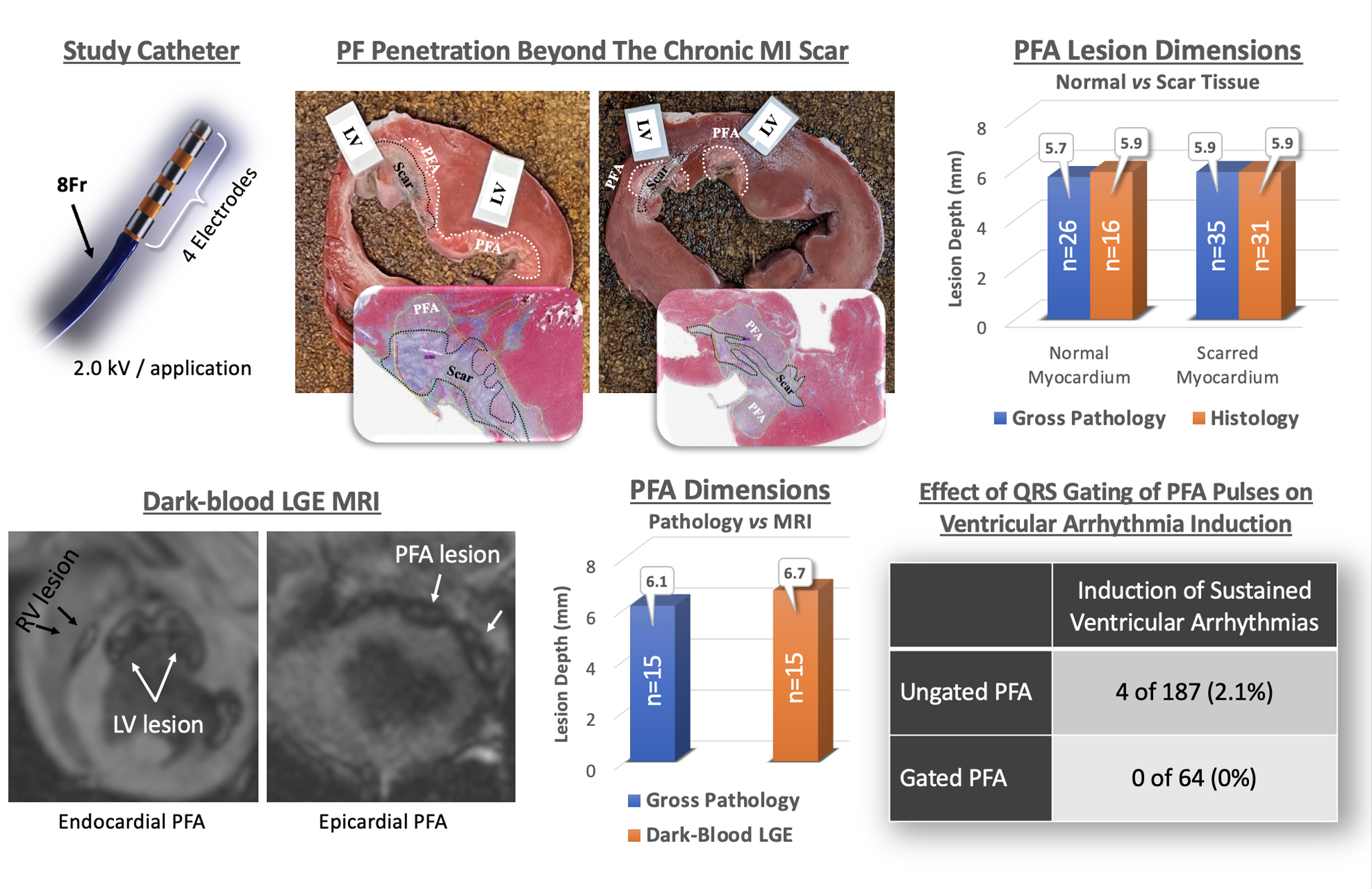
Pulsed field ablation (PFA) has recently been shown to penetrate ischemic scar, but details on its efficacy, risk of arrhythmias, and imaging insights are lacking. In a porcine model of myocardial scar, we studied the ability of ventricular PFA to penetrate scarred tissue, induce ventricular arrhythmias, and assess the influence of QRS gating during pulse delivery.
METHODS:
Of a total of 6 swine, 5 underwent coronary occlusion and 1 underwent radiofrequency ablation to create infarct scar and iatrogenic scar models, respectively. Two additional swine served as healthy controls. An 8 Fr focal PFA catheter was used to deliver bipolar, biphasic PFA (2.0 kV) lesions guided by electroanatomical mapping, fluoroscopy, and intracardiac echocardiography over both scarred and healthy myocardium. Swine underwent magnetic resonance imaging 2–7 days post-PFA.
RESULTS:
PFA successfully penetrated scar without significant difference in lesion depth between lesion at the infarct border (5.9±1.0 mm, n=41) and healthy myocardium (5.7±1.3 mm, n=26; P=0.53). PFA penetration of both infarct and iatrogenic radiofrequency abalation scar was observed in all examined sections. Sustained ventricular arrhythmias requiring defibrillation occurred in 4 of 187 (2.1%) ungated applications, whereas no ventricular arrhythmias occurred during gated PFA applications (0 of 64 [0%]). Dark-blood late-gadolinium–enhanced sequences allowed for improved endocardial border detection as well as lesion boundaries compared with conventional bright-blood late-gadolinium–enhanced sequences.
CONCLUSIONS:
PFA penetrates infarct and iatrogenic scar successfully to create deep lesions. Gated delivery eliminates the occurrence of ventricular arrhythmias observed with ungated porcine PFA. Optimized magnetic resonance imaging sequences can be helpful in detecting lesion boundaries.

留言 (0)