
記住我


Knee dislocation is defined as complete displacement of the tibia with respect to the femur, along with the disruption of ≥3 of the stabilizing ligaments and is a challenging traumatic condition affecting the knee. Multiligamentous knee injuries (MLI) have been classified using the Schenck classification system (Table 1). Injuries are classified by the number of ligaments injured (types 1 to 4) and multiligamentous injury plus presence of a periarticular fracture (type 5).1 Patients are at risk for popliteal artery disruption, nerve injury, and compartment syndrome.2 Morbidity associated with knee dislocations include laxity and stiffness. The literature has supported improved outcomes with operative treatment as compared with nonoperative treatment.3,4 Furthermore, the preponderance of data favors treating these injuries early, within 6 weeks of injury.5–8 However, the ideal method of treatment remains controversial.
 TABLE 1:
 Schenck Classification
For more than a decade, the senior author (W.T.O.) has advocated for early open surgical management of knee dislocations in which both cruciate ligaments are torn in combination with other structures such as the medial and lateral collateral ligaments, posterolateral corners (PLC), and posteromedial corners of the knee and the patellar tendon. Because of significant soft tissue disruption and associated injuries that are challenging to address with late reconstruction, open surgical treatment within 3 weeks after injury is pursued to repair or reconstruct all injured structures, restore joint stability, and enable early rehabilitation.
The purpose of our study was to present the senior author’s experience using a standard protocol of early operative treatment of severe MLIs and report clinical and functional outcomes at 1 year after surgery.
MATERIALS AND METHODSIn 2006 a prospective, IRB approved registry for patients admitted with an acute knee dislocation to our level-1 trauma center was initiated. Inclusion criteria are knee dislocations III to V (KD III-KD V) with repair and/or reconstruction within 3 weeks of injury and being available for a 12-month follow-up. Patients in this study had a variety of injuries, but the knee dislocation was the only injury of that extremity. Patients with vascular injuries were not included, as they often have a more prolonged course with compartment syndrome wounds that require skin grafting or delayed closure. Patient demographic information (Table 2) as well as injury and surgical details are recorded by a research coordinator using a web-based digital registry (RedCap, Nashville, TN). Patients are routinely evaluated at 3-month, 6-month, and 12-month intervals. At each follow-up visit, evaluator-based variables including range of motion measured with use of a standard goniometric technique and stability determined with the KT-1000 arthrometer (MEDmetric, San Diego, CA) are recorded by research assistants and then entered into a database. A physical examination of the operated knee is performed by the senior author and the results graded according to the International Knee Documentation Committee (IKDC) score.9 Furthermore, patients answer the subjective IKDC form to assess patient-based knee function. The IKDC score is based on a series of questions pertaining to symptoms, sports activities, and daily function. A summative score ranging from 18 to 100 is calculated, with 100 being asymptomatic with full return to sports and no limit in daily activities.9 The Lysholm knee score and Tegner activity scores were also obtained.10,11
 TABLE 2:
 Patient Demographics and Injuries
The present study focuses on the treatment of KD III-KD V knee dislocations treated with a standard protocol of early open surgical treatment and early range of motion. An open approach was utilized once all open wounds were closed and the skin condition was appropriate for an incision. An open approach allows accurate assessment and repair or reconstruction of all soft tissue injuries, as well as addressing any associated fractures of the distal femur, patella, or proximal tibia.
ProtocolAt admission, patients routinely obtain anteroposterior and lateral radiographs of the affected knee. Closed reduction under sedation is attempted to improve alignment and distal perfusion. Ankle-Brachial Index (ABI) is routinely obtained and considered normal for values >0.9. In the presence of an Ankle-Brachial Index <0.9 or hard signs of distal limb ischemia, computed tomography angiography is performed. In open-knee dislocations, injuries were examined at the time of initial surgical debridement and irrigation. Patients are placed in a long leg splint primarily. If the patient is unstable (ie, shows joint subluxation on knee x-rays in a splint, then external fixation is placed). Moreover, patients with severe soft tissue wounds requiring debridements or local wound care were treated with an external fixator and delayed ligament reconstruction. Injuries that presented after >3 weeks required soft tissue management with flap coverage or skin grafting because of soft tissue loss or compartment syndrome, which were repaired on a delayed basis with arthroscopic reconstruction of cruciates and collaterals as needed, and were not included in this cohort. Patients with associated compartment syndrome underwent lateral single-incision fasciotomy with either primary closure or split-thickness skin grafting. If possible, repair of collateral ligaments was performed at the time of fasciotomy closure, and arthroscopic reconstruction was performed on a delayed basis once wounds were stable. None of these patients were included in this analysis, as they had delayed definitive treatment. For preoperative planning, magnetic resonance imaging was obtained before definitive repair in all but 4 cases, wherein patients could not undergo magnetic resonance imaging, because of medical implants.
Surgical ManagementRepair and reconstruction is performed when patient stability, surgeon, and OR availability dictate. Open injuries and compartment syndrome incisions are closed before definitive stabilization. If compartment syndrome wounds require a skin graft, then patients receive delayed cruciate reconstruction. The medial and lateral structures are repaired as able and then arthroscopic cruciate reconstruction is scheduled once the wound has sealed.
Repair is performed if the cruciate is avulsed off of bone or a bony avulsion of the femur or tibia is present. Approximately 80% of posterior cruciate ligaments (PCLs) are avulsed, and 50% of anterior cruciate ligaments (ACLs) are avulsed in this patient population. Midsubstance tears were reconstructed with allograft posterior tibialis tendons (PTTs) doubled over with fixation in tunnels with resorbable screws.
With the patient placed in supine position on a radiolucent table, an examination under anesthesia is performed. The ACL is examined with Lachman’s test at 30 degrees of flexion. Integrity of the PCL is determined by the amount of medial tibial step-off at 90 degrees of flexion. Varus and valgus laxity are evaluated at full extension and at 30 degrees of flexion. When gross opening of the medial or lateral joint line is observed, ligament injury is considered to be complete. Each assessment is sequentially repeated during surgery and confirmed with intraoperative findings.
A midline anterior medial parapatellar arthrotomy is performed. In the presence of posterolateral injuries, an accessory anterolateral incision is completed when access is not feasible through the anterior approach. After identifying and confirming the injured structures, displaced structures such as patella tendon, hamstring tendons, menisci, or collateral ligaments are identified, tagged, and reduced. The cruciate ligaments are commonly avulsed or ruptured from the tibia or femoral attachments.12 If ligaments (ACL and PCL) are avulsed, they are primarily repaired with Krackow technique using number #2 braided polyester suture on the remaining stump. Using a 2.5-mm drill, an outside-in method is used to drill 2 holes from the anteromedial tibial metaphysis to the tibial footprint (for ACL tibial avulsions) or from the lateral femur (for ACL proximal avulsions) to the ACL origin (Figs. 1A–C). If an ACL reconstruction is indicated, the femoral ACL tunnel is placed as one would for a 2-incision technique for ACL reconstruction, and an allograft PTT is doubled over and is passed retrograde (Fig. 2). PCL avulsions are seen in ~80% of cases and reattached through the medial condyle to the femoral footprint (Fig. 3) or uncommonly through transtibial suture tunnels to the PCL tibial site of insertion (Fig. 4).
 FIGURE 1:
 A and B, Reattachment of avulsed ACL from tibial insertion. C, Reatttachment of avulsed PCL from tibial insertion. ACL indicates anterior cruciate ligament; PCL, posterior cruciate ligament.
 FIGURE 2:
 Passing an anterior cruciate ligament allograft.
 FIGURE 3:
 Repair of PCL femoral avulsion. PCL indicates posterior cruciate ligament.
 FIGURE 4:
 Reattachment of avulsed PCL from tibial insertion. PCL indicates posterior cruciate ligament.
PCL avulsions underwent primary repair. PCL tears in midsubstance had reconstruction with PTTs that were doubled over. The senior surgeon feels that doubled over PTT passes in bone tunnels easier than an Achilles allograft. The “double bundle” of a doubled over patella tendon allograft also allows an attempt to recreate the “double bundle” of native ligaments. The femoral tunnel is placed in a manner to reproduce the anterolateral component of the native ligament. The PCL tibia tunnel is drilled from a low anteromedial or anterolateral position to the PCL insertion site on the tibia using fluoroscopy for guide placement (Fig. 5). Similarly, an arthroscopic guide is used to place a guide wire from the anteromedial tibia to the central portion of the ACL footprint. The posterior cruciate femoral tunnel is drilled with an outside-in technique. A doubled over PTT or Achilles tendon allograft (Fig. 6) is prepared on the back table. The PCL graft is passed from the tibia into the notch and then into the femoral tunnel (Fig. 7). The grafts are initially secured on the femoral side with a bioresorbable interference screw and occasionally secured with an additional screw and washer or staple. Before definitively tying the stumps or completing tibial fixation of the grafts or the cruciate repairs, the collateral ligaments are addressed.
 FIGURE 5:
 Guidewire to direct drilling of a posterior cruciate ligament tibia tunnel for reconstruction.
 FIGURE 6:
 Posterior tibialis tendon allograft doubled over for posterior cruciate ligament reconstruction.
 FIGURE 7:
 Passing posterior cruciate ligament allograft.
Menisci may be torn at the meniscocapsular junction, within the body of the meniscus, or at the root. Meniscal and meniscocapsular junction tears are repaired with #1 nonabsorbable braided suture sewing the meniscus back to the capsule. Peripheral tears (meniscocapsular junction tears) are repaired back to the proximal tibia with suture anchors (Figs. 8A, B). Occasionally, the capsule is avulsed from the posteromedial or posterolateral attachment, and this is also repaired with suture anchors (Figs. 9A, B).
 FIGURE 8:
 A, Meniscal avulsion injury. B, Repair of peripheral meniscus tear to the capsule. ACL indicates anterior cruciate ligament.
 FIGURE 9:
 A, Repair of the posterior medial capsular avulsion from the distal femur. B, Radiograph of suture anchor posteromedial corner repair.
In the presence of gross valgus instability, medial collateral ligament (MCL) injuries are addressed most frequently using the anterior approach. Most frequently, a sleeve avulsion from the femur or tibia is found and repaired with staples (Figs. 10A, B, 11A, B). When midsubstance tears are identified, a primary repair with #2 fiberwire is performed with a locking running suture technique.
 FIGURE 10:
 A, Repair of the proximal and distal MCL. B, Radiograph of MCL staple repair. MCL indicates medial collateral ligament.
 FIGURE 11:
 A, Repair of the distal MCL insertion on tibia. B, Radiograph of MCL and lateral cruciate ligament avulsions repair. MCL indicates medial collateral ligament.
For PLC injuries the Laprade technique was used; a lateral incision over the lateral epicondyle is performed and extended distally just anterior to the fibula head.13 The peroneal nerve is identified and protected in every case. If a PLC reconstruction is needed, it follows previously described techniques to address the lateral collateral ligament, the popliteus tendon, and the popliteofibular ligaments.14 All but one of lateral-sided injuries were primarily repaired. The entire femoral attachment of the lateral cruciate ligament (LCL) and popliteus can be a “sleeve avulsion” and thus repaired with a staple (Fig. 11B). For distal avulsions, the LCL is repaired with Krackow suture technique and fibular head drill holes enabling direct repair to the LCL insertion site (Figs. 12, 13). A similar technique is utilized to repair a bicep tendon avulsion. If the entire fibular head is avulsed, it is repaired with an intramedullary screw and washer or #5 braided suture (Fig. 14). The popliteus may be avulsed from the musculotendonous junction of the muscle; in these instances, it is repaired to the meniscocapsular junction to function as a static rather than dynamic stabilizer (Fig. 15).
 FIGURE 12:
 Avulsion of distal attachment of the LCL. LCL indicates lateral cruciate ligament.
 FIGURE 13:
 Repair of the LCL back to the fibular head over bone tunnels. LCL indicates lateral cruciate ligament.
 FIGURE 14:
 Repair of fibular head avulsion with braided suture over bone tunnels. IT indicates iliotibial.
 FIGURE 15:
 Repair of popliteus muscle tendon junction avulsion to the posterior lateral capsule.
In general, all repairs are performed sequentially from deep to superficial and from posterior to anterior. Posterior meniscal root avulsions are repaired first with suture anchors. After cruciate ligaments have been proximally secured, menisci peripheral meniscal tears and collateral ligaments are repaired at 90 degrees of flexion; a medial tibial step-off of about 1 cm is reproduced. The repaired or reconstructed PCL is then tightened at 70 degrees with the knee joint centered and fixed distally with a resorbable screw in the tibial tunnel(s) and/or tied over a screw post. With the knee in 30 degrees of flexion, the tibial side of the repaired or reconstructed ACL is tensioned with posterior drawer pressure and secured. Associated meniscal injuries are addressed most frequently at the beginning of the repair as exposure is facilitated in the unstable knee. Patellar tendon avulsions and sleeve avulsion of the capsule and iliotibial band are fixed at the end of the procedure with a combination of sutures and staples (Figs. 16A, B). Knee stability and range of motion are assessed before wound closure.
 FIGURE 16:
 A and B, Repair of patellar tendon avulsion.
Postoperative RecoveryThe knee is placed in a hinged knee brace that can allow full extension and flexion, but motion is usually started at first postoperative visit once wounds are healed. Weight bearing is restricted to touch-down for the first 6 weeks. Exercises include passive knee extension and isometric quadriceps exercises with the knee in full extension. After 6 weeks, the brace is discontinued, and weight bearing as tolerated is initiated, and passive and active-assisted range of motion and/or stretching exercises are commenced, aiming to increase knee flexion.
Data AnalysisDescriptive statistics were used. Continuous variables were summarized with means, SDs, and ranges, whereas nominal data were presented with absolute values and percentages.
RESULTSSeventy-three patients with an acute traumatic knee dislocation were evaluated at our institution from 2006 to 2014. Each patient underwent acute multiligament repair or reconstruction and was followed-up for 12 months postoperatively.
In total, 64% (47/73) patients completed 1-year follow-up. Demographic data and results are summarized in Tables 2–4. In total, 83% of patients were male individuals. Average age was 35 years, ranging from 18 to 70 years. Average body mass index was 28.7 (range, 22 to 38.7) and average Injury Severity Score (ISS) 18 (range, 9 to 41). Time from admission to definitive surgery was on average 3.1 days (range, 0 to 18 d). All except 5 patients underwent definitive treatment within 7 days after injury. The remaining 5 patients underwent operative treatment within 18 days after injury. They were included in the analysis despite undergoing surgical treatment beyond the 1-week window, as it was felt that the time frame and protocol would be comparable with the remaining patients. No patient in this study underwent external fixation. Surgery was occasionally delayed because of associated injuries or surgeon’s schedule. Twenty-eight dislocations were classified as KD III, 16 as KD IV, and 3 as KD V. A PCL injury was present in 43/47 patients. In total, 42/47 patients had an ACL injury. One patient had a prior ACL tear, and 4 others had an intact ACL. The MCL or posteromedial capsule had a complete tear in 24/47 patients and the LCL or PLC was injured in 27/47 patients. The medial and lateral menisci were injured in 15 and 16 patients, respectively. In 6 cases both menisci were torn. The patellar tendon had been completely avulsed in 1 patient, and a patella fracture was present in 2 patients; these 3 cases accounted for all open dislocations in this series. One patient had a posteromendial tibia impaction fracture that had internal fixation with plate and screws. All fractures were stabilized at the time of ligament repair/reconstruction, 5 patients had a preoperative peroneal nerve palsy that manifested in a foot drop, and all peroneal nerves were found to be in anatomic continuity. None of these nerve palsies recovered, and 2 of the patients elected for a tendon transfer. No patient in this series had a compartment syndrome. Patients with a compartment syndrome or fasciotomies after vascular repair routinely have delayed skin graft or wound closure. These patients have routinely received more traditional late arthroscopic reconstruction to allow time for their wounds to heal to decrease the risk for an intra-articular deep infection. Primary ligamentous repair was possible in 21/42 ACLs, 36/43 PCLs, and all MCL and 26/27 LCL/PLC injuries. None of the included injuries required acute vascular repair.
 TABLE 3:
 Associated Ligament Injuries
 TABLE 4:
 Outcomes
At 1 year of follow-up, average extension was 0 degrees, and mean active and passive range of motion was 123 (range, 100 to 140) and 124 (range, 100 to 140) degrees, respectively. All but 1 patient achieved full extension, which developed a 5-degree flexion contracture. None of the 5 peroneal palsies recovered motor function. Two patients elected to have a tendon transfer. Anterior and posterior tibiofemoral translation of >5 mm measured with KT-1000 was present in 6/47 (13%) and 1 (2%) patient, respectively. The mean±SD IKDC scores were 53.3±26.7 (8 to 97.7). Using the IKDC scores, 16/47 (34%) knees were found to be normal, 13/47 (27.6%) near normal, 13 (27.6%) abnormal, and 5 (10.6%) severely abnormal (Table 5). The mean Lysholm knee score was 75.4±22.1 (range, 25 to 98). On the Tegner Activity Level Scale, mean±SD was 5.2±2.7 (range, 1 to 9). On the Tegner score, 17/47 (36%) patients reported their knee function to have a function between 7 and 10 (0 to 10 scale). No patients in the study had rotational instability at final follow-up with clinical assessment of posteromedial or posterolateral rotation (Dial test). Instability, swelling, and pain were equally frequent among patients with poor functional outcome. The mean Pain Visual Analog Scale (VAS) score at rest was 5.2±2.7 SD. Most patients reported some type of pain even with mild activity (Table 3).
 TABLE 5:
 Results by International Knee Documentation Committee Subjective Score Domains
Sixteen (34%) patients reported subjective IKDC scores of <50, whereas 21 (45%) had a score between 50 and 74 and 10 (21%) of ≥75. Of the 16 patients in the lowest scoring group, 3 had both medial and lateral meniscus tears. One patient in the mid-scoring group and 2 patients in the high-scoring group also had both medial and lateral meniscus tears. Furthermore, of the 3 open dislocations, 2 occurred in the lowest scoring group. Associated musculoskeletal and other injuries were equally found across all patients. A peroneal nerve palsy was identified in 2 patients in the lowest scoring group, in 2 patients in the mid-scoring group, and in 1 patient in the high-scoring group. Initial popliteal artery occlusion not requiring repair was found in 1 patient in the low-scoring group and 2 in the mid-scoring group.
Complications included 2 superficial infections requiring treatment with oral antibiotics alone, 1 patient with laxity and instability required revision arthroscopic reconstruction of a PCL and subsequent manipulation under anesthesia (MUA) (Table 6). Three other patients required either open lysis of adhesions or MUA. One of the patients underwent an MUA and also underwent concomitant removal of a prominent painful staple (Table 4).
 TABLE 6:
 Complications
DISCUSSIONWe had a low complication rate in our series. We had 2 superficial infections not requiring operative debridement. One patient had laxity and instability requiring revision arthroscopic reconstruction of the PCL and MUA. Three patients required MUA or open lysis because of stiffness.
One important finding in our study was that utilizing the IKDC, overall one-third of patients had a satisfactory subjective outcome, one-third had an acceptable outcome, and one-third a poor outcome. Scores from objective assessment mirror these results. Pain, instability, and swelling seem to contribute similarly to symptoms in patients with less favorable results.
The intent of this manuscript was to present the high-grade multiligament injuries; hence, KD II’s were not included. Historically, KD II’s are not as unstable, and typically present in a manner in which delayed management is an option.
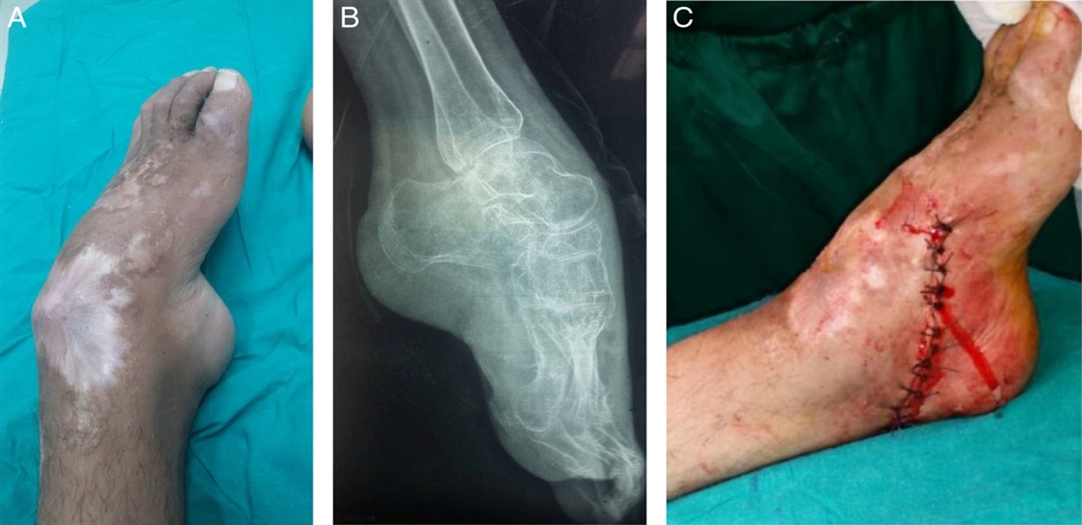
Some of these injuries represent the most severe knee dislocations in which an almost circumferential avulsion of the capsule occurs, leaving the meniscus devoid of peripheral attachments (Fig. 17). Six of 47 patients (13%) had circumferential avulsions. Similarly, one of the patients with a poor subjective outcome had a concomitant complete patellar tendon avulsion in the setting of an open dislocation. As seen in Figure 17, full thickness cartilage lesions further jeopardize long-term outcome, and may dictate final function but unlikely contributed to the outcomes in our studies, as our follow-up was 12 months. Typically, the effects of cartilage injuries are seen years later after injury.15
 FIGURE 17:
 Medial and lateral meniscus tear and associated cartilage lesion.
Our results show that with the presented protocol, good range of motion can be achieved. In our study all but 1 patient obtained full extension and an acceptable mean range of motion of 0 to 124 degrees. Only 4 patients (8.5%) required an additional procedure to address residual stiffness, and revision surgery to address instability was performed in only 1 patient (2%) at the time of final follow-up.
The soft tissue disruption present in these injuries does not permit safe arthroscopic management in the acute phase, because of the almost invariable presence of capsular disruption and resultant increased risk of compartment syndrome from fluid extravasation into the muscle compartments.16,17 The current literature on knee dislocations favors operative treatment compared with nonoperative management.3,4 In the meta-analysis by Dedmond and Almekinders,3 higher range of motion and Lysholm scores were found in the operative group compared with nonoperative management. The literature also favors early treatment (within 6 wk) over delayed reconstruction, with early treatment showing higher subjective knee scores and activity ratings.5–8 Several studies further support staged surgical treatment consisting of either acute collateral ligament repair or reconstruction and delayed ACL, and/or PCL reconstruction18 or early PCL reconstruction with delayed ACL and collateral ligament reconstruction,13–19 while other studies have shown that delayed reconstruction can also have good results.5,20–22
Open ligamentous repair or arthroscopic repair remains controversial. One concern of open repair is postoperative arthrofibrosis.23 However, a series with open repair and early aggressive modern rehabilitation demonstrated acceptable outcomes. In 2007 Owens et al7 reported a series of patients primarily repaired within 14 days of injury, who had a resultant mean arc of motion of 119.3 degrees and no incidence of late loosening requiring reconstruction. At 2-year follow-up, the mean Lysholm was 89 and Tegner score was 4.4 with arthrofibrosis requiring arthroscopic lysis of adhesions in 5 patients (19%).7 Our patients had an average range of motion arc of 124 degrees, which is comparable to the findings reported by Owen et al7 and other arthroscopic treatment protocols with only 4 additional operations because of stiffness and 3 of these were for a closed manipulation.1,18,21–24
Early stabilization of the PLC historically has been recommended by Shelbourne and Klootwyk.25 Because of risk of late loosening with staged procedures of repair of lateral or medial collaterals and delayed arthroscopic cruciate reconstruction, reconstruction has been recommended in 2 studies and in a JAAOS review article.26–28 One study showing higher failure rates in primary repair of the PLC was published by Stannard et al29 in 2005. This prospective cohort study included 57 PLC injuries in 56 patients of which 44 (77%) were MLIs. Patients were not randomized but were treated with either primary repair of the PLC (n=35) or reconstruction using allograft (n=22), and we found higher failure rates in the primary repair group (37% vs. 9%). Lysholm scores were similar in the repair and reconstruction cohorts of the patients who did not fail (88.2 vs. 89.6).
Levy et al26 also reported similar results in a recent study comparing primary repair versus reconstruction of the posterolateral corner. Both of these studies staged the collateral repair, and the arthroscopic cruciate reconstruction was delayed by an average of 115 days.
Stannard et al30 recently published a randomized control trial of delayed reconstruction of all ligaments with or without the use of a hinged fixator. The patients without the fixator had a significantly increased number of ligament failures (21% vs. 7%, P<0.001), but no difference in the overall percentage of patients who had ligament failure (29% vs. 15%, P=0.15). No differences between groups were seen in Lysholm, IKDC, Pain VAS, or ROM. The IKDC scores for patients in the Stannard study were nearly the same as this study (Ex fix, 56.8; No ex fix, 49.5; Acute repair, 50).
Engebretsen and colleagues have published the largest series (n=85 patients) of early treatment of patients with knee dislocations (KD III-KD V) within 2 weeks of injury with at least 2 years of follow-up. The ACL and PCL were reconstructed arthroscopically and collaterals were repaired if able. This group, which is similar to our cohort, had similar outcome scores as well.24
The efficiency of early surgical treatment and rehabilitation protocol allows 1 hospitalization and operation for the vast majority of patients. Less than 25% of our patients had a secondary surgery. A staged protocol requires additional hospitalizations and a potentially significant cost and morbidity of an external fixator. A staged protocol also makes it difficult to address many associated injuries, such as displaced menisci, patellar tendon tears, periarticular fractures, or quadriceps ruptures.
An important point of this series of patients is that a variety of injuries occur with this injury that can be treated simultaneously early in the postinjury period. Open injuries usually have an initial debridement and closure of the wound and then primary repair or reconstruction of all damaged structures. The patients who had a patellar fracture had the fracture and all ligaments repaired at the time of surgery, and range of motion was initiated at 4 weeks.
Our study has several limitations. We did not identify whether associated fractures and meniscal tears as prognostic indicators of outcome could be a focus of a future study. Because of the small size of our sample, it was not possible to perform a statistical analysis to determine whether predictive factors pertaining to patient demographics or injury pattern exist. Our sample size is relatively limited but larger than most all similar studies except the Engebretson and colleagues cohort. The lack of a control group does not allow for a comparative analysis with, for example, all arthroscopic or staged treatment. Our study included only high-grade multiligamentous injuries (KD III-KD V) that had at least 3 disrupted ligaments. Although there is some heterogeneity in the included group, it does offer a more homogeneous analysis than that offered by most studies, in which all types of knee dislocations are analyzed as a group. Our low follow-up of 64% could have skewed the data. Varus and valgus laxity was not formally recorded by stress radiographs in this study. It is important to note that even this was not formally recorded, and only 1 patient in this study needed revision because of laxity or instability. The KT-1000 was used to identify posterior laxity but has been shown to be only moderately reliable in identifying posterior laxity because of posterior sag.31 Furthermore, we performed a prospective follow-up of our patients using a standardized protocol and widely used and validated outcomes scores.
CONCLUSIONSOpen operative treatment within 3 weeks of injury with primary repair or reconstruction yields good outcomes in two-thirds of patients, as assessed by standardized knee outcome scores. Neither stiffness nor late laxity is a significant problem, and patients receive only one operation. On the basis of our study and other studies that promote a reconstruction, patients should be counselled that a significant loss of function should be expected.
ACKNOWLEDGMENTSThe authors acknowledge William Neterville, Scott Zuckerman MD for assistance with data collection and Basem Attum for manuscript preparation.
REFERENCES 1. Schenck RC Jr. The dislocated knee. Instr Course Lect. 1994;43:127–136. 2. Green NE, Allen BL. Vascular injuries associated with dislocation of the knee. JBJS. 1977;59:236–239. 3. Dedmond BT, Almekinders LC. Operative versus nonoperative treatment of knee dislocations: a meta-analysis. Am J knee Surg. 2000;14:33–38. 4. Richter M, Bosch U, Wippermann B, et al. Comparison of surgical repair or reconstruction of the cruciate ligaments versus nonsurgical treatment in patients with traumatic knee dislocations. Am J Sports Med. 2002;30:718–727. 5. Harner CD, Waltrip RL, Bennett CH, et al. Surgical management of knee dislocations. J Bone Joint Surg Am. 2004;86-a:262–273. 6. Liow R, McNicholas M, Keating J, et al. Ligament repair and reconstruction in traumatic dislocation of the knee. Bone Joint J. 2003;85:845–851. 7. Owens BD, Neault M, Benson E, et al. Primary repair of knee dislocations: results in 25 patients (28 knees) at a mean follow-up of four years. J Orthop Trauma. 2007;21:92. 8. Mariani PP, Santoriello P, Iannone S, et al. Comparison of surgical treatments for knee dislocation. Am J knee Surg. 1998;12:214–221. 9. Hefti E, Müller W, Jakob R, et al. Evaluation of knee ligament injuries with the IKDC form. Knee Surg Sports Traumatol Arthrosc. 1993;1:226–234. 10. Lysholm J, Gillquist J. Evaluation of knee ligament surgery results with special emphasis on use of a scoring scale. Am J Sports Med. 1982;10:150–154. 11. Tegner Y, Lysholm J. Rating systems in the evaluation of knee ligament injuries. Clin Orthop Relat Res. 1985;198:43–49. 12. Noyes FR, Barber-Westin SD. Reconstruction of the anterior and posterior cruciate ligaments after knee dislocation: use of early protected postoperative motion to decrease arthrofibrosis. Am J Sports Med. 1997;25:769–778. 13. Moatshe G, Dean CS, Chahla J, et al. Anatomic fibular collateral ligament reconstruction. Arthrosc Tech. 2016;5:e309–e314. 14. Geeslin AG, LaPrade RF. Outcomes of treatment of acute grade-III isolated and combined posterolateral knee injuries: a prospective case series and surgical technique. J Bone Joint Surg Am. 2011;93:1672–1683. 15. Potter HG, Jain SK, Ma Y, et al. Cartilage injury after acute, isolated anterior cruciate ligament tear: immediate and longitudinal effect with clinical/MRI follow-up. Am J Sports Med. 2012;40:276–285. 16. Ekman EF, Poehling GG. An experimental assessment of the risk of compartment syndrome during knee arthroscopy. Arthroscopy. 1996;12:193–199. 17. Belanger M, Fadale P. Compartment syndrome of the leg after arthroscopic examination of a tibial plateau fracture. Case report and review of the literature. Arthroscopy. 1997;13:646–651. 18. Levy BA, Krych AJ, Shah JP, et al. Staged protocol for initial management of the dislocated knee. Knee Surg Sports Traumatol Arthrosc. 2010;18:1630–1637. 19. Ohkoshi Y, Nagasaki S, Shibata N, et al. Two-stage reconstruction with autografts for knee dislocations. Clin Orthop Relat Res. 2002:169–175. 20. Fanelli GC, Orcutt DR, Edson CJ. The multiple-ligament injured knee: evaluation, treatment, and results. Arthroscopy. 2005;21:471–486. 21. Fanelli GC, Edson CJ. Surgical treatment of combined PCL-ACL medial and lateral side injuries (global laxity): surgical technique and 2- to 18-year results. J Knee Surg. 2012;25:307–316. 22. Cook S, Ridley TJ, McCarthy MA, et al. Surgical treatment of multiligament knee injuries. Knee Surg Sports Traumatol Arthrosc. 2015;23:2983–2991. 23. Shapiro MS, Freedman EL. Allograft reconstruction of the anterior and posterior cruciate ligaments after traumatic knee dislocation. Am J Sports Med. 1995;23:580–587. 24. Engebretsen L, Risberg MA, Robertson B, et al. Outcome after knee dislocations: a 2–9 years follow-up of 85 consecutive patients. Knee Surg Sports Traumatol Arthrosc. 2009;17:1013–1026. 25. Shelbourne KD, Klootwyk TE. Low-velocity knee dislocation with sports injuries: treatment principles. Clin Sports Med. 2000;19:443–456. 26. Levy BA, Dajani KA, Morgan JA, et al. Repair versus reconstruction of the fibular collateral ligament and posterolateral corner in the multiligament-injured knee. Am J Sports Med. 2010;38:804–809. 27. Levy BA, Fanelli GC, Whelan DB, et al. Controversies in the treatment of knee dislocations and multiligament reconstruction. J Am Acad Orthop Surg. 2009;17:197–206. 28. Stannard JP, Sheils TM, McGwin G, et al. Use of a hinged external knee fixator after surgery for knee dislocation. Arthroscopy. 2003;19:626–631. 29. Stannard JP, Brown SL, Farris RC, et al. The posterolateral corner of th
留言 (0)