
記住我


Many techniques describe reconstruction of the posterolateral corner (PLC) of the knee; the 2 main concepts include “anatomic” reconstruction or traditional “fibula sling” techniques utilizing a transosseous fibula tunnel combined with 1 or 2 isometric femoral tunnels to control varus and posterolateral rotatory laxity. Although anatomic techniques have shown biomechanical advantage in laboratory studies, there is yet to be any demonstratable clinical benefit over traditional techniques.1 All techniques have previously been described using a broad lateral approach using a single “hockey stick” incision to access the lateral femoral condyle and the fibula head, while also allowing direct visualization and protection of the common peroneal nerve (CPN), which is at risk of iatrogenic damage when drilling osseous tunnels in the fibula head. Although this extensive approach allows for excellent exposure, it potentially increases the risk of soft tissue trauma, hematoma, wound problems, and therefore subsequent infection.2,3
To the best of our knowledge, a minimally invasive 3-incision technique has yet to be described in the literature. The described approach allows for access to the fibula head, lateral femoral condyle, and identification of the CPN with minimal soft tissue dissection. We hope these simple modifications in the surgical approach will be of use, both to surgeons who regularly perform PLC reconstructions and to encourage surgeons who are currently apprehensive about using minimally invasive lateral approaches due to concerns about adequate exposure.
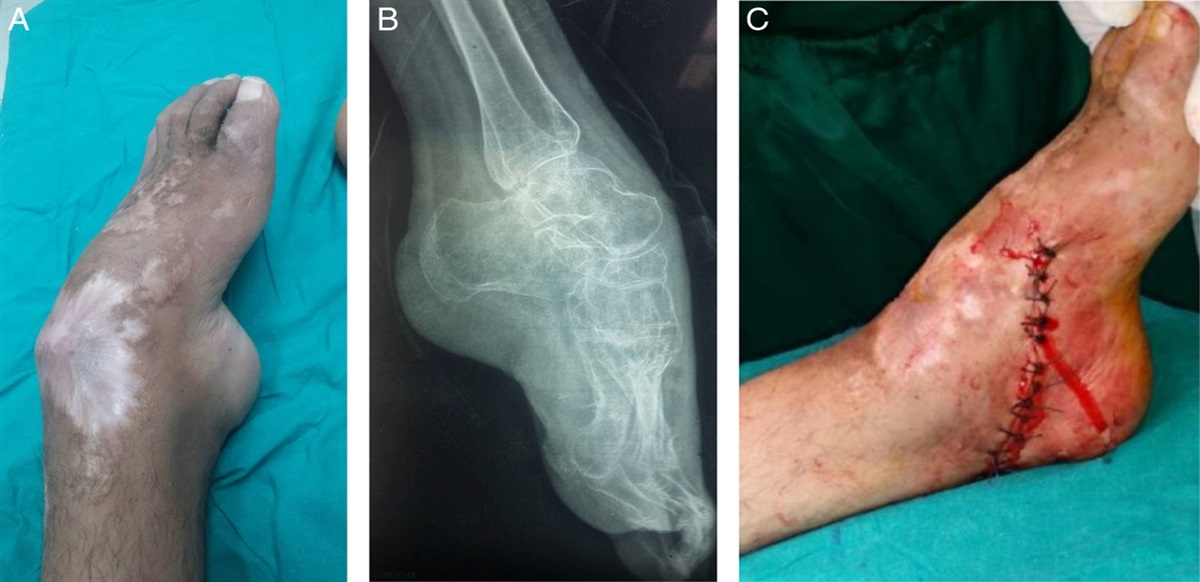
TECHNIQUEAll illustrations are from a single procedure in a 26-year-old patient who underwent a revision anterior cruciate ligament (ACL) reconstruction with combined PLC reconstruction and lateral extra-articular tenodesis (LET).
Patients are anaesthetized with a general anesthetic and prophylactic antibiotics are given. A high tourniquet is applied, patients are positioned supine with a single lateral side support and single foot bolster holding the knee in 90 degrees of flexion.
When performed alongside reconstruction of the ACL both meniscal surgery and preparation of the tibial and femoral ACL tunnels are performed before the PLC and LET reconstruction, this minimizes the risk of fluid extravasation around the posterolateral structures while allowing for direct tunnel visualization of any potential tunnel collision by viewing from the anteromedial portal.
Bony anatomical landmarks are marked out with a sterile skin marker including the lateral epicondyle, Gerdy’s tubercle on the anterolateral tibia, and the fibula head. The technique uses 3 small skin incisions (Figs. 1A, B). The incisions around the fibula head allow for both excellent access to the fibula head to safely drill a transosseous tunnel in the anterior-posterior direction while also allowing dissection and direct visualization of the CPN to ensure there is no risk of iatrogenic damage. To allow this, a small vertical incision is made anterior to the fibula head, and a small longitudinal incision (in line with the direction of the CPN) is made posterior to the fibula head. The third proximal incision is made longitudinally, inferior to the lateral epicondyle, this allows access to the femoral insertion point for the PLC reconstruction, and the LET can easily be performed through the same incision.
 FIGURE 1:
 A and B, The 3 incisions are based around the fibula head and the lateral epicondyle as shown. The diagram illustrates the anatomy encountered with each incision. The femoral incision allows for nonanatomic posterolateral corner reconstructions Larsons (blue), Arcerio (blue and green), plus the addition of a lateral extra-articular tenodesis (red).
The first stage of the approach uses the transverse incision posterior to the fibula neck to identify the CPN nerve both through palpation and direct visualization. The CPN can then be protected by placing a finger in the wound, ensuring that the nerve is always inferior to the drill (Fig. 2). The nerve is consistently found inferior to the tendon of biceps femoris. Extensive dissection and release of the nerve is not required in chronic PLC reconstructions. When the nerve has been identified the second stage of the approach is to make an incision anteriorly to the fibula head. Under direct vision, the anteroposterior transosseous tunnel can be drilled towards the surgeon’s finger placed on the back of the fibula head. A cannulated 4.5 mm drill bit is used to create this tunnel over an initial guide wire. A passing suture is then passed through the tunnel to allow easy passage of the ligament graft.
 FIGURE 2:
 Fingers can be placed in the wound to palpate the common peroneal nerve and feel the fibula head to allow safe drilling of the transosseous fibula tunnel.
The third stage of the approach is to make the proximal longitudinal incision over the lateral epicondyle. The length of the proximal incision at the lateral epicondyle can be varied depending on if an isolated PLC reconstruction is performed or a combined LET procedure is required. In this case, a 4 cm transverse incision is made inferior to the lateral epicondyle, a 1 cm×10 cm strip of the iliotibial band (ITB) is harvested with the leg in extension to ensure it is harvested in line with the ITB fibers and centered on Gerdy’s tubercle. The lateral collateral ligament remanent is identified and the strip of ITB is passed beneath it. The femoral tunnel for the PLC reconstruction is then made in the desired location depending on the technique used. Next, the ligament graft is passed through the fibula tunnel from the anteroposterior direction ensuring the CPN is protected. It is then passed beneath the ITB and secured in the femoral socket and tensioned with the leg in extension (Fig. 3). The LET can then be fixed to the femur at 30 degrees of knee flexion.
 FIGURE 3:
 The graft is passed through the tunnel in the fibula head, beneath the iliotibial band, and fixed into the femoral socket (red arrow). A lateral extra-articular tenodesis can be performed at the same time through the proximal incision (blue arrow).
Following the final fixation of the grafts the CPN is checked and the wounds are closed with absorbable sutures to subcutaneous tissues and clips to approximate the skin edges. Postoperative restrictions include a hinged knee brace and restricted weight bearing dictated by any concomitant meniscal surgery required.
The main apprehension and potential complication regarding this approach is limited exposure. However, the approach can be simply converted to the traditional lateral “hockey stick” incision by joining the proximal incision with the anterior fibula incision should one have any intraoperative concerns.
In the illustrations used a Larson PLC technique reconstruction is used but an Arcerio technique using 2 femoral attachments can also be performed when using this approach. This approach will not allow for an anatomic reconstruction technique that requires exposure to the posterior tibia.
EXPECTED OUTCOMESReconstruction of the PLC of the knee results in excellent postoperative outcomes, however, potential complications include damage to the CPN, hematoma, deep infection, and failure of the reconstruction. No single technique has been shown to be superior and reduce failure rates or improve clinical outcomes compared with others.1,4 One potential way to reduce the risk of hematoma, soft tissue trauma, and deep infection is minimally invasive surgery. Minimally invasive surgery has been shown to be advantageous in numerous other orthopedic procedures and safer when compared with open procedures.4
The authors use the described technique in all patients undergoing isolated PLC reconstruction, chronic multiligament reconstructions, and when performing combined PLC and LET procedures. If there are any concerns regarding exposure during the approach this technique is extensile and can be extended into a more traditional lateral approach at any point. This approach is not advised in acute knee dislocations or in previously high-energy knee dislocations which displayed a high-grade injury (bicep femoris tendon avulsion from the fibula head) to the PLC at the time of injury when the location of the CPN is unreliable.
The major concern with performing minimally invasive surgery on the lateral side of the knee is the potential risk of iatrogenic damage to the CPN due to inadequate exposure. The CPN is injured when drilling fibula-based transosseous tunnels. Arthroscopic techniques have been described for PLC reconstruction but these techniques are technically demanding, involve a significant learning curve, and can risk injury to the popliteal artery.5 A minimally invasive approach is therefore advantageous as it uses familiar techniques without additional risk.
This described 3-incision technique is valuable as it permits the benefits of minimally invasive surgery and it is our experience that this technique makes identification of the CPN simple, safe, and reproducible.
This technique allows for the easy addition of an LET procedure, we advise the addition of a LET in all patients at high risk of re-rupture (revision ACL, hyperlaxity, young age, high-grade pivot, return to cutting, and pivoting sports). It is perhaps sensible to make a larger incision than shown in the clinical images when adding a LET.
COMPLICATIONSWe are yet to encounter any complications with specific concern to exposure, injury to neurovascular structures, or fixation of the reconstructions when using this technique. We recommend careful identification of suitable patients for this technique (chronic low-energy PLC injuries) to avoid complications. When adopting a new technique it is always imperative to identify any complications and we would encourage surgeons to collect this data when adopting this technique.
CONCLUSIONSThe benefits of minimally invasive PLC reconstruction are advantageous and could reduce the risks of hematoma, wound problems, and infection. We therefore recommend performing minimally invasive reconstruction of the PLC in the first instance and reserving extensive approaches for high-energy acute repair/reconstructions or if any intraoperative concerns about adequate exposures are encountered.
REFERENCES 1. van Gennip S, van der Wal WA, Heesterbeek PJC, et al. Posterolateral corner reconstruction in combined injuries of the knee: improved stability with Larson’s fibular sling reconstruction and comparison with LaPrade anatomical reconstruction. Knee. 2020;27:124–131. 2. Moulton SG, Geeslin AG, LaPrade RF. A systematic review of the outcomes of posterolateral corner knee injuries, part 2: surgical treatment of chronic injuries. Am J Sports Med. 2016;44:1616–1623. 3. Chahla J, Murray IR, Robinson J, et al. Posterolateral corner of the knee: an expert consensus statement on diagnosis, classification, treatment, and rehabilitation. Knee Surg Sports Traumatol Arthrosc. 2019;27:2520–2529. 4. Rafaqat W, Ahmad T, Ibrahim MT, et al. Is minimally invasive orthopedic surgery safer than open? A systematic review of systematic reviews. Int J Surg. 2022;101:106616. 5. Hermanowicz K, Malinowski K, Góralczyk A, et al. Minimally invasive, arthroscopic-assisted, anatomic posterolateral corner reconstruction. Arthrosc Tech. 2019;8:e251–e257.
留言 (0)