
記住我


Cephalomedullary nail (CMN) fixation is a well-described and relatively successful strategy for the treatment of pertrochanteric hip fractures. Categorized as intramedullary fixation, this type of construct was originally proposed in the 1980s as a way of treating proximal femur fractures that were otherwise subject to high failure rates with extramedullary constructs. Although originally indicated for those proximal femur fractures with posteromedial comminution, subtrochanteric extension, questionable lateral cortex integrity, or reverse obliquity orientation, the indications for this fixation method have expanded to include most types of proximal femur fractures.
In the setting of pertrochanteric hip fractures (Fig. 1), these constructs function similarly to the sliding hip screws that were originally developed in the 1950s.1 Both constructs are capable of allowing for controlled linear collapse and compression across a fracture site, assuming proper orientation of the hip screw relative to the fracture line, which allows for improved dynamic osseous contact and fracture healing relative to “static” implants. While the CMN has undoubtedly revolutionized the way we treat hip fractures, all treatment modalities come with a potential downside, one of which for the CMN is the risk for symptomatic lateral hardware associated with fracture collapse (Fig. 2).
 FIGURE 1: AO/OTA type 31—A pertrochanteric hip fractures.1 Link to License at: https://creativecommons.org/licenses/by/2.5/legalcode. Available via license: CC BY 2.5.
 FIGURE 2:
 Anterior-posterior radiographs before and after the controlled collapse of a pertrochanteric hip fracture following dynamic cephalomedullary nail fixation. The radiographs on the left was taken on a postoperative day 1 after the index surgery and the radiograph on the right was taken 2 weeks before the blade exchange procedure for symptomatic lateral hardware.
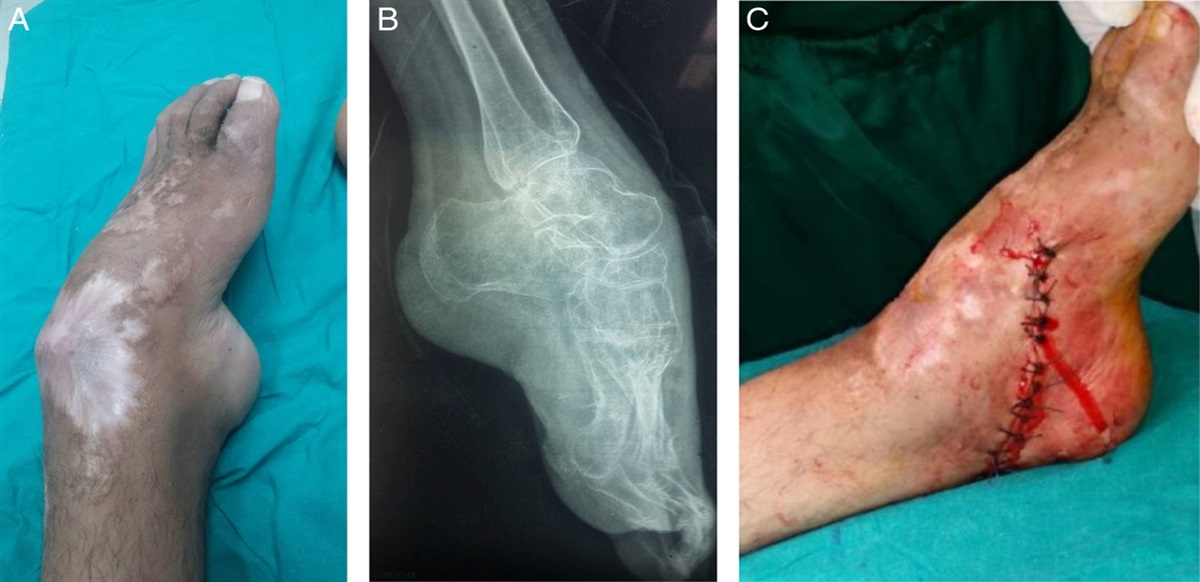
Often perceived as a postoperative complication, lateral hardware prominence, whether it is symptomatic or not, is an expected sequela of healing in patients treated with dynamic constructs. Even slight collapse and minimal prominence can cause symptoms considering the close proximity of the iliotibial band and trochanteric bursa. Furthermore, when patients present with complaints associated with hardware prominence it is often the most significant issue preventing them from returning to their baseline preinjury activities. Therefore, we present a simple yet elegant strategy for addressing this pain that does not subject our patients to an increased risk for future fracture, as is the case with hardware removal.2,3 We present 4 cases of symptomatic lateral hardware after CMN fixation of pertrochanteric hip fractures that were addressed by exchanging the prominent hip blade for a shorter version and engaging the static proximal locking mechanism, when available (Fig. 3).
 FIGURE 3:
 Anterior-posterior hip radiographs before and after the hardware exchange procedure for symptomatic lateral hardware following the controlled collapse of cephalomedullary nail construct.
TECHNIQUEAt the time of surgery, the patient is positioned supine on a radiolucent table with a soft bump of 1 or 2 blankets placed under the operative buttock in a manner that internally rotates the lower extremity into a more neutral position. The operative extremity is placed on a radiolucent ramp to facilitate lateral fluoroscopy of the hip and clearance of the nonoperative leg for adduction. The entire lower extremity and flank is prepped and draped in a standard sterile fashion.
The prior proximal skin incision and gluteus fascia are opened with a scalpel and the proximal aspect of the nail is identified. In rare circumstances, the incisions from the index procedure may not be optimally placed and therefore new incisions, with the assistance of fluoroscopy, may be required to facilitate the most efficient and least invasive approach. A flexible screwdriver is inserted to disengage the proximal locking screw. Next, attention is turned to the lateral skin incision overlying the proximal interlock/cephalomedullary implant. Again, a scalpel is used to incise the skin and underlying fascia so that the hip screw may be engaged with a screwdriver or extraction rod. The proximal interlock is removed. Fluoroscopy can be employed to aid in the localization of components. If difficulty is encountered while engaging the above screwdrivers, ensure there is no interposed soft tissue to prevent “stripping” of the implant. Once the proximal interlock is removed, its length is ascertained. A new proximal interlock is chosen, based upon either a preoperative template of the length of proximal interlock extending beyond the lateral cortex or the path is simply remeasured with a guidewire and rule. The new proximal interlock is placed flush or slightly recessed with the lateral cortex to ensure there is no remaining hardware prominence. Finally, the proximal interlock locking mechanism is engaged, which varies by manufacturer. This ensures that the new implant cannot slide in the nail interlock hole, as fracture collapse is no longer desired. Wounds are then irrigated and closed in a standard layered fashion.
EXPECTED OUTCOME AND COMPLICATIONSLateral hardware prominence following intramedullary fixation of pertrochanteric hip fractures can be a natural sequela of successful fracture management with certain dynamic compression constructs. Unfortunately, in the subgroup of patients who are symptomatic from this issue, the pain can often be debilitating enough to warrant operative intervention. Due to the relatively high risk of refracture, with estimates approaching 15% in one study, simply removing the prominent hardware at any postoperative timepoint is discouraged.2–4 With that being said, nonoperative strategies are also suboptimal. Therefore, we present a simple alternative to hardware removal in severely symptomatic patients—hardware exchange.
Although the incidence of secondary fracture following hardware removal varies based on the report, there are several clinical and biomechanical studies evaluating this issue.2–5 In a systematic review performed by Barquet et al,3 the incidence of femoral neck fracture following implant removal was 14.5%. More importantly, this review found that in almost 90% of these cases, the fractures occurred in the absence of any associated trauma.3 A potential reason for these atraumatic injuries was highlighted via a biomechanical cadaver study that evaluated the strength of the femoral neck after implant removal.5 This study found a 41% reduction in resistance to femoral neck fracture following CMN removal.5 While this study was performed on cadavers and is therefore limited in its clinical applicability, it does lend further support to the importance of implant retention at all postoperative timepoints in this cohort of patients.
In our small series of patients who have underwent this blade exchange procedure (n=4), there was a decrease in lateral hip pain with no associated intraoperative or postoperative complications. All patients tolerated the procedure without issue and were able to return home the same day without any opioid pain medications or weight-bearing restrictions. At the patient’s most recent follow-up appointment, which was on average 200 days after the exchange procedure, there were no cases of refracture. Although no complications were encountered from our procedure, it is important to recognize that all operative interventions come with risk, especially in this cohort of patients. The risks inherent to this procedure include the general risks associated with anesthesia, whether it is spinal or general endotracheal, secondary femoral neck fractures, screw/blade breakage due to the rotational torque required to remove the screw/blade, and wound complications. Therefore, proper patient education and shared decision-making are critical when deciding to undergo revision surgery based on the risks and benefits.
In conclusion, symptomatic lateral hardware following fracture collapse in the setting of dynamic CMN fixation of pertrochanteric hip fractures can be seen following successful fracture management. Although rare, if operative intervention is required, exchanging the prominent blade with a locked shorter blade is a reasonable strategy that is safe and does not seem to increase secondary fracture risk. Further investigation into this treatment strategy is warranted to validate the outcomes of our case series.
REFERENCES 1. Orthopaedic Trauma Association Classification, Database and Outcomes Committee. Fracture and dislocation classification compendium-2007. J Orthop Trauma. 2007;suppl:10. 2. Kovar F, Strasser E, Jaindl M, et al. Complications following implant removal in patients with proximal femur fractures—an observational study over 16 years. Orthop Traumatol Surg Res. 2015;101:785–789. 3. Barquet A, Giannoudis P, Gelink A. Femoral neck fractures after removal of hardware in healed trochanteric fractures. Injury. 2017;48:2619–2624. 4. Yang KH, Choi YW, Won JH, et al. Subcapital femoral neck fracture after removal of gamma/proximal femoral nails: report of two cases. Injury Extra. 2005;36:245–248. 5. Kukla C, Pichl W, Prokesch R, et al. Femoral neck fracture after removal of the standard gamma interlocking nail: a cadaveric study to determine factors influencing the biomechanical properties of the proximal femur. J Biomech. 2001;34:1519–1526.
留言 (0)