
記住我


Pes cavus is a foot deformity characterized by an increase in the normal plantar concavity, which presents as a spectrum of foot deformities that includes forefoot adduction and pronation, plantar flexed first ray, and hindfoot varus or high calcaneal pitch. Cavovarus deformity severity ranges from a subtle and flexible cavovarus foot to a severe and fixed cavovarus deformity.1,2 Adult-onset foot cavovarus deformity is a highly disabling deformity that seriously interferes with the patient's daily activities.3 Most foot deformities in skeletally mature patients include mainly cavus, varus, and equinus components, and they are attributed to a variety of etiologies, such as Charcot-Marie-Tooth (CMT), poliomyelitis, cerebral palsy, peripheral neuropathy, and idiopathic pes cavus.4,5 However, an underlying neurological abnormality is the main causative factor, which represents around 80% of these deformities. The most common one is CMT disease, which is a hereditary sensory and motor neuropathy.6 In patients with CMT, a flexible foot deformity develops during adolescence and later progresses to a fixed deformity. Therefore, the deformities worsen, and surgical intervention must be considered as early as possible to prevent the progression to a fixed and symptomatic deformity.7 Various treatment options have been described for the treatment of these foot deformities, and procedures commonly aim to restore a plantigrade foot.8 The current concept in most centers is that soft tissue procedures are most useful for correcting flexible deformity, especially in younger patients, whereas bony procedures should be performed for rigid deformity in skeletally mature patients.9,10 Our hypothesis is that, in adult-onset rigid foot deformity, first, we have no structural bony anomalies, and the problem is in the soft tissues, so there is no need for a bony procedure. Second, these deformities are multiaxial deformities, and the osteotomies done are not in the center of the deformity, so excessive bone translation is needed, which cannot realign the relationship between the tarsal bones and does not reach complete correction. Third, when performing osteotomies, complications are not uncommon, and nonunion, malunion, hardware failure, and wound dehiscence are all reported complications. Therefore, osteotomy is not to be taken lightly, especially when it leads to major alterations in the architecture of the foot and even shortening. Fourth, the fusion procedure will end with a stiff foot.11–14
PATIENTS AND METHODSWe reviewed 30 feet (25 patients) with adult-onset cavovarus deformities consecutively treated with extensive posteromedial soft tissue release (EPMSTR; Figs. 1 and 2) between March 2017 and March 2019. Neurological work-up was done for all the patients upon demonstration by a specialized neurologist to determine the etiology of the foot deformity. It was CMT disease (peroneal muscle atrophy) in all cases. All the patients presented to us with rigid cavovarus deformities evident clinically by the Coleman block test.
 FIGURE 1:
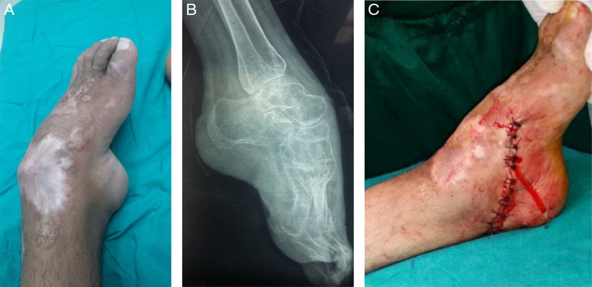
 A, Preoperative clinical photo. B, Preoperative radiograph. C, Immediate postoperative photo of a patient with unilateral severe pes cavovarus deformity.
 FIGURE 2:
 A, Preoperative clinical photo. B, Preoperative radiograph. C and D, Immediate postoperative photos of a patient with bilateral pes cavovarus deformity.
All our patients were ambulant with leg and foot muscle weakness more pronounced in the tibialis anterior and peroneus brevis, with fair motor function and they had intact peripheral sensation.
We excluded patients with significant arthritis (subtalar or ankle), severe instability, significant joint contractures, or chronic ulcers.
Our incision is transverse and centered over the tip of the medial malleolus to avoid making the incision scar a deforming force after healing. Isolate the neurovascular bundle, then, EPMSTR done, including the ligamentous attachments of the talus with the complete release of the subtalar, talonavicular, as well as the naviculocuniform joints to provide the necessary malleability to make the freed head of talus as the axis of the corrective rotation. Rendering the head of the talus an axis around which we release all the soft tissues for correcting the deformity is the main principle applied in the surgical interventions of this technique. Posteromedial release was done including tibialis posterior, flexor digitorum, flexor hallucis (tenotomies), and Achilles tendons (by percutaneous or open tenotomy) but no interventions were conducted on the peronei or the tibialis anterior tendons. Finally, we do percutaneous plantar fascia release. No osteotomies were performed, and no fixation modalities were used (Fig. 3).
 FIGURE 3:
 Intraoperative photos of the surgical procedure. A, Incision. B and C, Tendons exposure and tenotomy under the flexor retinaculum. D and E, Tendo-Achilles release. F and G, Talonavicular joint release. H and I, Subtalar joint release. J and K, Navicular-cuneiform joint release. L, Plantar fascia release.
Direct postoperative casting fixed the foot in the new corrected position (Fig. 4). In 5 feet (16.6%), the deformity was very severe such that there was a risk of protrusion of the freed talus from the surgical incision if direct postoperative correction and casting was performed. In these cases, the foot was left without being placed in the full corrected position in a slab postoperatively for a 2-week interval to allow time for the wound to heal, after which the foot was placed in cast in the corrected position.
 FIGURE 4:
 Postoperative casting in a patient with bilateral deformity.
All patients had cast removal after 4 weeks from its placement and were recommended to apply night braces for a period of 3 months after cast removal to prevent the recurrence of the deformity by contractures of the soft tissues. A staged rehabilitation protocol is of utmost importance postoperatively to prevent significant scar tissue formation to maximize the functional outcome in the operated foot. Stage 1 (0 to 4 wk) includes pain management, wound care, and protected weight-bearing in cast is allowed using crutches or a walker. Gentle range of motion exercises and edema control measures are also included. Stage 2 (4 to 12 wk) joint mobilization, scar massage, myofascial release, stretching, proprioceptive and balance exercises are incorporated. Modalities, such as ultra sound therapy or low-level laser therapy, are used to promote scar remodeling and tissue healing. Stage 3 (after 12 wk) includes muscle strengthening and endurance exercises. Gait, balance, and proprioceptive retraining were also included. Functional activities (such as single-leg stance, heel-to-toe walking, and stair climbing) are incorporated to facilitate the transition to normal walking and life-demand patterns.
All the patients were available for follow-up (Figs. 5 and 6), the mean age at the time of surgery was 20.4 (range: 20 to 23) years, and the mean follow-up period was 36 (range: 33 to 42) months.
 FIGURE 5:
 Follow-up at 6 weeks postoperatively in a patient with bilateral pes cavus deformity.
 FIGURE 6:
 Follow-up at 32 months postoperatively in a patient with unilateral pes cavus deformity.
Clinical evaluation was done using the Foot Function Index (FFI) for the pain, disability, and limitation of movement. The radiologic assessment was done by measurement of the latera talo-first metatarsal angle, the lateral calcaneal-first metatarsal angle, and the lateral tibio-calcaneal angle in standing lateral foot radiographs.
RESULTSAll patients were followed for a mean of 36 (range: 33 to 42) months postoperatively. The mean FFI for pain improved from 51.46 (range: 46 to 56) points preoperatively to a mean of 23.65 (range: 20 to 28) points postoperatively at the final follow-up. The mean FFI for disability improved from 47.06 (range: 42 to 53) points preoperatively to a mean of 21.88 (range: 20 to 25) points postoperatively. Finally, the mean FFI for limitation improved from 22.6 (range: 20 to 26) points preoperatively to a mean of 10.2 (range: 9 to 12) points at the final postoperative follow-up.
Radiologic evaluation was assessed by obtaining standing lateral foot radiographs, the following angles were measured:
The mean lateral talo-first metatarsal (Meary) angle improved from 28.9 degrees (range: 24 degrees to 34 degrees) preoperatively to a mean of 7.55 degrees (range: 5 degrees to 11 degrees) at the final follow-up visit. The mean lateral calcaneo-first metatarsal angle improved from 132.4 (range: 130 degrees to 135 degrees) preoperatively to a mean of 117.65 degrees (range: 115 degrees to 120 degrees) at the final follow-up visit. The mean lateral tibio-calcaneal angle improved from 62.35 degrees (range: 59 degrees to 65 degrees) preoperatively to a mean of 50.35° (range: 48 degrees to 53 degrees) at the final follow-up visit.
No wound infection or skin complications were recorded except in one foot (3.33%) in the form of wound dehiscence, management was in the form of daily dressing and the wound healed within 5 weeks.
DISCUSSIONAdult cavus foot deformity has 4 primary causes: neuromuscular, traumatic, idiopathic processes, and the presence of a residual clubfoot.15,16 Approximately two-thirds of adults with symptomatic cavus foot have an underlying neurological abnormality.15,17 CMT is the most common etiology associated with cavus foot. Patients with CMT experience symmetrical, slowly progressive distal motor neuropathy, usually starting in the first to third decade and resulting in weakness and atrophy in the leg muscles. The prevalence of CMT is 78% in patients with bilateral cavus feet.18 As the disease progresses, foot deformities become stiffer. Patients develop discomfort, loss of strength and endurance, painful callosities, and ankle instability. Furthermore, they complain about unsteady gait and loss of balance secondary to sensory deficiency, weakness, and deformity. It is advised by several authors that early intervention is obligatory to alleviate the muscle imbalance to decrease the loss of function and long-term morbidity.19
The treatment of the cavus foot is challenging because the progressive nature of the muscle imbalance is likely to cause relapse, even after adequate surgical repair and even after triple arthrodesis.18 It is now generally recognized that the cavus deformity is multifactorial and there is no single procedure that would be universally applicable to correct it. The goal of surgical treatment is to produce a plantigrade stable foot, relieve symptoms, correct the deformity, and prevent recurrence. Surgical treatment should leave the foot in a normal position or slightly overcorrected because an iatrogenic flat foot is better tolerated than a residual cavus deformity.17
Large numbers of surgical procedures and their modifications have been described for pes cavus treatment. The severity of deformity in pes cavus may vary greatly and routine use of the same procedure for all such deformities does not seem appropriate. In fact, unless the surgery is tailored precisely to address the underlying pathology, it will invariably fail.20
Surgical treatment requires meticulous preoperative planning. Plain radiographs are essential, not only to identify the deformity site but also to quantify the required correction degree and to decide whether to perform an osteotomy or an arthrodesis.21 Clinical examination aims to assess the rigidity of the cavovarus deformity and to identify the underlying etiology. A complete neurological examination of both limbs is needed. Neurological investigations are best performed by a neurologist and electro-diagnostic studies can be considered to confirm hereditary motor sensory neuropathies.22
Surgical techniques can be divided into soft tissue procedures, osteotomies, and arthrodesis. Soft tissue procedures include soft tissue release or lengthening and tendon transfers. Osteotomies may be done on the calcaneus, midfoot, and metatarsus. Generally, interventions involve only the soft tissues for mild flexible deformities, whereas osteotomies are used when greater deformity correction is needed or when the cavus foot has started to become rigid. Arthrodesis (subtalar, midtarsal, or triple) is indicated in severe rigid cavus foot or in degenerative cases18
To the extent of our knowledge, few studies were performed on pure soft tissue procedures, especially EPMSTR to correct the rigid cavovarus deformity in skeletally mature patients.
Kozanek and colleagues (2014) evaluated the medial and lateral arches of the foot following EPMSTR in 25 patients with cavovarus deformity. The authors reported significant improvements in the arch angles, the arch indices, and the naviculo-cuboid overlap ratio at the last follow-up, which was 2 years on average.23
A review article by Huang and colleagues (2019) discussed various surgical techniques for the management of cavus foot deformity, including EPMSTR. The authors reported that EPMSTR can effectively correct the hindfoot varus and midfoot cavus deformity and provide a more plantigrade foot.24
Vahdatpour and colleagues (2021) reported the outcomes of EPMSTR in 16 patients with rigid pes cavus deformity. The authors reported significant improvements in the American orthopedic foot & ankle society score, the foot & ankle ability measure, and the visual analogue scale for pain at the last follow-up, which was 16 months on average. The authors also reported a high rate of patient satisfaction and a low rate of complications.25
Ding and colleagues (2021) compared the outcomes of EPMSTR alone versus those combined with calcaneal osteotomy in 50 patients with rigid pes cavus deformity. The authors reported no significant difference in the American orthopedic foot & ankle society score, the foot & ankle ability measure, or the visual analogue scale for pain between the two groups. However, the combined group had a higher rate of complications, including delayed wound healing and deep infection.26
In terms of long-term outcomes, Kim and colleagues (2021) reported on a study that evaluated patients who underwent EPMSTR for rigid pes cavus deformity with a mean follow-up of 6.8 years. They found that the procedure led to significant improvement in foot function and pain, with maintenance of correction over time.27
In our study, we reviewed 30 CMT feet with rigid cavovarus deformities consecutively treated with EPMSTR. The mean age was 20.4 (range: 20 to 23) years and the mean follow-up period was 36 (range: 33 to 42) months.
We used the FFI scoring system which is the only validated and most objective scoring system, which can be used.11 The improved FFI scores for pain, disability, and limitation in this study could be attributed to the sparing of the foot joints and not using any sort of internal fixation modalities to preserve the correction with an aim more centered on the patient’s foot function rather than the postoperative radiologic appearance. It is difficult to compare our results with the others because of the varieties and combinations of surgical procedures, as well as the lack of homogeneity in outcome criteria.
As for the reported complications, we experienced wound dehiscence during follow-up in one foot (3.33%) in a patient with CMT which was managed conservatively with dressing and healed in a 5-week period. No recurrence of deformity or overcorrection was noted throughout the follow-up period.
In summary, we support the use of EPMSTR as an effective treatment option for selected cases in skeletally mature patients with rigid pes cavus deformity. The procedure can lead to significant improvement in foot function and pain, with maintenance of correction over time. Although combined procedures may offer better correction of deformity, they come with an increased risk of complications and longer operative time.
REFERENCES 1. Nery C, Raduan F, Del Buono A, et al. Management of cavus foot deformity: a review of the literature. Revista Brasileira de Ortopedia. 2015;50:135–141. 2. Stavlas P, Roberts A, Xaltiropoulos A, et al. The evolution of foot and ankle surgery: a historical review. EFORT Open Rev. 2017;2:343–353. 3. Shenaq DS, Halim A, Patel K. Adult onset pes cavovarus: a review. Foot. 2014;24:71–77. 4. Shibata T, Saito M, Nakamura S, et al. Overview of foot and ankle disorders and their management. J Orthop Sci. 2018;23:13–22. 5. Huang J, Wang C, Zhang Y, et al. Osteotomies for multiplanar deformities in adult cavovarus foot: a retrospective study. BMC Musculoskelet Disord. 2017;18:61. 6. Leigheb M, Pirola E, Bizzini M, et al. Adult acquired cavus foot: clinical and radiological analysis of conservative and surgical treatment. Musculoskelet Surg. 2015;99(suppl 1):S53–S60. 7. Vardi M, Salamon A, Lidar Z, et al. Long-term outcomes of calcaneo-cuboid-cuneiform osteotomy for correction of multiplanar deformities of the adult acquired foot. J Bone Joint Surg Am. 2016;98:815–823. 8. Mann RA, Coughlin MJ. Surgery of the Foot and Ankle, 9th edn. St. Louis, MO: Mosby; 2012. 9. Lee KB, Cho JH, Park JH, et al. Clinical outcomes of soft-tissue procedures with calcaneal osteotomy for cavovarus foot deformity. Foot Ankle Int. 2015;36:540–548. 10. Donnan L, Refshauge K, Vicenzino B. Rehabilitation of the foot and ankle following sports injury. Sports Med. 2006;36:585–609. 11. Budiman-Mak E, Conrad KJ, Roach KE. The Foot Function Index: a measure of foot pain and disability. J Clin Epidemiol. 1991;44:561–570. 12. Hsu AR, Davis WH, Cohen BE, et al. Arthroereisis of the subtalar joint. Foot Ankle Clin. 2014;19:481–491. 13. Hill RS. Neurological disorders of the lower extremities Canale ST, Beaty JH. Campbell’s Operative Orthopaedics, 12th edn. Philadelphia, PA: Mosby Elsevier; 2013:3157–3158. 14. Kennedy JG, Johnson SM, Collins AL, et al. Adult acquired flatfoot deformity: treatment of dysfunction of the posterior tibial tendon. Instr Course Lect. 2015;64:443–455. 15. Rosenbaum AJ, Lisella J, Patel N, et al. The cavus foot. Med Clin North Am. 2014;98:301–312. 16. Di Fabio R, Lispi L, Santorelli FM. Idiopathic pes cavus in adults is not associated with neurophysiological impairment in the lower limbs. Neurol Sci. 2015;36:2287–2290. 17. Maynou C, Szymanski C, Thiounn A. The adult cavus foot. EFORT Open Rev. 2017;2:221–229. 18. Faldini C, Traina F, Nanni M. Surgical treatment of cavus foot in Charcot-Marie-tooth disease: a review of twenty-four cases: AAOS exhibit selection. J Bone Joint Surg. 2015;97:e30. 19. Leeuwesteijn AE, De Visser E, Louwerens JW. Flexible cavovarus feet in Charcot-Marie-Tooth disease treated with first ray proximal dorsiflexion osteotomy combined with soft tissue surgery: a short-term to mid-term outcome study. Foot Ankle Surg. 2010;16:142–147. 20. Nogueira MP, Farcetta F, Zuccon A. Cavus foot. Foot Ankle Clin. 2015;20:645–656. 21. Aminian A, Sangeorzan BJ. The anatomy of cavus foot deformity. Foot Ankle Clin. 2008;13:191–198. 22. Choi JK, Cha EJ, Kim KA, et al. Effects of custom-made insoles on idiopathic pes cavus foot during walking. Biomed Mater Eng. 2015;26:S705–S715. 23. Kozanek M, Liu F, Honaker JA, et al. Evaluation of the medial and lateral arches of the foot following extensive release for cavovarus deformity. Foot Ankle Int. 2014;35:1293–1303. 24. Huang Y, Li H, Liang X, et al. The surgical management of cavus foot deformity: a review. Int Orthop. 2019;43:547–555. 25. Vahdatpour B, Zeinalizadeh M, Mohtadi A, et al. Extensive posteromedial soft tissue release for treatment of rigid pes cavus deformity. Arch Bone Jt Surg. 2021;9:173–178. 26. Ding Y, Zhang X, Li Y, et al. Comparison of extensive posteromedial soft tissue release alone versus combined with calcaneal osteotomy for rigid pes cavus. BMC Musculoskelet Disord. 2021;22:1–7. 27. Kim TW, Kim JH, Oh JK. Long-term outcomes of extensive posteromedial soft tissue release in rigid pes cavus deformity. Foot Ankle Int. 2021;42:315–320.
留言 (0)