
記住我


Corynebacterium ulcerans is a zoonotic pathogen isolated from various livestock and wild animals. Corynebacterium ulcerans infections have been reported in many countries worldwide, including Japan.1, 2 Here, we report a case involving a patient suspected to have contracted C. ulcerans infection from her domestic cats.
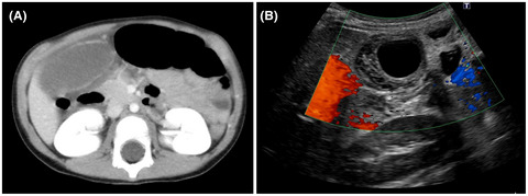
Case PresentationA 63-year-old woman with a history of diabetes and hypertension developed acute dyspnea and was rushed to a hospital, where emergency endotracheal intubation was carried out due to respiratory failure and cyanosis. She was transferred to our hospital for further evaluation and management. She had no relevant travel history and lived with her husband and seven cats. Ten days before admission, she had developed cold-like symptoms and forehead pain. At the time of transfer, her Glasgow Coma Scale score was 11. Her vital signs were as follows: pulse rate, 119 b.p.m.; blood pressure, 128/99 mmHg; respiratory rate, 20 breaths/min; and body temperature, 37.4°C. Her oxygen saturation was 100% on 10 L/min oxygen therapy. Laryngoscopy revealed mild edema in the right arytenoid region and epiglottis. Computed tomography showed no peritonsillar abscess. She was placed on ventilator support, and ampicillin/sulbactam (ABPC/SBT) antibacterial therapy was initiated. Gram staining of a tracheal aspirate on admission showed Gram-positive rods (Fig. 1). On day 2 of hospitalization, laryngoscopy revealed a small amount of a white substance in the interarytenoid region and mild epiglottis edema. We inferred that this caused temporary clogging near the glottis, resulting in asphyxia. She was extubated on day 2 of hospitalization. She underwent reintubation within 4 h due to dyspnea and stridor. Tracheal aspirate revealed an abnormal membrane-like structure (Fig. 2). After reintubation, her respiratory function stabilized, and she was extubated on day 4 of hospitalization.

Gram staining of a sputum sample aspirated from the endotracheal tube of a 63-year-old woman with Corynebacterium ulcerans infection and airway obstruction on the first day of hospitalization. The image shows Gram-positive rods. Magnification, ×1,000.

Membrane-like abnormal structure expectorated from the endotracheal tube of a 63-year-old woman with Corynebacterium ulcerans infection and airway obstruction.
Sputum cultures submitted on the day of admission showed colonies specific to the genus Corynebacterium. Laryngeal deposits and membranous structures retrieved from the endotracheal tube were identified as pseudomembranes (Fig. 2). Antibiotic susceptibility testing showed that the minimum inhibitory concentration for ABPC was 0.06 μg/ml or less and ABPC/SBT treatment was continued. On day 7 of hospitalization, the causative microorganism was identified as C. ulcerans. According to a report from the National Institute of Infectious Diseases, the isolated strain tested positive for a toxin gene on polymerase chain reaction (PCR), and diphtheria-like toxin production was confirmed by a Vero cell cytotoxicity test. The isolate showed a ribotype identical to the previously isolated 0102 strain.2 On day 16 of hospitalization, the patient was transferred to a local medical institution for rehabilitation.
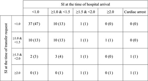
We investigated whether the strain was present in the patient’s domestic cats. Seventeen swab samples from the eyes and nasal and oral cavities of six cats were collected. Isolates of the same species were identified from four of the cats and five of 17 swab samples; all samples tested positive for the toxin gene by PCR and Vero cell cytotoxicity tests. Moreover, multilocus sequence typing (MLST) revealed the presence of the ST337 strain in all six samples, including a sample collected from the patient (Table 1).
Table 1. Summary of laboratory tests carried out on samples collected from a 63-year-old woman with Corynebacterium ulcerans infection and her domestic cats Strain name Origin Identification (MALDI-TOF or rpoB sequence) Toxin gene PCR Vero cell cytotoxicity MLST type Dokkyo1603 Sputum of the patient C. ulcerans Positive Positive 337 DokkyoCat3-N Cat no. 3 nose swab C. ulcerans Positive Positive 337 DokkyoCat4-N Cat no. 4 nose swab C. ulcerans Positive Positive 337 DokkyoCat5-E Cat no. 5 eye swab C. ulcerans Positive Positive 337 DokkyoCat5-N Cat no. 5 nose swab C. ulcerans Positive Positive 337 DokkyoCat6-E Cat no. 6 eye swab C. ulcerans Positive Positive 337 MALDI-TOF, matrix-assisted laser desorption ionization–time-of-flight mass spectrometry; MLST, multilocus sequence typing; PCR, polymerase chain reaction; rpoB, RNA polymerase B. DiscussionCorynebacterium ulcerans is a Gram-positive rod that causes zoonotic disease. In recent years, the frequency of human transmission has increased, and the infection is borne not only by livestock but also by various domesticated animals.1 This microorganism produces the diphtheria toxin and causes symptoms similar to those caused by Corynebacterium diphtheriae.3 Human-to-human transmission of C. ulcerans infection has not been reported. Diphtheria caused by C. diphtheriae is a category II infectious disease under the Infectious Diseases Control Law of Japan. However, notifying cases of C. ulcerans infection is not mandatory. According to the World Health Organization, C. ulcerans infections should be treated the same as C. diphtheriae and Corynebacterium pseudotuberculosis infections and is a subject of epidemiological interest.4
In Japan, vaccination reduced the incidence of C. diphtheriae infections. However, the prevalence of protective diphtheria antibody titers in Japan in 2018 was just under 50% among adults aged over 40 years except their 50s’.5 Our patient’s diphtheria vaccination history is unclear. As routine vaccination began in Japan in 1968, she might not have been vaccinated. This is the 17th reported case in Japan. Corynebacterium ulcerans is sensitive to various antibacterial agents, including penicillins.6
As the diphtheria toxoid antibodies cannot neutralize absorbed diphtheria toxin in tissue, prompt concurrent treatment with diphtheria antitoxin and antibacterial agents is recommended at diphtheria onset.7 In our case, diphtheria antitoxin was not given because diphtheria-like illness from toxigenic C. ulcerans had already developed, thus limiting treatment effectiveness.
Ribotyping of the isolate revealed a 0102-type pattern, which was first isolated in Japan.2 In Japan, this strain has been associated with zoonotic infections transmitted from pets. Corynebacterium ulcerans strain 0102 has been genomically sequenced, and is one of the three identified prophages that carry the diphtheria toxin gene.8
Isolates from the patient and her cats were positive for the toxin gene on PCR testing, and the strain was found to be toxicogenic, possessing diphtheria toxin activity. König et al.9 developed an MLST scheme for C. ulcerans to facilitate epidemiological research. They found that if sequence types originating from animals were found in human isolates, then MLST indicated zoonotic transmission of C. ulcerans. Similarly, in our case, MLST indicated the presence of the same ST337 strain in both the patient and her cats (Table 1). Therefore, we concluded that the patient’s domestic cats were the source of the infection.
Disease severity may be reduced with appropriate antibiotic treatment, timely diphtheria antitoxin administration, and airway and respiratory management. Thus, physicians should consider the possibility of diphtheria-like symptoms when treating infectious diseases accompanied by respiratory symptoms.
ConclusionWe reported a case of C. ulcerans infection with airway obstruction. Considering PCR findings indicating the presence of a toxin gene and MLST of samples from the patient and her domestic cats, cat-to-human transmission is likely. Future epidemiological studies are needed to confirm this route of transmission.
AcknowledgmentThis report was supported in part by AMED under grant numbers 20fk0108097j0702 and 21fk0108097j0703.
DisclosureApproval of the research protocol: N/A.
Informed consent: Informed consent was obtained from the subject for publication of this report and the images.
Registry and registration no. of the study/trial: N/A.
Animal studies: N/A.
Conflict of interest: None.
Previous PresentationThis case report was presented in part at the 45th Annual Meeting of the Japanese Association for Acute Medicine in October 2017.
References
1Dias AA, Santos LS, Sabbadini PS, et al. Corynebacterium ulcerans diphtheria: an emerging zoonosis in Brazil and worldwide. Rev. Saude. Publica 2011; 45: 1176– 91. 2Komiya T, Seto Y, De Zoysa A, et al. Two Japanese Corynebacterium ulcerans isolates from the same hospital: ribotype, toxigenicity and serum antitoxin titre. J. Med. Microbiol. 2010; 59: 1497– 504. 3Tiwari TS, Golaz A, Yu DT, et al. Investigations of 2 cases of diphtheria-like illness due to toxigenic Corynebacterium ulcerans. Clin. Infect. Dis. 2008; 46: 395– 401. 4 World Health Organization. WHO Vaccine-Preventable Diseases Surveillance Standards. Diphtheria [updated September 5, 2018; cited July 1, 2021]. Available from: https://www.who.int/immunization/monitoring_surveillance/burden/vpd/WHO_SurveillanceVaccinePreventable_04_Diphtheria_R2.pdf. 5 National Institute of Infectious Diseases, Japan. National Epidemiological Surveillance of Vaccine-Preventable Diseases [updated June 11, 2021; cited July 1, 2021]. Available from: https://www.niid.go.jp/niid/ja/y-graphs/8789-diphtheria-yosoku-serum2018.html. 6Berger A, Huber I, Merbecks SS, et al. Toxigenic Corynebacterium ulcerans in woman and cat. Emerg. Infect. Dis. 2011; 17: 1767– 9. 7Begg N, World Health Organization. Manual for the Management and Control of Diphtheria in the European Region. WHO Regional Office for Europe, Copenhagen, 1994 [cited July 1, 2021]. Available from: https://apps.who.int/iris/handle/10665/108107. 8Sekizuka T, Yamamoto A, Komiya T, et al. Corynebacterium ulcerans 0102 carries the gene encoding diphtheria toxin on a prophage different from the C. diphtheriae NCTC 13129 prophage. B.M.C. Microbiol. 2012; 12: 72. 9König C, Meinel DM, Margos G, Konrad R, Sing A. Multilocus sequence typing of Corynebacterium ulcerans provides evidence for zoonotic transmission and for increased prevalence of certain sequence types among toxigenic strains. J. Clin. Microbiol. 2014; 52: 4318– 24.
留言 (0)