
記住我


This study was approved by Taiwan’s Food and Drug Administration (TFDA) and the Institutional Review Board of BLINDED INFORMATION (BLINDED INFORMATION; reference number: 104-4660A) and was conducted in accordance with the Declaration of Helsinki Ethical Principles and Good Clinical Practices. Each subject gave a written informed consent form. The datasets used and analyzed during the current study were available from the corresponding author on reasonable request.
ArcBlate focused ultrasound ablation systemThe first prototype of the ArcBlate focused ultrasound ablation system was developed by the Department of Biomedical Engineering in the National Health Research Institute (NHRI), which initially tested the prototype in 7 mini pigs between 2009 and 2011, then conducted a pilot study involving 6 patients with uterine fibroids in BLINDED INFORMATION in 2015 (NCT02283502). EpiSonica undertook subsequent development and manufacture from 2014 and finished this study.
In contrast to other commercially available MRgHIFU systems, the ArcBlate focused ultrasound ablation system (100 M, manufactured by EpiSonica Corporation, Hsinchu, Taiwan) consists of three parts; a special portable arc for anchoring the ultrasound detector, a control cabinet, and a control console. Figure 1 shows the placement of the ArcBlate system. The portable ARC is designed for easy attachment to the MR patient table and can perform a three-dimensional movement of 20 cm horizontally along the patient table (longitudinal axis), allowing for a ≤ 20° shift along the arc (horizontal axis) and ≤ 20° rotation, in combination with a one-dimensional ArcBlate transducer to allow vertical movements to depths of 6–20 cm. A 15-channel annular array transducer integrated with a flexible water bag was designed for mounting on the portable ARC for generating high-intensity focused ultrasound energy. The flexible water bag is filled with degassed water and is in direct contact with the patient’s skin to enable propagation of the ultrasound beam to the target tumor. The positioning of the portable ARC and the ultrasonic energy delivered by the HIFU transducer is controlled by the console through the control cabinet.
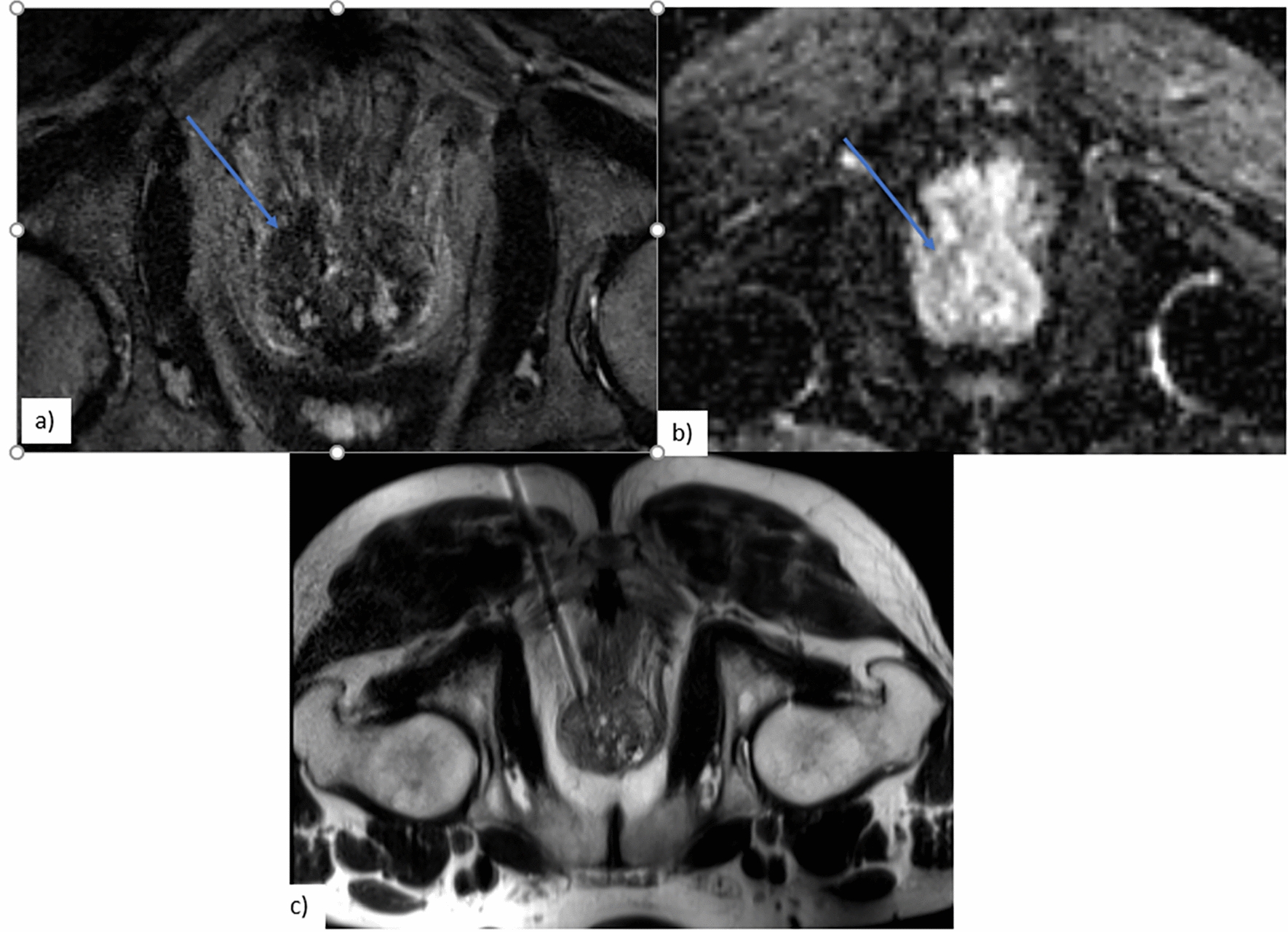
Fig. 1
Placement of the ArcBlate system. A MRI. B Patient table. C MRI console. D Portable ARC. E, F HIFU transducer and the water bag. G HIFU console. H Emergency stop
PatientsTen patients, including nine with uterine fibroids and one with adenomyosis, were enrolled. Inclusion criteria were (1) benign uterine tumor, such as uterine fibroids, adenomyoma, or adenomyosis; (2) tumor size between 4 and 15 cm or an adenomyosis area larger than 4 cm; (3) age > 20 years and scheduled for a hysterectomy; (4) abdominal circuit under 100 cm. Exclusion criteria were (1) pregnancy; (2) hemoglobin below 9 g/dL; (3) other pelvic diseases; (4) unsuitable HIFU beam path; (5) MRI contraindications; (6) medical history of calcified gynecologic tumor; (7) co-existing diseases including heart, vascular, or renal; (8) malignant uterine tumors identified by MRI; (9) failure to satisfy study enrollment criteria after an assessment by an expert gynecologist and/or radiologist.
The diagnostic criteria for adenomyosis using MRI included (1) low signal intensity of myometrium, with indistinct margins and (2) diffuse or focal junctional zone exceeding 12 mm [20].
Pretreatment screening and preparationBefore treatment, all subjects underwent routine blood testing and quality of life assessment by the 36-Item Short Form 36 Health Survey (SF-36) questionnaire. MRI was performed within 30 days of these testing procedures using the 3 T Trio MRI scanner (Siemens Healthcare, Erlangen, Germany).
Standard T2-weighted imaging using three orthogonal planes (i.e., coronal, sagittal, and transverse) assigned the Funaki classification and evaluated the size, volume, location, and status of the benign uterine tumor. Standard T1-weighted imaging identified the benign or malignant uterine tumor after the intravenous administration of a gadolinium-based contrast agent (Magnevist, Bayer Pharma AG, Berlin, Germany; dose: 0.2 mL/kg). Other pre-procedural preparations included an 8-h fasting period and enema administrations to exclude air and other possible obstacles (e.g., bowel loops anterior to the uterus), and abdominal and pubic hair shaving to allow the skin to connect with the ultrasound beam closely to prevent the skin burn [21,22,23]. Bladder filling was conducted only if the fibroid location was close to the intestinal tract.
MRgHIFU treatmentThe patient was placed on the MRI table in the supine position. The portable ARC was put on the MRI table and the water bag was placed in direct contact with the patient’s abdominal skin. Sterile ultrasound gel was applied to the contact area to eliminate air gaps between the water bag and the patient’s skin surface. A quick T2-weighted MR image in the sagittal plane was acquired to confirm the absence of air bubbles between the water bag and the patient’s skin surface.
Subsequently, high-resolution MR images in three orthogonal planes for treatment planning were acquired using a T2-weighted turbo-spin echo sequence with the following parameters: variable TR depending on different planes and/or different patients, TE = 120 ms, flip angle = 120 degrees, matrix size = 512 × 358, field of view (FOV) = 384 × 384 mm2, and slice thickness = 4 mm without gaps. These MR images were displayed on the ArcBlate user control GUI, software contained inside the control console designed to assist the physician with establishing the treatment plan based on the tumor characteristics and to identify critical organs such as the bladder, bowel, and spine surrounding the targeted fibroids.
At the treatment planning stage, a single-point treatment pattern, six square-shaped treatment patterns consisting of treatment points (i.e., 4 and 9 points), and gap distances (i.e., 3, 6, or 10 mm gap) were manually selected and inserted in the coronal plane of the T2-weighted MR images. These different treatment patterns helped the physician establish the treatment plan for each patient. Moreover, the power (i.e., 120–300 W), ablation time (i.e., 10–60 s), and cooling time (i.e., 5–60 s) were defined for each treatment pattern. Based on the individual variability, several test sonications with low energy using a single-point treatment pattern were performed in the center of the targeted fibroid on the deeper MR coronal plane to verify the location of heating and to determine the power level for ablation. The treatment plan for each patient was established slice-by-slice from a deeper MR coronal plane, and the planned treatment pattern in each slice was put within a 10-mm distance away from the margin of the targeted fibroid. The planned treatment tumor volume was up to 50% of the fibroid according to safety considerations defined by the TFDA and treatment time was kept to within 1–2 h.
The treatment dose was measured with the following formula:
$$}\left( J \right) = \sum }\left( W \right) \times }\;}\;}\;}\;}\;}\;}\;} \times }\;}\;\left( \right)$$
During the treatment, the ArcBlate transducer was mounted under the portable ARC and automatically moved to the planned treatment location mapped out in the patient’s treatment plan. Real-time thermal monitoring was also performed in three adjacent coronal slices, using a fast spoiled gradient-recalled-echo sequence with the following parameters: TR = 11 ms, TE = 7 ms, flip angle = 30 degrees, matrix size = 128 × 128, FOV = 384 × 384 mm2, slice thickness = 4 mm with a 4 mm gap, number of slices = 3 with the center slice placed at the planned treatment plane, and temporal resolution = 4.2 s/dynamic. Following thermal imaging scanning, 3 dynamics were performed and then the sonications began. Thermal imaging continued to scan more than 10 dynamics after the sonication stopped. These temperature maps were calculated from raw data centered in the k-space matrix and based on the proton resonance frequency shift [24, 25], and then displayed on the ArcBlate user control GUI to assist the physician to monitor temperature changes in the focal, near, and far fields along the ultrasound beam axis.
No patients required sedation, anesthesia, a urinary catheter, or MRI contrast agent. At the end of the treatment, patients were monitored for at least one hour for the occurrence of any acute adverse events.
Post-treatment follow-upAfter discharge, patients were followed-up by a telephone call at 1 week for the assessment of postoperative adverse events. At 1 and 3 months after treatment, quality of life was assessed with the SF-36 questionnaire and tumor volume was determined by MRI.
Assessments and data analysisClassification of the benign uterine tumor by fibroid (i.e., subserosal, intramural, submucosal, or adenomyosis) and Funaki type was based on the signal intensity of the T2-weighted MR images [26], in the judgment of one of the investigators (G.G.L., with 15 years of experience in analyzing gynecological MRI images). The Funaki classification evaluates uterine fibroids as either type 1 (low; comparable to that of skeletal muscle), type 2 (intermediate; lower than that of the myometrium and higher than that of the skeletal muscle), or type 3 (high; equal to or higher than that of the myometrium). This classification has been adopted in clinical practices at most institutions performing MR-guided HIFU ablation.
Tumor volume was calculated with the following formula:
$$}\;}\left( }^ } \right) = \frac\pi \cdot d_ \left( }} \right) \cdot d_ \left( }} \right) \cdot d_ \left( }} \right)$$
where \(\pi\) equals 3.14, and \(_\), \(_\), and \(_\) equal the longest diameter in three orthogonal planes (i.e., coronal, sagittal, and transverse) of the benign uterine tumor measured on the T2-weighted MR images. Treatment effect was measured by fibroid shrinkage using T2-weighted MR images.
Shrinkage of the tumor volume was calculated with the following formula:
$$}~\left( \% \right) = \frac}\;}\;}\;}\left( }^ } \right) - }\;}\;}\;}\left( }^ } \right)}}}\;}\;}\;}~\left( }^ } \right)}} \times 100$$
The SF-36 questionnaire was used for evaluating quality of life as described in the previous research. Higher scores indicated better quality of life [27].
留言 (0)