
記住我
Surgery remains the cornerstone of ovarian cancer treatment, with the primary goal being the complete resection of the tumor. The quality of surgery and the surgeon's expertise are critical to patient outcomes and survival. With advances in surgical techniques and a growing focus on improving patient perioperative outcomes, minimally invasive surgery (MIS) has become increasingly important in managing gynecological malignancies. However, its application in the treatment of ovarian cancer remains a subject of on-going debate.
Current guidelines recommend performing cytoreductive surgery for ovarian cancer via midline laparotomy, even in its early stages (1). However, to date, no randomized controlled trials have directly compared MIS with open surgery for the treatment of early-stage ovarian cancer and borderline ovarian tumors (BOTs) (2). Several studies suggest the feasibility and safety of MIS approaches for the management of early-stage ovarian cancer, appearing to be non-inferior to laparotomy (3, 4) and presenting with lower rates of surgical complications (5, 6).
Transvaginal Natural Orifice Transluminal Endoscopic Surgery (vNOTES) is an innovative, minimally invasive approach that combines laparoscopy and vaginal surgery (7). This approach has proven its feasibility and safety for treating several benign gynecological conditions, being a valuable option for performing hysterectomies, myomectomies, adnexal procedures, and pelvic organ prolapse treatments with a short learning curve (7–12). In addition, vNOTES has shown promising results in managing early-stage endometrial cancer, allowing complete surgical staging, including sentinel lymph node biopsies, lymphadenectomies, and omentectomies (10, 13–16). However, although increasing evidence supports the use of vNOTES approaches to manage early-stage endometrial cancer and to perform benign adnexal surgeries, little is known about the feasibility and safety of performing vNOTES oncological staging for tubo-ovarian malignancies (14, 17–19). Hereby, we report our initial experience performing vNOTES surgical staging for early-stage ovarian cancer and BOTs.
2 Materials and methods 2.1 Patient selection, data collection, and methodsvNOTES was implemented in our institution in May 2020. Since January 2022, we have collected retrospectively and prospectively data concerning patients who underwent vNOTES procedures to create an institutional database using the Research Electronic Data Capture (REDCap) software. The project received approval from the local ethical committee (Commission cantonale d'éthique de la recherche sur l'être humain, CER-VD), with registration number 2021-02346, and all patients gave written informed consent.
From this database, we retrospectively identified all patients diagnosed with borderline ovarian tumors (BOTs) or early-stage ovarian cancer between May 2020 and August 2024. At our institution, exclusion criteria for a vNOTES approach include confirmed ovarian cancer, active genital tract infections, history of rectovaginal endometriosis, rectal surgery, pelvic radiotherapy, severe pelvic inflammatory disease and mesh sacrocolpopexy. All patients underwent pelvic ultrasound, thoraco-abdominal computed tomography, and tumor marker levels. In addition, pelvic magnetic resonance imaging (MRI) was performed when further characterization of ovarian lesions was necessary according to the recommendations of the European Society of Gynecologic Oncology (ESGO) (20). No patients showed evidence of advanced ovarian disease at the preoperative workup.
Demographic features, as well as clinical and perioperative information, were collected and analyzed. Intraoperative parameters included total operative time (from catheterization of the bladder to vaginal closure), vNOTES port insertion time (from incision to intrabdominal CO2 insufflation), estimated blood loss, intraoperative complications (including transfusion-requiring bleeding or iatrogenic organ injury), and the necessity for conversion to conventional laparoscopy or laparotomy. Postoperative assessments comprised pain evaluation using the visual analog scale graded from 0 to 10 at 12-, 24-, and 48-h post-surgery, opioid analgesic use, duration of hospital stay, and postoperative complications within 8 postoperative weeks, graded according to the Clavien-Dindo classification (CD) (21). In addition, we recorded histopathological results, the timing and type of any adjuvant therapies, and the patient status at the last follow-up.
Continuous variables were expressed as median and range, while dichotomous variables were represented as absolute numbers and percentages (%). No statistical inter-group comparisons were undertaken. Statistical analyses were performed using IBM SPSS version 29.0.2.0.
2.2 Surgical techniqueAll interventions were performed by the same oncogynecological surgeon (DH). Patients received a single dose of clindamycin vaginal cream 2% (5 g of cream with 100 mg of clindamycin) the day before the surgery, and 2–4 h before the intervention, in addition to cefuroxime 1.5 g (3 g for patients weighing more than 80 kg) and metronidazole 500 mg intravenously at induction of anesthesia. Under general anesthesia and muscular relaxation, patients were positioned in a horizontal dorsal lithotomy position, and a bladder catheter was placed.
Access was gained with a posterior 2 cm colpotomy through Douglas's pouch to perform interventions limited to the adnexa. If hysterectomies were performed, access to the abdominal cavity was achieved through anterior and posterior colpotomies, with the transvaginal uterosacral ligaments section when developing the posterior access. A vNOTES port (GelPoint vPath, Applied Medical, Rancho Santa Margarita, CA, USA) with an adapted diameter (7 cm for adnexectomies and 9.5 cm for hysterectomies) was placed in the abdominal cavity through the anterior and/or posterior colpotomies. Carbon dioxide was insufflated to create a pneumoperitoneum with an intraperitoneal pressure of 8–15 mmHg. Three 10 mm trocars were used to insert a 10-mm rigid 30° scope, 5-mm instruments such as Johan and bipolar graspers, and sealing devices. If necessary, a 4th 12 mm supplementary trocar was added (Figure 1).
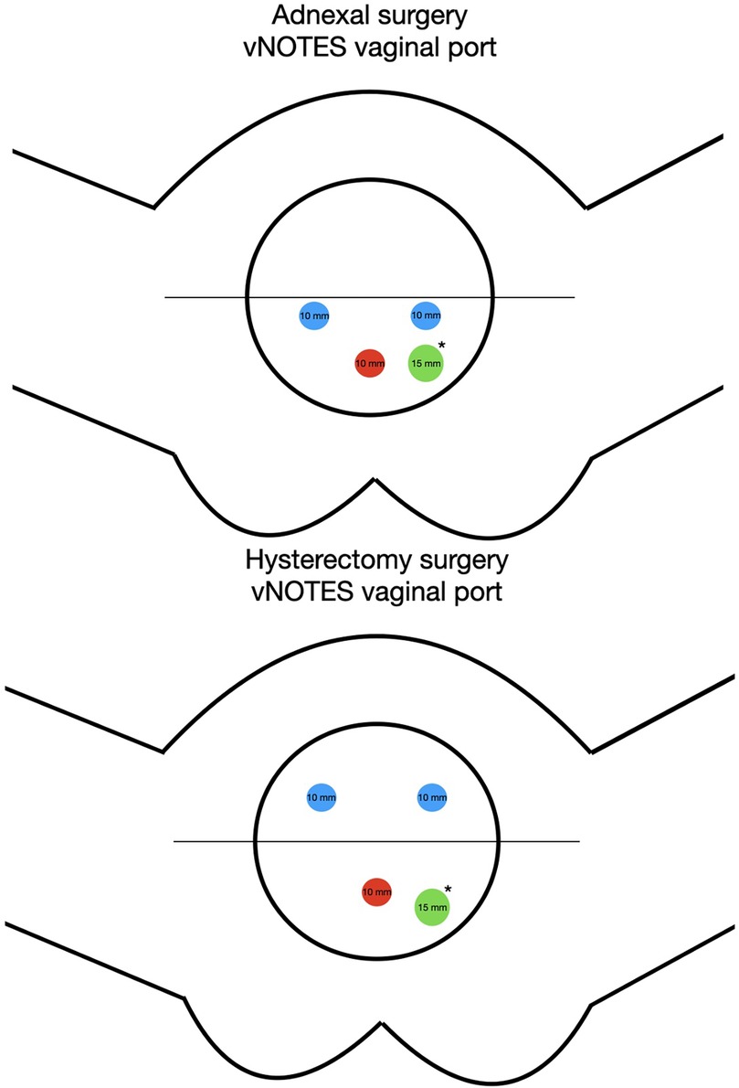
Figure 1. Adnexal surgery: blue = 10 mm trocar for instruments, Red = 10 mm optical trocar. The instrument trocars are inserted below the medial line for better access to the adnexa. Hysterectomy surgery: Blue = 10 mm trocar for instruments, Red = 10 mm optical trocar. The instrument trocars are inserted over the medial line. Green* = A 12 mm accessory trocar can be added as needed for exposure or insertion of an endobag.
Surgical staging included peritoneal washing, uni or bilateral salpingo-oophorectomy, abdominal cavity inspection, peritoneal biopsies, infracolic omentectomy, and total hysterectomy. In selected cases, fertility-sparing approaches with unilateral salpingo-oophorectomy or cystectomy and uterus preservation were performed. To perform hysterectomies, the uterine vessels, broad ligaments, and round ligaments were sealed and cut from caudal to cranial. Salpingo-oophorectomies were always performed after correctly visualizing the ureters, the fallopian tubes, and the infundibulopelvic ligaments, with utmost care to avoid spillage.
All specimens have been extracted vaginally. To avoid intraabdominal spillage, large adnexal lesions were retrieved into an Inzii Retrieval System of 10 or 15 cm of diameter, or Alexis Contained Extraction System of 14 or 17 cm (Applied Medical, Rancho Santa Margarita, CA, USA). Intraoperative frozen section analysis was performed in cases with suspicious adnexal masses. Omentectomies were performed with an articulating sealing device, as we previously described (14).
At the end of the procedure, the colpotomy was closed under direct visualization with a continuous Vicryl 0 suture, incorporating the anterior and posterior peritoneal folds and vaginal layers into the single running suture. Postoperatively, patients received a single dose of clindamycin vaginal cream 2% (5 g of cream with 100 mg of clindamycin) once a day during the first seven postoperative days.
3 ResultsFrom October 2021 to August 2024, 7 patients with early-stage tubal or ovarian cancer and 4 patients with BOTs underwent surgical staging by vNOTES at Valais Hospital (Sion, Switzerland).
The median age was 47 (27–81) years, with a median body mass index of 28.1 (22.4–39.2) kg/m2. Seven patients (63.6%) were classified as American Society of Anesthesiologists score (ASA) II and four (36.4%) as ASA III. Table 1 provides an overview of patient characteristics and their perioperative outcomes.
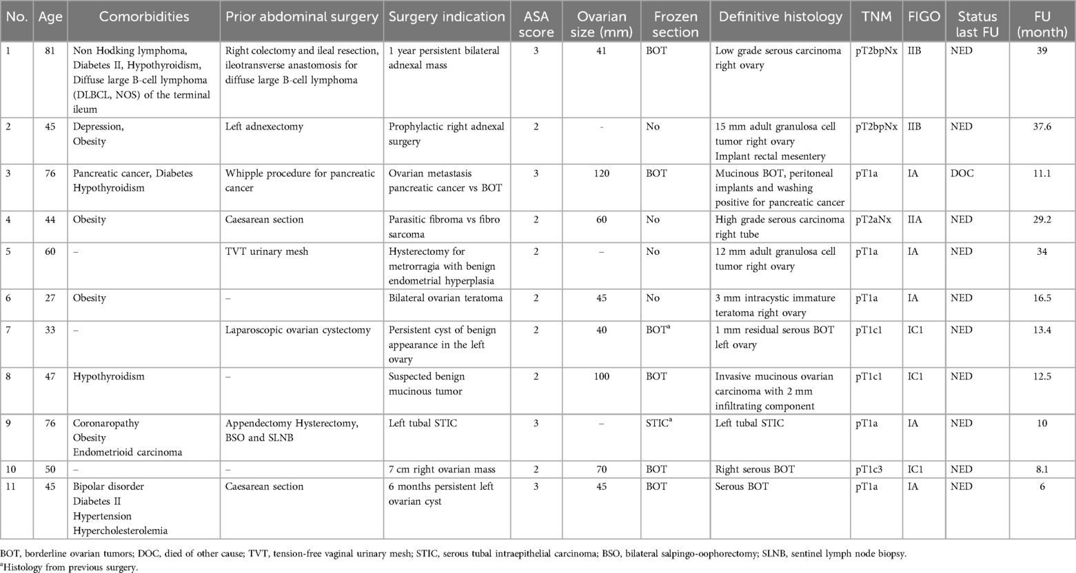
Table 1. Patient characteristics and perioperative outcomes.
Bilateral salpingo-oophorectomy was performed in five patients (45.5%), while six patients (54.5%) underwent fertility-sparing surgery with preservation of at least one ovary and the uterus. Table 2 summarizes the surgical procedures performed to complete surgical staging. The median operating time was 70 (35–138) min, with a median blood loss of 50 (10–100) ml. No conversion to conventional laparoscopy or laparotomy was necessary, and all procedures were performed as planned. In a patient with one suspicious pelvic implant, a hybrid approach was used to explore the utero-vesical peritoneum. All surgical material was extracted vaginally with an endobag. No intraoperative complications were reported, except for one case involving a minimal pelvic ovarian spillage during extraction in the retrieval system (9.1%). The percentage of patients with positive peritoneal cytology was 54.5% (Table 2).
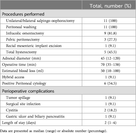
Table 2. Surgical procedure, operative characteristics, and perioperative outcomes.
Post-operative complications were reported in three patients (27.3%). These included one surgical site infection (9.1%) and two cases of cystitis (18.2%). All three postoperative complications were graded as CD grade 2 and treated with antibiotics. The median hospital stay was 48 (24–96) h. After the final histological results, four patients underwent a second intervention to complete the surgical staging, three by subsequent vNOTES and one by conventional laparoscopy (Table 3).
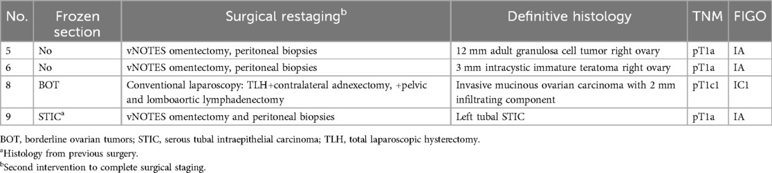
Table 3. Patients with restaging surgery.
Adjuvant chemotherapy was administered in four patients (36.4%), one of whom received palliative chemotherapy for relapsed pancreatic disease. The median time from surgery to adjuvant therapy was 23 (19–31) days. In this series, no evidence of recurrence was observed, with a median follow-up time of 13.4 (6–39) months. One patient (9.1%) died of metastatic pancreatic cancer one year after the surgery. The final histopathological diagnoses are summarized for each patient in Table 1.
4 DiscussionThe role of MIS in gynecological oncology has undergone progressive development. This has involved introducing both conventional and robot-assisted laparoscopic techniques, which have demonstrated their feasibility and efficacy in staging and treating uterus-confined endometrial cancer (13, 22, 23). In the case of early-stage ovarian cancer, the latest international guidelines recommend midline laparotomy as the standard procedure. The rationale behind this is that the open surgery allows an accurate abdominal exploration and a reduced risk of rupture of the primary tumor. Nevertheless, the laparoscopic and robotic approaches are often used worldwide for surgical staging of BOTs and early-stage ovarian cancer, and some studies have shown better surgical outcomes and no difference in recurrence rates or survival for those who received minimally invasive vs. open surgical staging (5, 24–27). However, the oncologic outcomes remain a topic of debate, lacking sufficient high-quality evidence to change current guidelines (2, 4, 27, 28). To date, only a few publications report a vNOTES approach in the management of ovarian cancer (14, 17, 18).
According to the current guidelines of the European Society of Gynaecological Oncology (ESGO), surgical management of Stage I–II ovarian cancer must include a total hysterectomy and bilateral salpingo-oophorectomy or fertility-sparing surgery (unilateral salpingo-oophorectomy) in selected patients desiring fertility. Peritoneal washings or cytology, taken before manipulation of the tumor, and peritoneal biopsies with at least infracolic omentectomy are also recommended (1). Since omentectomy via vNOTES has been proven to be feasible (14, 18), in the case of intraoperative diagnosis of BOTs or early-stage ovarian cancer, surgical staging through the same vaginal incision is possible.
Many early-stage ovarian cancer diagnoses are made postoperatively on lesions initially presumed benign (29). The accuracy of frozen section varies between 82% and 88% for BOTs and malignant tumor with most discordancy encountered for younger, premenopausal women, early-stage ovarian malignancies and mucinous histology (30). In our series, five patients have a BOTs identified intraoperatively through frozen section analysis. Two out of five patients were reclassified upon final postoperative histopathology as stage IIB low-grade serous ovarian carcinoma and stage IC1 invasive mucinous ovarian carcinoma. We diagnosed one case of serous tubal intraepithelial carcinoma (STIC) during the surgery for endometrioid endometrial carcinoma. One serous BOT diagnosis was made prior to referral to our institution. The remaining four cases were diagnosed postoperatively. Three had small intraovarian non-epithelial cancers, including one immature teratoma and two adult granulosa cell tumors, while one patient was diagnosed with high-grade serous carcinoma.
A further challenge of the MIS approaches in early-stage ovarian cancers is the ability to perform a complete pelvic and paraaortic lymphadenectomy. For clinical stage I and low-risk invasive ovarian tumors such as mucinous, malignant germ cell, and sex cord-stromal tumors as well as for BOTs and STIC, the systematic lymphadenectomy is not recommended (31). The survival benefit of complete staging with lymphadenectomy in early-stage epithelial ovarian cancer has not been confirmed in prospective trials (29), though it is known that 10%–15% of cases are upstaged due to nodal involvement (32) and require adjuvant treatments. Lymphadenectomy might be subsequently omitted if an occult positive lymph node will not influence the adjuvant treatment allocation. We performed only one subsequent retroperitoneal pelvic and paraaortic lymphadenectomy for a stage IA mucinous invasive carcinoma due to a minimal infiltrative invasive component in a majoritarian expansile tumor.
We hypothesize that both the vNOTES technique for pelvic lymphadenectomy and paraaortic lymphadenectomy can be successfully applied to early-stage ovarian malignancies. The vNOTES approach for pelvic lymphadenectomy was first described in 2014, with further validation by other authors (33–35). Additionally, in 2024, a hybrid technique combining vNOTES with a single-port retroperitoneal approach for pelvic and infrarenal paraaortic lymphadenectomy was reported (36). For patients diagnosed intraoperatively with early-stage invasive ovarian cancer requiring both pelvic and paraaortic lymphadenectomy, a vNOTES hybrid approach, may be an option (36–38). If restaging is required, the absence of an abdominal peritoneal scar after retroperitoneal vNOTES can simplify the successive procedures.
In patients with complex surgical history and suspected severe intra-abdominal adhesions vNOTES approach allows pelvic exploration, hysterectomy and adnexectomy if needed, with less risk of organ damage. In one of our patients, we diagnosed a mucinous BOT and a peritoneal relapse of pancreatic cancer after a previous Wipple procedure. Less than 3 weeks after the surgery the patient started palliative chemotherapy.
One limitation of VNOTES is the restricted accessibility of certain anatomical regions, including the posterior costodiaphragmatic recesses, the Morrison's pouch, the lesser omentum and the mesenteric root. Nevertheless, these regions are challenging to explore also by conventional laparoscopy. Furthermore, the visualization of the vesico-uterine peritoneum during VNOTES adnexal surgery is hindered by the presence of the uterus. A hybrid approach with a transabdominal trocar can effectively address this challenge. Ghezzi et al. emphasized that isolated metastases in these specific areas are extremely rare (39). Despite these limitations, several studies have shown no significant differences in surgical outcomes, recurrence rates or survival between patients undergoing minimally invasive vs. open surgical staging for patients with early-stage ovarian cancer (4–6).
The duration of vNOTES and standard laparoscopic procedures for early stages of adnexal malignancies seems equivalent. Data in the literature is heterogeneous, comparing the time of the open vs. MIS approach with no clear advantage for one or another. The surgeons' experience may be the main factor influencing the operating time (4, 5, 28, 40). The blood loss reported in our series is low and consistent with existing literature (4, 5, 28, 40).
In our series, perioperative complication rates were low. No intraoperative complications were noted, except for one case of minimal ovarian spillage during adnexal extraction in endobag (9.1%). Tumor spillage might occur even in large laparotomies, raising the possibility that aggressive biology associated with more adherent and fragile tumors may be responsible for rupture more than the surgical approach (29). This is crucial as spillage can lower survival rates and is associated with an upstaging of the tumor (40–42). Some studies suggest a higher risk of cyst rupture with laparoscopic cystectomy, which may be reduced if adnexectomy is performed rather than cystectomy (43). Evidence on spillage risk with vNOTES is scarce but appears similar to the laparoscopic approach (8, 44, 45).
Lower rates of all types of complications have been reported with the vNOTES approach in benign indications, ranging from 2.5% to 4.1% (46–48). Laparoscopy has been demonstrated to significantly reduce the duration of hospitalization compared to laparotomy (49), a finding consistent with our series, which had a median hospital stay of 48 (24–96) h. Postoperative complications related to surgery were minimal in our cohort, with one case surgical site infection (9.1%) and two cases of cystitis (18.2%), all successfully treated with antibiotics.
In the vNOTES approach, the single vaginal scar can improve the post-operative recovery. Fast recovery is particularly important for the management of malignant cases, allowing for the earlier administration of adjuvant treatments. In our series, the median time from surgery to adjuvant therapy was 23 (19–31) days. Present recommendations endorse starting adjuvant treatments 28–42 days after the surgery (50).
Abdominal port site metastases has been an important concern in MIS for intra-abdominal malignancies. The vNOTES approach offers an advantage, particularly in patients requiring hysterectomy, by eliminating the need for additional abdominal incisions. For adnexal surgery, vNOTES limits this concern by the presence of a single vaginal incision. Furthermore, vNOTES allows extraction of masses of up to 6–7 cm without the need for morcelation or puncture. For bigger sizes, adnexal mass extraction after puncture in surgical bags is possible with the same approaches as with conventional laparoscopy (8). Baekelandt and al. have recently described a technique for bagging a 20 cm BOTs via vNOTES without spillage (19).
With vNOTES access, vaginal post-site metastasis becomes a significant concern for patients with ovarian cancer. As described by Chitrathara et al., the vaginal vault is a potential site of recurrence in ovarian cancer following laparoscopic treatment and is associated with a worse prognosis compared to other port-site recurrences (51). In their retrospective series, all specimens were extracted vaginally without an endobag for presumed benign lesions. In our series, all patient underwent surgery through a transvaginal access platform protecting both the vaginal wall and the colpotomy site. If morcellation or puncture of ovarian mass were necessary, we used additional layers of protection provided by Inzii retrieval system or Alexis contained extraction system to minimize the risk of contamination.
We acknowledge limitations in this study, notably the small sample size, which limits comparative analysis with other methods. Additionally, case heterogeneity and short follow-up as well as single-center setting with only one oncogynecological surgeon reduce generalizability. However, our focus on early surgical outcomes supports the feasibility of vNOTES for highly selected early-stage ovarian cancer patients, with potential benefits in perioperative morbidity and quality of life.
In conclusion, despite the limited cohort size, our findings indicate the technical feasibility of vNOTES for highly selected patients with early-stage ovarian cancer. Further research with larger cohorts and extended follow-up is needed to assess the long-term oncological outcomes and safety of this technique.
Data availability statementThe original contributions presented in the study are included in the article/Supplementary Material, further inquiries can be directed to the corresponding author.
Ethics statementThe studies involving humans were approved by Commission cantonale d'éthique de la recherche sur l'être humain (CER-VD), registration no 2021-02346. The studies were conducted in accordance with the local legislation and institutional requirements. The participants provided their written informed consent to participate in this study. Written informed consent was obtained from the individual(s) for the publication of any potentially identifiable images or data included in this article.
Author contributionsGK: Conceptualization, Methodology, Writing – original draft, Formal Analysis. JN: Conceptualization, Formal Analysis, Methodology, Writing – original draft, Writing – review & editing. YH: Writing – review & editing. DH: Conceptualization, Data curation, Investigation, Methodology, Project administration, Supervision, Validation, Writing – original draft, Writing – review & editing.
FundingThe author(s) declare financial support was received for the research, authorship, and/or publication of this article. Open access funding by University of Geneva.
AcknowledgmentsAn earlier version of this article has been made available as a preprint and can be accessed at preprints.org (52).
Conflict of interestThe authors declare that the research was conducted in the absence of any commercial or financial relationships that could be construed as a potential conflict of interest.
Generative AI statementThe author(s) declare that no Generative AI was used in the creation of this manuscript.
Publisher's noteAll claims expressed in this article are solely those of the authors and do not necessarily represent those of their affiliated organizations, or those of the publisher, the editors and the reviewers. Any product that may be evaluated in this article, or claim that may be made by its manufacturer, is not guaranteed or endorsed by the publisher.
References1. Querleu D, Planchamp F, Chiva L, Fotopoulou C, Barton D, Cibula D, et al. European Society of Gynaecological Oncology (ESGO) guidelines for ovarian cancer surgery. Int J Gynecol Cancer. (2017) 27(7):1534–42. doi: 10.1097/IGC.0000000000001041
PubMed Abstract | Crossref Full Text | Google Scholar
2. Falcetta FS, Lawrie TA, Medeiros LR, Da Rosa MI, Edelweiss MI, Stein AT, et al. Laparoscopy versus laparotomy for FIGO stage I ovarian cancer. Cochrane Database Syst Rev. (2016) 2022(1):CD005344. doi: 10.1002/14651858.CD005344.pub4
Crossref Full Text | Google Scholar
4. Ghezzi F, Cromi A, Uccella S, Bergamini V, Tomera S, Franchi M, et al. Laparoscopy versus laparotomy for the surgical management of apparent early stage ovarian cancer. Gynecol Oncol. (2007) 105(2):409–13. doi: 10.1016/j.ygyno.2006.12.025
PubMed Abstract | Crossref Full Text | Google Scholar
5. Chi DS, Abu-Rustum NR, Sonoda Y, Ivy J, Rhee E, Moore K, et al. The safety and efficacy of laparoscopic surgical staging of apparent stage I ovarian and fallopian tube cancers. Am J Obstet Gynecol. (2005) 192(5):1614–9. doi: 10.1016/j.ajog.2004.11.018
PubMed Abstract | Crossref Full Text | Google Scholar
6. Colomer AT, Jiménez AM, Bover Barceló MI. Laparoscopic treatment and staging of early ovarian cancer. J Minim Invasive Gynecol. (2008) 15(4):414–9. doi: 10.1016/j.jmig.2008.04.002
PubMed Abstract | Crossref Full Text | Google Scholar
7. Ferro R, Hurni Y, Seidler S, Huber D. Transvaginal natural orifice transluminal endoscopic surgery (vNOTES) in gynecological emergencies. Eur J Obstet Gynecol Reprod Biol X. (2023) 20:100261. doi: 10.1016/j.eurox.2023.100261
PubMed Abstract | Crossref Full Text | Google Scholar
8. Baekelandt J, De Mulder P, Le Roy I, Mathieu C, Laenen A, Enzlin P, et al. Adnexectomy by vaginal natural orifice transluminal endoscopic surgery versus laparoscopy: results of a first randomised controlled trial (NOTABLE trial). BJOG. (2021) 128(11):1782–91. doi: 10.1111/1471-0528.16838
PubMed Abstract | Crossref Full Text | Google Scholar
9. Huber D, Hurni Y. Sentinel node biopsy for endometrial cancer by retroperitoneal transvaginal natural orifice transluminal endoscopic surgery: a preliminary study. Front Surg. (2022) 9:907548. doi: 10.3389/fsurg.2022.907548
PubMed Abstract | Crossref Full Text | Google Scholar
10. Huber D, Hurni Y. Anatomical distribution of sentinel lymph nodes harvested by retroperitoneal vNOTES in 34 consecutive patients with early-stage endometrial cancer: analysis of 124 lymph nodes. J Minim Invasive Gynecol. (2024) 31(5):438–44. doi: 10.1016/j.jmig.2024.02.007
PubMed Abstract | Crossref Full Text | Google Scholar
11. McCulloch P, Altman DG, Campbell WB, Flum DR, Glasziou P, Marshall JC, et al. No surgical innovation without evaluation: the IDEAL recommendations. Lancet. (2009) 374(9695):1105–12. doi: 10.1016/S0140-6736(09)61116-8
PubMed Abstract | Crossref Full Text | Google Scholar
12. Mereu L, Pecorino B, Ferrara M, Siniscalchi M, Garraffa G, D’Agate MG, et al. Cumulative sum analysis of learning curve process for vaginal natural orifice transluminal endoscopic surgery hysterectomy. J Minim Invasive Gynecol. (2023) 30(7):582–6. doi: 10.1016/j.jmig.2023.03.013
PubMed Abstract | Crossref Full Text | Google Scholar
13. Lee CL, Liu HM, Khan S, Lee PS, Huang KG, Yen CF. Vaginal natural orifice transvaginal endoscopic surgery (vNOTES) surgical staging for endometrial carcinoma: the feasibility of an innovative approach. Taiwan J Obstet Gynecol. (2022) 61(2):345–52. doi: 10.1016/j.tjog.2022.02.026
PubMed Abstract | Crossref Full Text | Google Scholar
14. Hurni Y, Huber D. Omentectomy for oncological surgical staging by transvaginal natural orifice transluminal endoscopic surgery (vNOTES): a preliminary study. Front Surg. (2023) 10:1224770. doi: 10.3389/fsurg.2023.1224770
PubMed Abstract | Crossref Full Text | Google Scholar
15. Huber D, Hurni Y. Sentinel lymph node mapping by retroperitoneal vNOTES for uterus-confined malignancies: a standardized 10-step approach. Cancers (Basel). (2024) 16(11):2142. doi: 10.3390/cancers16112142
PubMed Abstract | Crossref Full Text | Google Scholar
16. Şimşek E, Yıldız ÖA, Gündüz S, Karakaş S, Yaşar L. vNOTES scarless and painless endometrial cancer staging surgery. J of Obstet and Gynaecol. (2024) 50(10):1965–70. doi: 10.1111/jog.16083
PubMed Abstract | Crossref Full Text | Google Scholar
17. Hurni Y, Romito F, Huber D. Transvaginal natural orifice transluminal endoscopic surgery for surgical staging of early-stage ovarian cancers: a report of two cases. Front Surg. (2022) 9:833126. doi: 10.3389/fsurg.2022.833126
PubMed Abstract | Crossref Full Text | Google Scholar
18. Lowenstein L, Matanes E, Lauterbach R, Boulus S, Amit A, Baekelandt J. Transvaginal natural orifice transluminal endoscopic surgery (vNOTES) for omentectomy—a case series. Surg Oncol. (2020) 34:186–9. doi: 10.1016/j.suronc.2020.04.018
PubMed Abstract | Crossref Full Text | Google Scholar
19. Baekelandt J, Van Camp J, Vercammen L, Stuart A. Too big to bag?—technique for bagging very large adnexal masses via vNOTES. J Gynecol Obstetr Hum Reprod. (2024) 53(9):102857. doi: 10.1016/j.jogoh.2024.102857
Crossref Full Text | Google Scholar
20. Timmerman D, Planchamp F, Bourne T, Landolfo C, Du Bois A, Chiva L, et al. ESGO/ISUOG/IOTA/ESGE consensus statement on pre-operative diagnosis of ovarian tumors. Int J Gynecol Cancer. (2021) 31(7):961–82. doi: 10.1136/ijgc-2021-002565
PubMed Abstract | Crossref Full Text | Google Scholar
21. Dindo D, Demartines N, Clavien PA. Classification of surgical complications: a new proposal with evaluation in a cohort of 6336 patients and results of a survey. Ann Surg. (2004) 240(2):205–13. doi: 10.1097/01.sla.0000133083.54934.ae
PubMed Abstract | Crossref Full Text | Google Scholar
22. Deng L, Liu Y, Yao Y, Deng Y, Tang S, Sun L, et al. Efficacy of vaginal natural orifice transluminal endoscopic sentinel lymph node biopsy for endometrial cancer: a prospective multicenter cohort study. Int J Surg. (2023) 109(10):2996–3002. doi: 10.1097/JS9.0000000000000551
PubMed Abstract | Crossref Full Text | Google Scholar
23. Wang Y, Deng L, Tang S, Dou Y, Yao Y, Li Y, et al. vNOTES hysterectomy with sentinel lymph node mapping for endometrial cancer: description of technique and perioperative outcomes. J Minim Invasive Gynecol. (2021) 28(6):1254–61. doi: 10.1016/j.jmig.2021.01.022
PubMed Abstract | Crossref Full Text | Google Scholar
24. Radosa JC, Radosa MP, Schweitzer PA, Juhasz-Boess I, Rimbach S, Solomayer EF. Report of the survey on current opinions and practice of German society for gynecologic endoscopy (AGE) members regarding the laparoscopic treatment of ovarian malignancies. Arch Gynecol Obstet. (2018) 297(5):1255–64. doi: 10.1007/s00404-018-4709-5
PubMed Abstract | Crossref Full Text | Google Scholar
25. Brockbank EC, Harry V, Kolomainen D, Mukhopadhyay D, Sohaib A, Bridges JE, et al. Laparoscopic staging for apparent early stage ovarian or fallopian tube cancer. First case series from a UK cancer centre and systematic literature review. Eur J Surg Oncol. (2013) 39(8):912–7. doi: 10.1016/j.ejso.2013.05.007
PubMed Abstract | Crossref Full Text | Google Scholar
26. Lu Q, Qu H, Liu C, Wang S, Zhang Z, Zhang Z. Comparison of laparoscopy and laparotomy in surgical staging of apparent early ovarian cancer: 13-year experience. Medicine (Baltimore). (2016) 95(20):e3655. doi: 10.1097/MD.0000000000003655
PubMed Abstract | Crossref Full Text | Google Scholar
27. Park HJ, Kim DW, Yim GW, Nam EJ, Kim S, Kim YT. Staging laparoscopy for the management of early-stage ovarian cancer: a metaanalysis. Am J Obstet Gynecol. (2013) 209(1):58.e1–8. doi: 10.1016/j.ajog.2013.04.013
PubMed Abstract | Crossref Full Text | Google Scholar
28. Park JY, Bae J, Lim MC, Lim SY, Seo SS, Kang S, et al. Laparoscopic and laparotomic staging in stage I epithelial ovarian cancer: a comparison of feasibility and safety. Int J Gynecol Cancer. (2008) 18(6):1202–9. doi: 10.1111/j.1525-1438.2008.01190.x
PubMed Abstract | Crossref Full Text | Google Scholar
29. Somasegar S, Anastasio MK, Karam A, Rossi EC, Obermair A. Controversies in the surgical management of gynecologic cancer: balancing the decision to operate or hesitate. Am Soc Clin Oncol Educ Book. (2024) 44(3):e438550. doi: 10.1200/EDBK_438550
PubMed Abstract | Crossref Full Text | Google Scholar
30. Shen H, Hsu HC, Tai YJ, Kuo KT, Wu CY, Lai YL, et al. Factors influencing the discordancy between intraoperative frozen sections and final paraffin pathologies in ovarian tumors. Front Oncol. (2021) 11:694441. doi: 10.3389/fonc.2021.694441
PubMed Abstract | Crossref Full Text | Google Scholar
31. Armstrong DK, Alvarez RD, Bakkum-Gamez JN, Barroilhet L, Behbakht K, Berchuck A, et al. Ovarian cancer, version 2.2020, NCCN clinical practice guidelines in oncology. J Natl Compr Canc Netw. (2021) 19(2):191–226. doi: 10.6004/jnccn.2021.0007
PubMed Abstract | Crossref Full Text | Google Scholar
32. Morice P, Joulie F, Camatte S, Atallah D, Rouzier R, Pautier P, et al. Lymph node involvement in epithelial ovarian cancer: analysis of 276 pelvic and paraaortic lymphadenectomies and surgical implications. J Am Coll Surg. (2003) 197(2):198–205. doi: 10.1016/S1072-7515(03)00234-5
PubMed Abstract | Crossref Full Text | Google Scholar
33. Oh SH, Park SJ, Lee EJ, Yim GW, Kim HS. Pelvic lymphadenectomy by vaginal natural orifice transluminal endoscopic surgery (vNOTES) for early-stage endometrial cancer. Gynecol Oncol. (2019) 153(1):211–2. doi: 10.1016/j.ygyno.2019.01.003
PubMed Abstract | Crossref Full Text | Google Scholar
34. Baekelandt JF. New retroperitoneal transvaginal natural orifice transluminal endoscopic surgery approach to sentinel node for endometrial cancer: a demonstration video. J Minim Invasive Gynecol. (2019) 26(7):1231–2. doi: 10.1016/j.jmig.2019.05.002
PubMed Abstract | Crossref Full Text | Google Scholar
35. Lee CL, Wu KY, Tsao FY, Huang CY, Han CM, Yen CF, et al. Natural orifice transvaginal endoscopic surgery for endometrial cancer. Gynecology and Minimally Invasive Therapy. (2014) 3(3):89–92. doi: 10.1016/j.gmit.2014.08.004
留言 (0)