Source of bacteria
It has been demonstrated that human stool is a significant source of microbial variety with the capacity to develop new bioactive substances such as beneficial bacteria present in the gut environment that can produce bacteriocins and prevent pathogenic microbes from colonization [40]. Various genera of probiotic bacteria can produce exopolysaccharides (EPSs) in large quantities [41]. Recently, microbial exopolysaccharides (EPSs) have a lot of attention due to their health benefits [42]. The exopolysaccharides from lactic acid bacteria have immunostimulatory activity, antitumor effects, antioxidant activity, and blood cholesterol-lowering ability [43]. Also, probiotic bacteria can carry out various metabolic activities due to their production of some enzymes such as lipases, esterases, and amylases [44]. Many bacteria produce B-group vitamins, that are soluble in water and absorbed into the intestine [45]. So, our findings demonstrated that bacterial isolates that generate L-glutaminase might potentially be found in the human feces environment, which encourages microbial diversity with exceptional, powerful enzymatic capacities [22]. Human feces samples included more L-glutaminase-producing bacteria than sewage and canal water samples.
Screening and identification
The screening of the isolates was based on: the qualitative method described by Gulati et al. [27]. The change in the color of the medium from yellow to pink is an indication of extracellular L-glutaminase production by the colony. This color change can be due to a change in the pH of the medium, due to the breakdown of the amide bond in L-glutamine which liberates ammonia by the L-glutaminase enzyme. Phenol red at an acidic pH is yellow, and at an alkaline pH it turns pink; So, a pink zone is formed around the microbial colonies producing L-glutaminase. This results in agreement with Bacillus sp. DV2-37 [23]. Another method involves the isolation of microorganisms by routine isolation procedures from certain environments, which are then screened for enzymatic activity. However, the use of selective media and the presence of antibiotics, NaCl, and pH indicators make the MGA medium suitable for both direct and selective isolation of L-glutaminase-producing organisms [46].
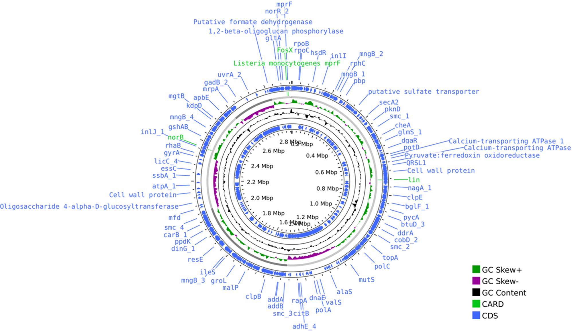
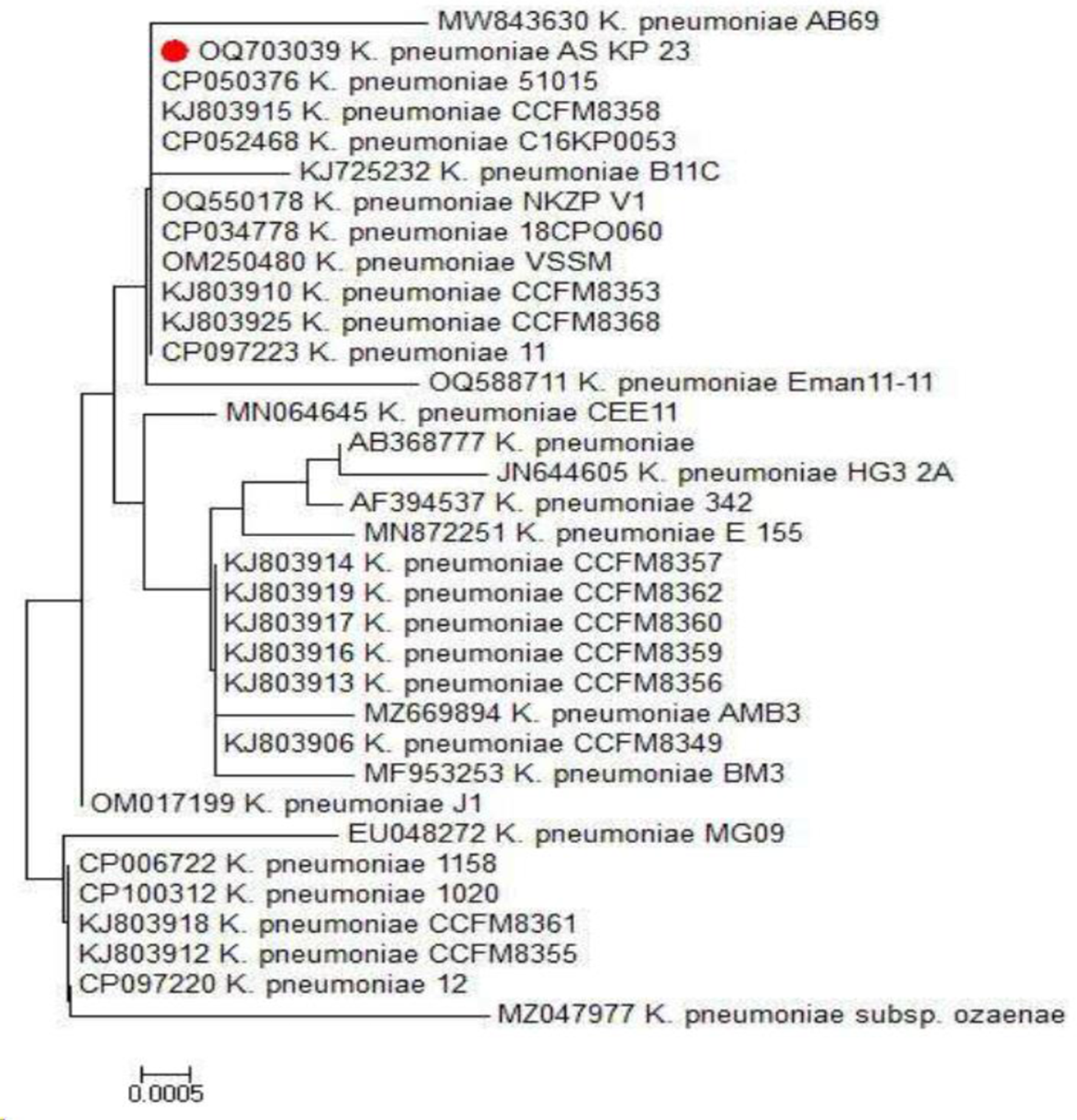
AS KP 23 was grown into nutrient agar and MacConkey agar, respectively, by following Edwards and Ewing [26]. From cultural and gram staining tests, gram-negative, and rod-shaped, several different biochemical tests were performed to identify the isolate. The most common genomic indicator designed for confirmation of the identified bacteria molecularly is the 16 S rRNA gene, and it is thought to be a more precise way than more traditional ones. The 16 S rRNA gene was 100% identical to the sequence from Klebsiella pneumoniae and was submitted to GenBank under accession number OQ703039. Thus, this strain was named Klebsiella pneumoniae AS KP 23. The isolate that produced the greatest L-glutaminase, was AS KP 23. This bacterium species may have biotechnological uses that have previously been documented [27]. It may be possible to discover novel anticancer enzyme-producing microbial strains that have features that lessen the immunological response brought on by treatment. The 16 S rRNA gene is used to identify some bacterial strains that manufacture L-glutaminase such as Bacillus cereus MTCC 1305, Aeromonas veronii, Acinetobacter calcoaceticus PJB1 and Halomonas [47, 48]. The results show that the enzyme activity is 415.07 U/mL, the total protein content is 2824 mg, and the specific activity is 587.9 U/mg, which is considered to be the initial recovery of the enzyme activity after centrifugation of the extract and 100% assumed for subsequent studies.
The optimization of L-glutaminase production parameters, such as pH, temperature, incubation period, and nutrient sources, plays a crucial role in enhancing its potential as an anticancer agent. L-glutaminase has garnered significant interest due to its ability to deplete L-glutamine, an essential amino acid for the proliferation of cancer cells, particularly in glutamine-dependent tumors. The enzyme catalyzes the hydrolysis of L-glutamine to L-glutamate and ammonia, effectively starving cancer cells of a critical nutrient for their growth and survival. The synthesis of enzyme metabolic processes and the bioavailability of trace minerals are significantly influenced by the initial pH of the media [22]. In this study, we found that pH 8.0 is optimal for L-glutaminase production by Klebsiella pneumoniae, which aligns with the optimal pH range (7.0 to 8.5) reported for enzyme activity in other microorganisms [49]. The influence of pH on bacterial growth and enzyme production is crucial, as it directly affects the availability of metabolic ions and cell membrane permeability, both important factors in enzyme biosynthesis and stability. These findings are relevant to anticancer applications, as the stability and efficacy of L-glutaminase at physiological pH (close to 7.4) can enhance its therapeutic potential when administered in clinical settings. The optimal temperature for L-glutaminase production in this study was 40 °C, which is in agreement with other reports [48], and suggests that the enzyme retains significant activity at temperatures close to physiological conditions (37 °C). This is particularly important for anticancer applications, as maintaining enzyme activity within the human body’s temperature range ensures its effectiveness during therapeutic use. Due to the mesophilic character of the species, enzyme production is best at a temperature of 40 °C and decreases when microbial cultures are grown at temperatures above their maximum. The outcomes match those of Bacillus subtilis at 37 °C and pH 7.24 [50]. The observed enzyme production peak after 96 h of incubation indicates that extended fermentation can optimize yield before nutrient depletion and enzyme inactivation occur. Efficient production of L-glutaminase is critical for its use in large-scale biotechnological applications, including drug manufacturing for cancer therapy. After optimizing the fermentation process, the enzyme productivity of L-glutaminase of Klebsiella pneumoniae increased by 1.33 times. Optimum L-glutaminase production was recorded at pH 8.0, at a temperature of 40 °C, and after 96 h. of incubation. While L-glutaminase from Brevundimonas’ displayed its highest activity at 40 °C and pH 8.0 and after 28 h., maximal L-glutaminase synthesis started to decline [51], 48 h. of incubation of Streptomyces griseus [52] and 40 h. of Bacillus cereus MTCC 1305 [50]. As a result of the efficiency of bacterial growth and catalytic suppression of the finished product, glutamate, a lengthening of the fermentation time lowers enzyme production [53]. Nitrogen and carbon sources also significantly influenced L-glutaminase production. In our study, beef extract proved to be the best nitrogen source, which is consistent with other findings, and fructose was the most effective carbon source. The ability to enhance enzyme yield with specific nutrients supports the scalability of L-glutaminase production for pharmaceutical purposes [54, 55]. These optimized conditions for L-glutaminase production contribute to its potential application as an anticancer agent by ensuring high yield, stability, and activity in conditions relevant to therapeutic contexts. Further studies on its mechanism of action in cancer cells, along with clinical trials, are essential to fully harness the therapeutic potential of L-glutaminase in anticancer treatment.
In the case of Bacillus sp. DV2-37, glucose was found to be the best carbon source, L-glutamine was observed to enhance L-glutaminase synthesis (36.12 U/ml), the optimum L-glutaminase production was recorded at pH 7.0, and maximum L-glutaminase production was noticed at a temperature of 37 °C [23].
In the case of Halomonas meridiana, pH 8.0 was the most favorable for enzyme production (41.30 U/mL) and enzyme productivity, the optimum temperature for enzyme production was spotted up to 37 °C., glucose exhibited an enhanced effect for bacterial growth and enzyme production, and the yeast extract was the favored nitrogen source that enhanced L-glutaminase production [31].
In the case of Halomonas hydrothermalis B-15-9-2, the beef extract increases the enzyme activity from 3.45 ± 0.2 U/mL to 4.55 ± 0.4 U/mL and fructose is the most preferable carbon source which increases the enzyme activity [56].
The activity of L-glutaminase produced by Fusarium solani-melongenae was best at pH 8.0, with 0.980 U/mL, Starch was found to be a good carbon source compared to others resulting in maximum activity of 0.797 U/mL and maximum enzyme production on 7th day with activity of 0.665 U/mL [57].
Enzyme purification
The purification protocol in this work includes successive analytical techniques to exclude undesirable proteins, as follows: precipitation of crude proteins by salting out with ammonium sulphate; dialysis via cellophane bag; gel filtration using column chromatography; a Sephadex G-200 column; and a Superose 12 h. column. Sodium dodecyl sulphate-polyacrylamide gel electrophoresis (SDS-PAGE) was performed, and the molecular weight of L-glutaminase was determined using standard molecular weight markers. In the same manner, L-glutaminase was partially purified by using the 70% ammonium sulphate saturation method, followed by dialysis, and purified by the column chromatography method using Sephadex G100 [58, 59]. Other investigators used ion-exchange chromatography for the purification of L-glutaminase, such as that produced by Alcaligenes faecalis KLU102 [60], and Bacillus subtilis OHEM11 [12], 75% ammonium sulphate was used [61], and 80% ammonium sulphate was used [62]. Also, gel filtration with a G-75 column was used [63]. Through the use of dialysis, Sephadex-200, and ammonium sulphate precipitation, the purity of the enzymes steadily improved [64].
SDS-PAGE has been used by many researchers to detect the purity of the L-glutaminase enzyme. Based on the source of the microorganisms (such as Bacillus subtilis OHEM11 (54.8 kDa) and Bacillus cereus LC13 (35.0 kDa), the optimal pH level and temperature of L-glutaminase activity are equivalent to the physical conditions of the human body, and K. pneumoniae L-glutaminase (kDa) has a variable molecular weight that is measured by SDS-PAGE examination [47]. On the other hand, the molecular weight of the glutaminase enzyme was 40 kDa [59], the molecular weight of L-glutaminase from Streptomyces avermitilis was 50 KDa [65], and the marine species Halomonas meridian produced L-glutaminase with a molecular weight of 57 kDa [31]. Most L-glutaminase enzymes have molecular weights ranging from 30 to 60 kDa [65].
Characterization of enzyme activity
In the present study, the optimal substrate concentration for the activity of L-glutaminase was found to be 0.04 M and had the highest specific activity (942.9 u/mg protein). In the same way, the optimum substrate concentration for L-glutaminase activity was observed at 0.04 M [65, 66]. On the other hand, the optimum substrate concentration for maximum L-glutaminase activity was observed at 0.05 M [12].
In many investigations, the ideal temperature for maximal L-glutaminase activity was around 40–50 °C [59, 67, 68]. In our study, L-glutaminase also demonstrated thermal stability throughout a temperature range of 30 to 50 °C, maintaining 90% of its activity at that temperature. In the same way, L-glutaminase from Bacillus sp. DV2-37. showed thermal stability over a temperature range of 30–50 oC and retained 90% of its activity at 60 °C [23]. In the case of Bacillus subtilis NRRL 1315, the enzyme still retained more than 58.44% of its optimal activity at elevated reaction temperatures from 45 to 60 °C, pointing out its good heat thermostability [66].
Additionally, according to the relationship between enzyme activity and pH, the glutaminases that have an optimum pH above neutral are appropriate for therapeutic use [69]. The purified Klebsiella pneumoniae AS KP 23 L-glutaminase activity peaked at pH 8.0 (1166 u/mg protein). Similarly, other studies of the optimal pH of the L-glutaminase enzyme were found to be in the alkaline range [31, 70, 71]. On the contrary, the optimum pH value of the purified L-glutaminase enzyme was 7.0 [23, 59].
Cytotoxicity and Anticancer Potential
One of the most hazardous illnesses is cancer. It is the second-most prevalent illness among people [72]. There are various treatments, but the most efficient treatment is thought to be enzyme therapy. Enzymes are useful in cancer treatment because they are non-toxic, low-molecular-weight proteins with specialized actions. Enzymatic techniques have also been said to be more effective in treating cancer [73]. Enzymes can bind and work on their targets with remarkable affinity, then convert and catalyze a large number of target molecules into the desired products. Because of these two aspects, they are extremely particular and effective medications that can perform a specific therapeutic role in the body that other molecules cannot [74]. Furthermore, amidases deprive tumor cells of L-glutamine, resulting in the selective death of tumor cells that depend on L-glutamine [75].
The use of L-glutaminase-dependent deprivation treatment, which hydrolyzes L-glutamine into glutamate and ammonia and specifically suppresses tumor development by preventing the synthesis of new proteins, also raises levels of oxidative stress superoxides and encourages cancer cells’ demise [31]. As a result, it might be a candidate for enzyme treatment. In a variety of cancer types, L-glutaminase is being researched more and more as a potential antileukemia agent [12].
In this study, with an IC50 of 305.78 ± 11.42 µg/ml against HepG-2 and 400.51 ± 14.93 µg/ml against MCF-7, the isolated bacterium Klebsiella pneumoniae produced L-glutaminase with potential anticancer properties across all examined cell lines, showing promising activity compared to the untreated control. Since breast cancer is the second most frequent cancer globally, ongoing research focuses on identifying and screening potential therapeutic agents for its treatment [76].
Comparative analysis with previous studies
The current study showed that the L-glutaminase-induced effects of Klebsiella pneumoniae are high in cell lines of hepatocarcinoma (HepG-2) and breast cancer (MCF-7) and associated with suppressing the progression of tumor cells. Our findings were in line with Streptomyces rochei SAH2_CWMSG L-glutaminase, which suppressed HepG2, MCF-7, and HCT-116 growth with IC50 of 279.7, 405.1, and 354.2 µg/ml, respectively [77], and Streptomyces canarius L-glutaminase anticancer activity, which was very active on the HepG2 (IC50, 6.8 g/mL) cell line, but ineffective against MCF7 cells [78]. L-glutaminase from Bacillus cereus MTCC gradually reduced the proliferation of hepatocellular carcinoma (Hep-G2) cell lines in the presence of various doses of L-glutaminase (10–100 µg/l) with an IC50 value of 82.27 µg/ml [69]. Furthermore, L-glutaminase of Aspergillus flavus displayed significant cytotoxicity against the two cell lines Hela and Hep G2, and the IC50 values for them were about 18 and 12 µg/ml, respectively, while the IC50 values for HCT-116 and MCF7 cells were 44 and 58 µg/ml, respectively [70]. The variations in cytotoxic effects among different L-glutaminases produced from different strains are related to the variation in glutamine levels in plasma throughout therapy. The level of glutamine reduction in cancer cells results in their death with varying efficacy, and this depends on the biological half-life of the animal, the kinetic properties of the enzymes, and the rate at which the amino acid enters circulation [78]. L-glutaminase from Halomonas meridiana showed the most potent cell apoptosis towards colorectal adenocarcinoma cells (LS 174T) with IC50 7 µg/mL. Concerning colorectal carcinoma cells (HCT 116), the enzyme showed a significantly promising cytotoxicity effect with IC50 13.2 µg/ml [31]. L-glutaminase produced by Bacillus sp. DV2-37 showed a potent cytotoxic activity of all cell lines in a dose-dependent manner. The results showed that MCF-7, HepG-2, and HCT-116 cell proliferation were significantly inhibited by L-glutaminase with IC50 values of 3.5, 3.4, and 3.8 µg/ml, respectively [23]. The toxic activity of Pseudomonas sp. RAS123 L-glutaminase against HCT-116 cells was significant with 83.51% inhibitory effect (IC50 value of 122 µg/ml), which was greater than the impact on HepG-2, MCF-7 (IC50 values of 175, 195, µg/ml, respectively), and a moderate inhibitory effect against HeLa (IC50 = 306 µg/ml), and a weak effect against CCL-86 (IC50 > 500 µg/ml) cells was noticed [79].
Few reports have been observed for the antimicrobial activity of L-glutaminase. L-glutaminase enzyme from Klebsiella pneumoniae has poor antibacterial activity, as a crude enzyme preparation of a concentration of 1000 µg/ml gives an inhibition zone with a diameter of 2 mm. On the contrary, the antibacterial activity of L-asparaginase and L-glutaminase-producing isolates with potent activity was examined, and the maximum antibacterial activity was observed against Staphylococcus aureus, Pseudomonas aeroginosa, Shigella flexneri, Salmonella typhi, and Vibrio cholerae [71]. In addition, the antimicrobial effect of Pseudomonas sp. RAS123 L-glutaminase showed activity against only bacterial strains (S. aureus, B. subtilis, Streptococcus mutants, Enterobacter cloacae, and E. coli) with inhibitory diameter zones ranging from 14 to 36 mm and no activity against fungal strains [79]. The antimicrobial activity of Streptomyces griseorubens was evaluated against different bacterial and fungal pathogens. Data showed that the enzyme has promising antimicrobial activity against F. oxysporium, A. flavus, C. albicans, and S. aureus. On the contrary, the enzyme showed no antimicrobial activity against E. coli [80]. The purified L-glutaminase from Lactobacillus gasseri BRLHM possessed significant antimicrobial activity against Pseudomonas aeruginosa isolates (p < 0.05), and the antibiofilm formation activity of the purified L-glutaminase was stronger than the antibiofilm activity of the referral standard drug, gentamicin (P < 0.05) [81].
To fully comprehend the therapeutic effectiveness of enzymes, more studies are required, including those on kinetic parameters, antigenic property reduction, half-life determination tests, and studies on enzyme stability, optimization of production conditions, pharmacologic profiles in animals, in vivo studies, potential clinical applications, and human clinical trials.


留言 (0)