
記住我
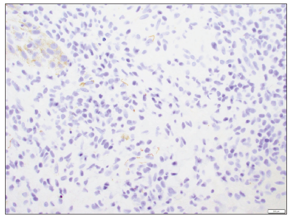
A 42-year-old man presented with several occasionally itchy and painful lesions on his trunk and extremities for six months. Examination revealed lesions of varying morphologies, including violaceous ovoid macules, depressed erythematous plaques, and crusted ulcers surrounded by multiple discrete and clustered pink, scaly, or oedematous papules [Figure 1]. Histopathology showed superficial and deep mixed inflammatory cell infiltrate, and immunohistochemical staining for Treponema pallidum highlighted numerous spirochetes [Figure 2]. A positive syphilis enzyme immunoassay and reactive plasma reagin test, along with this unique bombshell-like configuration were diagnostic of a rare form of secondary syphilis known as corymbiform syphilis.

Export to PPT
Export to PPT
留言 (0)