
記住我
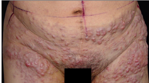
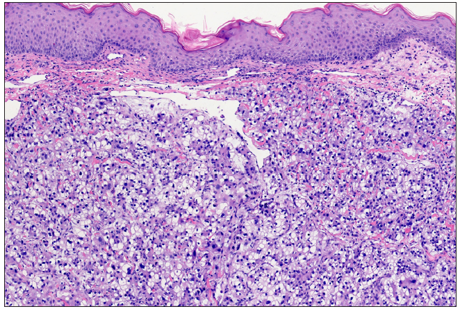
A 47-year-old woman presented with a two-month history of skin lesions on the lower abdomen and thighs. Physical examination revealed multiple, erythematous, painful nodules with a hard consistency, poor mobility, partial surface erosions, and marginal red infiltrating patches [Figure 1]. A biopsy of the nodule from the patient’s right thigh showed polygonal and cuboidal cells of varying sizes with clear cytoplasm in the dermis [Figure 2]. Immunohistochemistry results were positive for hepatocyte nuclear factor (HNF)-1β, Napsin A, paired box (PAX)-8, and Wilm’s tumour gene (WT)-1, while negative for estrogen receptor (ER). She was undergoing treatment for stage IIIC ovarian clear cell carcinoma and was diagnosed with cutaneous metastases from the carcinoma.
Export to PPT
Export to PPT
留言 (0)