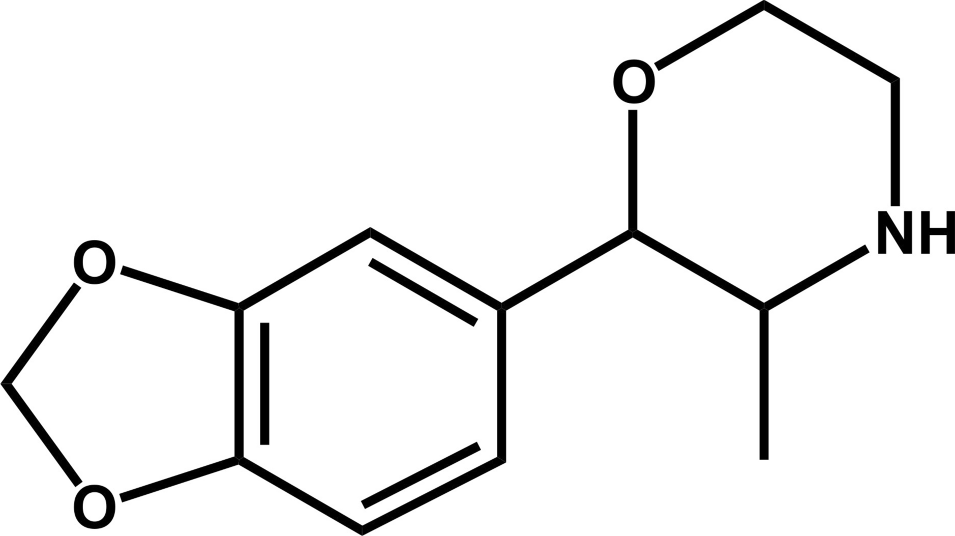
Chemicals and reagents
MDPM was provided by the University of Applied Sciences Kaiserslautern, Germany. Identity was confirmed with a purity > 98% using infrared spectroscopy and HPLC–MS. A 1 mg/mL stock solution of MDPM was prepared in methanol. Alpha-naphthoflavone, amodiaquine 2HCl, bupropion HCl, diclofenac, nicotinamide adenine dinucleotide phosphate (NADP+), omeprazole, phenacetin, sulfaphenazole, and trimethoprim were obtained from Sigma-Aldrich (Steinheim, Germany), dextromethorphan from Roche (Grenzach, Germany), fluconazole from Pfizer (Berlin, Germany), quinidine from Chininfabrik Buchler (Braunschweig, Germany), and testosterone from Fluka (Neu-Ulm, Germany). 3′-Phosphoadenosine-5′-phosphosulfate (PAPS), acetylcarnitine, acetyl coenzyme A (AcCoA), carnitine acetyltransferase, dimethyl sulfoxide (DMSO), dipotassium hydrogen phosphate (K2HPO4), dithiothreitol (DTT), isocitrate, isocitrate dehydrogenase, magnesium chloride (MgCl2), potassium dihydrogen phosphate (KH2PO4), reduced glutathione (GSH), S-(5′-adenosyl)-l-methionine (SAM), superoxide dismutase, tris HCl, verapamil HCl, and two-chambered Centrifree devices were purchased from Merck KGaA (Darmstadt, Germany). UDP-glucuronic acid 25 mM (UGT reaction mixture solution A), 250 mM Tris HCl, 40 mM MgCl2, and 125 μg/mL alamethicin (UGT reaction mixture solution B) were obtained from Corning (Amsterdam, Netherlands). Water was purified using a Millipore filtration unit. Pooled human liver microsomes (20 mg microsomal protein/mL, 25 donors), pooled human liver S9 fraction (20 mg microsomal protein/mL, 30 donors), baculovirus-infected insect cell microsomes (Supersomes) containing human cDNA-expressed CYP isoforms CYP1A2, CYP2B6, CYP2C8, CYP2C19, CYP2D6, CYP3A4, CYP3A5 (1 nmol/mL); CYP2A6, CYP2C9, and CYP2E1 (2 nmol/mL), and flavin-containing monooxygenase 3 (FMO3, 5 mg/mL) were purchased from Discovery Life Sciences (Huntsville, LA, USA). All enzyme containing preparations were thawed at 37 °C after delivery, aliquoted, snap-frozen in liquid nitrogen and stored at − 80 °C until use. Trimipramine-d3 was from LGC (Wesel, Germany). Williams E medium, HPRG670 supplement, GlutaMAX, penicillin, streptomycin, cryopreserved and differentiated HepaRG cells, and type I collagen-coated 96-well plates were purchased from Life Invitrogen (Darmstadt, Germany). Sertraline, acetonitrile, methanol, formic acid (LC–MS grade each), ammonium formate (analytical grade), and all other reagents and chemicals (analytical grade) were from VWR (Darmstadt, Germany). 96-Well plates were purchased from Sarstedt (Nümbrecht, Germany).
Plasma protein binding
PPB of MDPM was investigated based on published procedures with minor adjustments (Gampfer et al. 2020), (Mardal et al. 2016). A volume of 450 µL fresh human blood plasma was spiked with 50 µL of a 5 µM methanolic solution of MDPM. The mixture was incubated for 30 min at 37 °C. Plasma aliquots of 100 µL and 400 µL were transferred to a new reaction tube (global approach, GA) and onto two-chambered Centrifree devices from Merck (Darmstadt, Germany), respectively. The Centrifree devices were centrifuged for 40 min and 1600×g to obtain 100 µL of ultrafiltrate (UF). UF and GA were precipitated using 50 µL acetonitrile (− 20 °C). The mixture was vortexed, cooled for 30 min at − 20 °C, and centrifuged for 3 min at 18,407×g. A volume of 100 µL of the supernatant was transferred into an autosampler vial and 10 µL were injected onto the LC-HRMS/MS system. Experiments were done in triplicate. Fraction unbound (fu) and PPB was determined by comparing the area ratios of MDPM and trimipramine-d3 in the UF and GA using the following equations:
$$_=\frac\left(\frac}_}}}_}}\right)}\left(\frac}_}}}_}}\right)}$$
(1)
$$\text[]=\left(1-_\right)*100$$
(2)
Lipophilicity (logP) of all compounds were calculated with ChemDraw version 23.1.1 (PerkinElmer, Waltham, MA, USA).
Incubations using pooled human liver microsomes
Incubations with pHLM were in accordance with previous publications and minor modifications (Richter et al. 2017). First, 2.5 mM isocitrate, 0.8 U/mL isocitrate dehydrogenase, 100 U/mL superoxide dismutase, 0.6 mM NADP+, 2.5 mM Mg2+ and pHLM (1 mg microsomal protein/mL) were preincubated for 10 min at 37 °C. Reaction was started by adding 25 µM MDPM or 25 µM verapamil (positive control). Substances were incubated for 60 and 120 min at 37 °C. All concentrations are final concentrations, and incubations were done in duplicates. 50 µL aliquots were transferred to a new reaction tube at both timepoints. The reaction was stopped by adding 30 µL acetonitrile (− 20 °C) with 2.5 µM trimipramine-d3 as an internal standard. Negative control samples without enzymes and blank samples without substrates were performed to identify not metabolically formed compounds and to confirm the absence of interfering compounds. Organic solvent in the incubation mixture was below 1% (Chauret et al. 1998). Afterwards the samples were vortexed and centrifuged for 2 min at 18,407×g. 70 µL of the supernatant were transferred to autosampler vials and 10 µL were injected onto the LC-HRMS/MS system.
Incubations using pooled human liver S9 fraction
Incubations with pHLS9 were performed in accordance with previous publications and minor adjustments (Richter et al. 2017). First, 25 μg/mL alamethicin (UGT reaction mix B), pHLS9 (2 mg microsomal protein/mL), 0.1 mM AcCoA, 2.3 mM acetylcarnitine, 8 U/mL carnitine acetyltransferase, 2.5 mM isocitrate, 0.8 U/mL isocitrate dehydrogenase, 100 U/mL superoxide dismutase, 0.6 mM NADP+ and 2.5 mM Mg2+ were preincubated for 10 min at 37 °C. Then, 2.5 mM UDP-glucuronic acid (UGT reaction mix A), 40 µM PAPS, 1.2 mM SAM, 1 mM DTT and 10 mM GSH were added. The reaction was started by adding the respective substrate MDPM, MDMA, or quetiapine (25 µM each). MDMA and quetiapine were incubated as positive controls. Negative control samples without enzymes and blank samples without substrates were incubated to identify not metabolically formed compounds and to confirm the absence of interfering compounds. Incubations were done in duplicate and organic solvent in the incubation mixture was below 1% (Chauret et al. 1998). Aliquots (50 µL) were transferred to a new reaction tube after 60 and 360 min. The reaction was stopped with 30 µL acetonitrile (− 20 °C) containing 2.5 µM trimipramine-d3 as an internal standard. Samples were vortexed, stored at − 20 °C for 30 min, and centrifuged for 2 min at 18,407×g. 70 µL of the supernatant were transferred to autosampler vials and 10 µL were injected onto the LC-HRMS/MS system.
Incubations to investigate in vitro half-life were performed under the same incubation conditions as described above and done in duplicate. Thirty µL aliquots were taken after 0, 15, 30, 45, 60, 75, 90 and 180 min and were terminated with 20 µL acetonitrile (− 20 °C) containing 2.5 µM trimipramine-d3 as an internal standard. 40 µL of the supernatant were transferred to autosampler vials and 10 µL were injected onto the LC-HRMS/MS system. A t-test was performed to compare absolute peak areas of the incubation group and the control group at 0 min. The following parameters were used: unpaired; two-tailed; significance level, 0.05; confidence intervals, 99%.
Incubations using HepaRG cells
Cell incubations were performed in a monolayer assay as previously described (Richter et al. 2019) and in accordance with the manufacturer’s instructions. HepaRG cells were thawed and resuspended in 13.5 mL (~ 740,000 cells/mL) thaw and seed medium at 37 °C. Thaw and seed medium consisted of Williams E medium, 100 U/mL penicillin, 100 µg/mL streptomycin, GlutaMAX and HPRG670 supplement. Cells were handled under sterile conditions using a laminar flow bench class II (Thermo Scientific, TF, Schwerte, Germany). Aliquots of 100 µL cell suspension were seeded on collagen-coated 96-well plates (~ 74,000 cells/well). Evaporation was minimized by filling the outer wells with 100 µL thaw and seed medium. Cells were preincubated for 4 h in an incubator (Binder, Tuttlingen, Germany) at 37 °C, 95% air humidity, and 5% CO2. After preincubation, 50 µL of the thaw and seed medium was replaced with 50 µL MDPM solution (25 µM and 250 µM final concentrations), followed by an incubation for 24 h at 37 °C, 95% air humidity, and 5% CO2 atmosphere. MDMA or quetiapine (25 µM and 250 µM final concentrations, respectively) were incubated as positive controls. All incubations were done in triplicates. Each well contained 0.5% (v/v) DMSO. After incubation, 50 µL of the medium supernatant was precipitated in a new reaction tube with 30 µL acetonitrile (− 20 °C) containing 2.5 µM trimipramine-d3 as an internal standard. A negative control sample without HepaRG cells and a blank sample without substrate were performed to identify not metabolically formed compounds and to confirm the absence of interfering compounds. The mixture was vortexed, cooled for 30 min at − 20 °C, and centrifuged for 3 min at 18,407×g. 70 µL of the supernatant was transferred into an autosampler vial and 10 µL were injected onto the LC-HRMS/MS system.
Monooxygenase mapping
As described elsewhere (Wagmann et al. 2016) and with minor modifications, MDPM (25 µM) was incubated with 2.5 mM isocitrate, 0.8 U/mL isocitrate dehydrogenase, 100 U/mL superoxide dismutase, 0.6 mM NADP+, 2.5 mM Mg2+ and CYP1A2, CYP2A6, CYP2B6, CYP2C8, CYP2C9, CYP2C19, CYP2D6, CYP2E1, CYP3A4, CYP3A5 (50 pmol/mL each), or FMO3 (0.25 mg protein/mL) for 90 and 270 min at 37 °C. For incubations with CYP2A6 or CYP2C9, phosphate buffer was replaced with 90 mM Tris buffer, according to the manufacturer’s guideline. Thirty µL aliquots were taken at both timepoints. The reaction was stopped using 20 µL acetonitrile (− 20 °C) with 2.5 µM trimipramine-d3 as an internal standard. All incubations were done in duplicates and concentrations are final. Verapamil (25 µM) was incubated as a positive control. Negative control samples without enzymes and blank samples without substrate were incubated to identify not metabolically formed compounds and to confirm the absence of interfering compounds. The amount of organic solvent in the incubation mixture was below 1% (Chauret et al. 1998). Afterwards the samples were vortexed and centrifuged for 2 min at 18,407×g. 40 µL of the supernatant were transferred to autosampler vials and 10 µL were injected onto the LC-HRMS/MS system.
CYP inhibition studies
A modified dual-cocktail based method (Dinger et al. 2014b) was used. Substrate cocktail A consisted of 8.9 µM dextromethorphan (CYP2D6), 86 µM testosterone (CYP3A4), 3.5 µM diclofenac (CYP2C9), and 12 µM phenacetin (CYP1A2). Substrate cocktail B included 30 µM bupropion (CYP2B6), 2 µM amodiaquine (CYP2C8) and 21 µM omeprazole (CYP2C19). Substrate concentrations were near their respective Michaelis–Menten constant (Km) values (Dinger et al. 2014a) and all concentrations are final. Enzyme inhibition was investigated using a positive control, a test group, a control group, an interference group, and a negative control. In the test group, specific inhibitors were replaced by MDPM (20 µM). Instead of inhibitors, phosphate buffer was used in the control group. Enzymes were replaced with phosphate buffer in the negative control group. The interference group was incubated like the control group, but the reaction was stopped with 20 µM MDPM in acetonitrile, also containing 2.5 µM trimipramine-d3. All groups were incubated with a NADPH-regenerating system, consisting of 5 mM isocitrate, 2 U/mL isocitrate dehydrogenase, 5 mM Mg2+, and 1.2 mM NADP+. Further, substrate cocktail A or B, 90 mM phosphate buffer (pH 7.4), and 200 U/mL superoxide dismutase were added to each group. The positive control included specific inhibitors (20 µM each, except for 100 µM trimethoprim) of the respective investigated enzymes (cocktail A or B). Specific inhibitors in cocktail A were quinidine (CYP2D6), verapamil (CYP3A4), alpha-naphthoflavone (CYP1A2), and sulfaphenazole (CYP2C9). In cocktail B, sertraline (CYP2B6), trimethoprim (CYP2C8), and fluconazole (CYP2C19) were used. All groups were preincubated for 10 min at 37 °C. Then, pHLM (0.4 mg microsomal protein/mL) was added to each group except the negative control. Incubations were done for 15 min at 37 °C. Therefore, 50 µL of acetonitrile (− 20 °C) containing 2.5 µM trimipramine-d3 as internal standard was added. Incubations were done in triplicates and organic solvent in the incubation mixture was below 1% (Chauret et al. 1998). Afterwards the samples were vortexed and centrifuged for 2 min at 18,407×g. 70 µL of the supernatant were transferred to autosampler vials and 10 µL were injected onto the LC-HRMS/MS system. Enzyme inhibition was evaluated by metabolite formation. A t-test was performed to investigate significant inhibition in the positive control and test group compared to the control group (rejection of null hypothesis) or to prove similarity in results in the interference group and negative control (acceptance of null hypothesis) compared to the control group. The following settings were used: unpaired; one-tailed; significance level, 0.05; confidence intervals, 99%.
LC-HRMS/MS apparatus and conditions
A TF (Dreieich, Germany) Dionex UltiMate 3000 RS pump consisting of a degasser, a quaternary pump, and an HTC prep and load (PAL) autosampler, coupled to a TF Q Exactive system equipped with a heated electrospray ionization (HESI)-II source were used. A mass calibration was done according to the manufacturer's recommendations using external mass calibration prior to analysis. Gradient elution was performed according to a previous study (Helfer et al. 2015) on a TF Accucore PhenylHexyl column (100 mm × 2.1 mm, 2.6 μm) with a 2 mM aqueous ammonium formate solution containing 0.1% (v/v) formic acid (pH 3, eluent A) and 2 mM ammonium formate solution in acetonitrile/methanol (50:50, v/v) containing 0.1% (v/v) formic acid, and 1% (v/v) water (eluent B). The gradient was stepped as follows: 0–2.5 min hold 99% A, 2.5–8 min to 1% A, 8–9.5 min hold 1% A, and 9.5–11.5 min hold 99% A. Initial flow rate from 0–9.5 min was 500 μL/min and final flow rate was 800 μL/min from 9.5–11 min. Injection volume was 10 μL for every sample. Mass spectrometry was performed using full scan data and a subsequent data-dependent acquisition (DDA) with priority to mass-to-charge ratios (m/z) of parent compounds and their expected metabolites. The inclusion list contained m/z values of likely formed metabolites such as O-demethylenyl and hydroxy metabolites (phase I) as well as sulfates, glucuronides, methoxy metabolites and combinations thereof (phase II). Chemdraw 23.1.1 was used to draw structures of expected metabolites and for exact mass calculations. TF Xcalibur Qual Browser software version 4.6 (Dreieich, Germany) was used for data handling. Mass deviations of the parent compound were accepted up to 5 ppm. Plasma protein binding samples were measured only in positive ionization mode, all other samples were measured in positive and negative ionization mode.
Instrument settings, data generation settings, and data evaluation settings are shown in Table S1 in the supporting information.


留言 (0)