The case study concerns a 23-year-old Caucasian male of Polish nationality who was employed in one of the countries on the east coast of Africa. A review of the medical documentation and case files revealed that prior to travelling abroad, the patient had not been diagnosed with any significant diseases or chronic conditions and was not taking any medications on a regular basis. Prior to his departure, the patient was provided with medical advice at the Clinic of Tropical Diseases, which included guidance on malaria prophylaxis and the prevention of traveller’s diarrhoea. It was recommended that the patient use repellents (Mugga 50%) and Malarone (proguanil hydrochloride/atovaquone), among other measures. The patient was instructed to commence treatment with Malarone one day prior to departure and to continue taking it for a period of seven days following the conclusion of their stay.
In the eighth month of his stay, he presented with symptoms of illness, fatigue, and respiratory infection. The patient presented with a fever and abdominal discomfort. Over the following days, his condition deteriorated significantly, necessitating hospital admission. Upon admission, the patient was unconscious and febrile. Imaging diagnostics (chest X-ray and head CT) were performed but did not show any pathology. Laboratory tests confirmed the presence of severe malaria in the patient, caused by the parasite Plasmodium falciparum. Parasitemia was found to be 1.8% of red blood cells. Furthermore, laboratory tests yielded abnormal results, indicating the presence of severe intravascular hemolysis (anaemia, hyperbilirubinemia, elevated LDH), coagulopathy (thrombocytopenia, prolonged APTT), electrolyte imbalance (hypercalcemia, hyperkalemia, hypermagnesaemia, hyperphosphatemia, hyponatremia), and evidence of severe renal and hepatic failure, hyperuricemia. A summary of the selected test results is presented in Table 1.
The antimalarial treatment was initiated with artezunat. The patient remained unconscious (Glasgow Coma Scale score of 7/15) and subsequently developed severe respiratory failure shortly after admission to the hospital. Additionally, an episode of upper gastrointestinal bleeding and convulsions were observed during the initial hours of hospitalization. The treatment regimen was augmented with the addition of anticonvulsants, gastroprotective agents, antibiotics, and high-flow oxygen therapy. Despite the administration of treatment, the patient’s condition continued to deteriorate. On the second day of hospitalization, the patient experienced a sudden cardiac arrest, and resuscitation was unsuccessful.
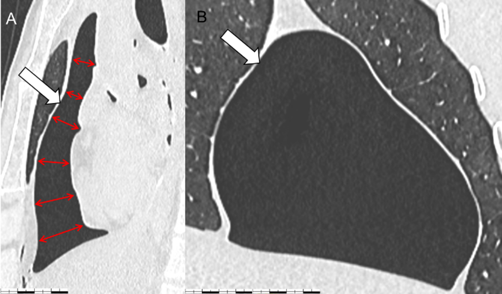
A forensic medical autopsy was conducted on the day of the patient’s death. No injuries were identified, apart from the typical consequences of medical rescue operations. The autopsy report, which was included in the case files, was cursory in nature, yet it did describe several abnormalities. The presence of pleural and peritoneal effusions was documented, along with the diagnosis of bronchopneumonia, cardiomegaly, hepatomegaly, and acute fatty pancreatitis. The spleen was enlarged and exhibited the hallmark features of tropical splenomegaly syndrome (TSS). The analysis of the blood sample revealed a significant presence of malaria parasites.
The cause of death was determined to be severe malaria caused by a Plasmodium falciparum infection, accompanied by uremic encephalopathy, TSS, cardiomegaly, hepatomegaly, acute fatty pancreatitis, severe anaemia and acute renal failure.
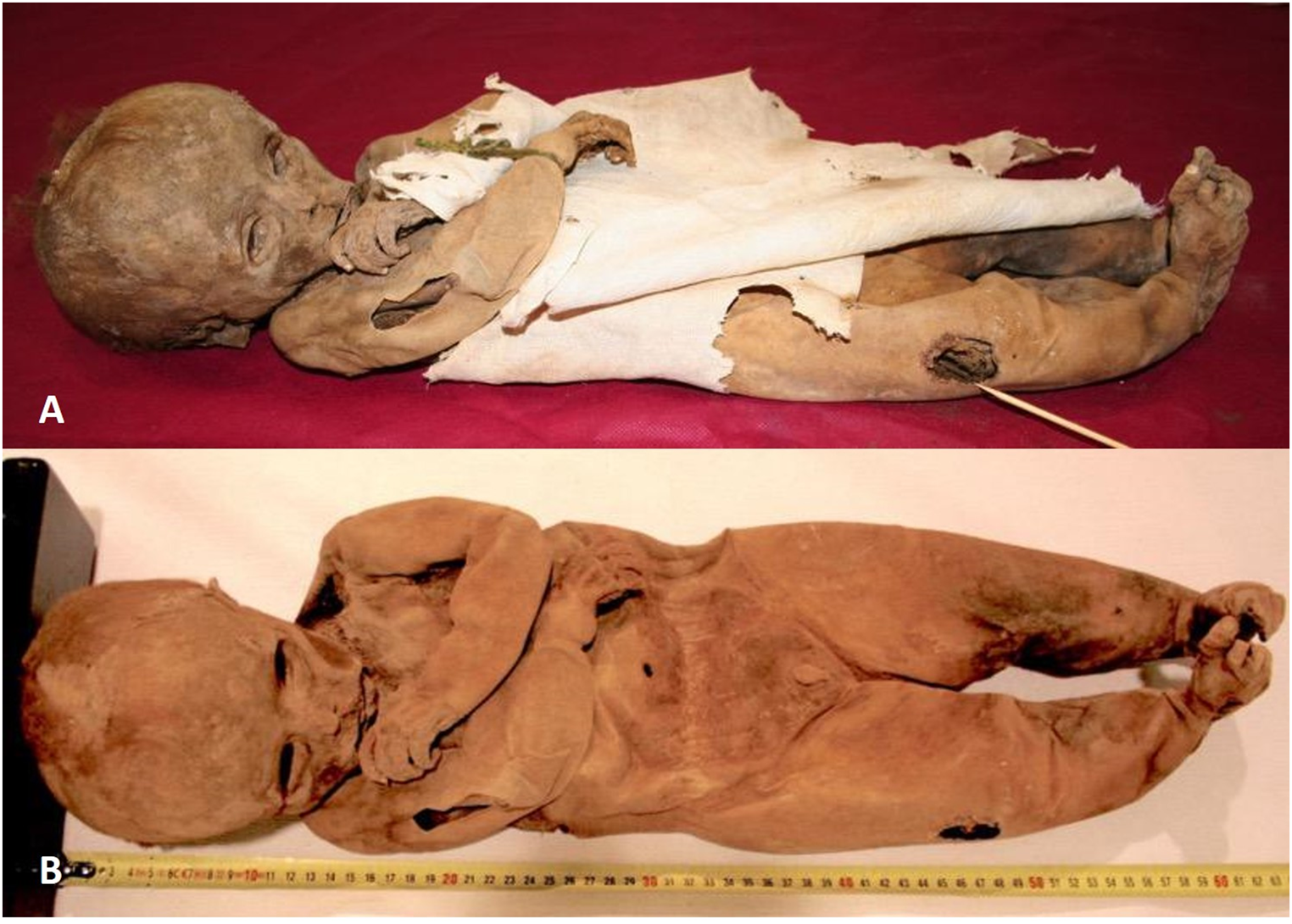
Following the autopsy, the corpse was embalmed and stored in cold storage for 8.5 months in the African country due to logistical challenges associated with transporting the body to Poland. The specific embalming conditions remain unknown.
Following the transfer of the corpse to Poland, the prosecutor decided that a further autopsy was required to confirm results of the first postmortem examination.
The body exhibited indications of advanced putrefaction and exhibited the hallmarks of a prior autopsy, in addition to vascular access points utilized for the embalming of the corpse through infusion.
The cranial cavity had not been previously dissected, thereby allowing for an assessment of the brain structures. However, due to the advanced state of decomposition, a thorough examination was not possible. It was only possible to make a general statement that no focal discolourations were present. The macroscopic evaluation of individual organs was significantly limited by the effects of decomposition, embalming and the number of previous dissections. The only confirmed finding was splenomegaly. The atria and ventricles of the heart and the large blood vessels were empty, and it was not possible to obtain blood samples.
The main challenge for the autopsy was to re-confirm parasite infection. Due to the advanced stage of decomposition, immunochromatographic tests were ruled out, and the lack of blood made it impossible to perform standard parasitological tests. A large number of samples were taken from internal organs for toxicological and genetic examinations.
Liver and kidney samples were subjected to toxicological analysis for the presence of ethanol, methanol, isopropanol and acetone using gas chromatography (GC) with flame ionisation detection (FID). The toxicological examination revealed the presence of ethyl alcohol in concentration 0.3‰ (liver) and 0.2‰ (kidney). This finding could be fully explained by the advanced post-mortem processes and did not provide sufficient evidence to assume that the deceased was under the influence of ethyl alcohol at the time of death. Furthermore, the post-mortem material that was secured contained methyl alcohol (0.7‰ in the liver and 1.1‰ in the kidney), which is frequently employed for the embalming of corpses. However, the documentation did not include any information regarding the composition of the preservative agents used. A review of the medical records from the hospital treatment did not provide any evidence to suggest that the patient had suffered from methanol poisoning.
Toxicological analysis of internal organ samples was conducted using the LC–ESI-MS–MS method according to standard protocol using in our Toxicological Departament and previously described by Rojek et al. [12] The internal organ samples (liver and kidney) revealed the presence of substances with anticonvulsant (diazepam), analgesic (acetaminophen, dipyrone), antimalarial (quinine) and antiarrhythmic (lidocaine) properties, which corresponded to the records in the submitted medical documentation.
Toxicological analysis of hair samples of the deceased’s hair was conducted using the LC–ESI-MS–MS method previously described by Klys et al. [13]. The drugs were identified in the analysed segment of hair, along with the additional presence of tramadol. No antimalarial drugs were identified. The main objective of the multiparameter analysis was to identify the presence of antimalarial drugs, including atovaquone, proguanil, quinine, and a comprehensive range of antibiotics (e.g. doxycycline). On the other hand, it should be emphasisedthat toxicological analysis of hair samples is aimed to prove long-time intake, rather than to identify a one-time intake. The absence of any antimalarial medications in the analysed hair samples ruled out that the patient did used the pharmacological prophylaxis of malaria in the final months of his life. As indicated in the literature, antimalarial drugs are incorporated into the hair structure and can be successfully detected using the LC-MS-MS method [14].
In the present case, the most pertinent information was derived from molecular tests. Post-mortem samples from a range of organs, including the heart, lung, brain, kidney, liver and spleen, were subjected to examination. The total genomic DNA was extracted using a silica-based method with a Sherlock AX kit (A&A Biotechnology, Poland), and the DNA amplification was performed on a QuantStudio 5 Real-Time PCR System (Applied Biosystems, USA) with the real-time PCR (RT-PCR) method using a Malaria TaqMan PCR (Norgen Biotek Corp., Canada) kit, in accordance with the manufacturer’s protocols. The Malaria TaqMan PCR Kit is designed for the detection of malaria-specific DNA using a real-time PCR based on the use of a 5’-nuclease assay of a TaqMan reporter probe. The molecular testing for malaria parasite infection revealed the presence of malaria parasite DNA in the lung, brain, kidney, liver and spleen sections. Conversely, no malaria parasite DNA was detected in a sample obtained from the heart. The results of the duplicate reactions for each tissue section were found to be consistently accurate. The correct result for both the positive and negative standards was obtained, as well as the internal control, in each diagnostic reaction. This was done to ensure the quality of the isolated DNA sample. The result of the molecular test was unequivocal, confirming a severe infection with the malaria parasite in its most severe form, cerebral malaria. Figure 1.


留言 (0)