
記住我


With the increasing number of molecularly targeted therapies for thyroid carcinoma, the accurate identification of gene mutations and fusion genes is necessary to maximize therapeutic efficacy [4, 11, 12]. Particularly in thyroid carcinoma, some patients may exhibit late recurrence and/or distant metastasis [3, 13, 14]. Therefore, it is important to obtain long-term tissue samples that retain a high nucleic acid quality. In this study, we evaluated the nucleic acid quality of thyroid carcinoma specimens and investigated the utility of using separately fixed tumor samples. Given the typically high nucleic acid quality resulting from the effective formalin fixation of biopsy specimens [7], we prepared separately fixed tumor samples from the thyroid glands and lymph nodes immediately upon arrival at the Department of Pathology. Separately fixed tumor samples showed significantly higher values of DIN and S/L Ct ratio, the indicators of DNA quality, than conventionally processed thyroid tumors and lymph node metastases. These samples also showed significantly higher levels of DV200, an indicator of RNA quality. Considering their size of 3–5 mm and regulated fixation time in formalin, immediate and adequate fixation may have contributed to the preservation of nucleic acid quality by ensuring thorough and even formalin penetration without over- or under-fixation. Furthermore, the preparation of such samples can be seamlessly integrated into the routine workflow of pathological examinations, making it a highly valuable method for preserving nucleic acid quality in specimen samples, particularly in hospitals lacking frozen storage facilities. However, Hatanaka et al. reported higher DIN and DV200 values [8] than observed in the present study. This result may reflect differences in specimen characteristics and processing conditions at our specialized oncology hospital, which frequently handles advanced cases and large lymph node metastases.
Evaluation of the effect of formalin fixation time on nucleic acid quality in the present study showed that DNA quality (DIN and S/L Ct ratio) in thyroid gland tumors tended to decrease with longer fixation times. This finding aligns with those of the previous reports showing that excessive fixation negatively affects DNA quality [15, 16]. Such degradation can be attributed to the susceptibility of DNA to fragmentation during extended fixation periods, primarily due to cross-linking and chemical modifications that compromise the structural integrity of the DNA molecules [15]. These findings highlight the importance of optimizing fixation time to preserve DNA quality for subsequent genomic analyses. In contrast, no significant difference was observed between formalin fixation time and nucleic acid quality in lymph node metastasis. This lack of variation could be attributed to the anatomical and procedural differences between thyroid gland tumors and lymph nodes. For thyroid gland tumors, our fixation process involved a biopsy punch for lesions, followed by gauze insertion into the hollowed-out area, formalin injection, and subsequent submersion in formalin by vacuum-assisted fixation. While these methods aim to enhance formalin penetration, extended fixation times may result in over fixation, potentially affecting DNA quality. Conversely, lymph nodes did not undergo extensive processing in this study. Additionally, the presence of adipose tissue and capsules around the lymph nodes could impede the penetration of formalin solution [17]. These procedural and anatomical differences could explain why extended formalin fixation time alone did not lead to significant variation in nucleic acid quality in lymph node metastases.
In addition, we examined the quality of RNA in relation to specimen fixation time, which is known to promote the inactivation of RNases [18] and ensure more complete tissue fixation [19]. An appropriate fixation time can prevent RNA degradation and potentially improve RNA integrity preservation in properly fixed samples. In some cases, this may require longer fixation times than those optimal for DNA [19]. However, our findings reveal a more complex picture. The RIN values for the separately fixed tumor samples were lower than those for the other samples, in contrast to the parameters observed for DNA quality. Moreover, further analysis of histological features could not fully explain this discrepancy, suggesting that additional factors may be involved in determining the RNA quality in these samples. Although the RIN value is widely used as an indicator of RNA quality [20,21,22], it should be noted that separately fixed tumor samples exhibited the highest DV200 values, despite their lowest RIN values. Additionally, the RIN values of samples obtained from FFPE tissues were generally very low, indicating extended fragmentation. In contrast, high DV200 values, which represent the percentage of RNA fragments that are 200 bases or longer, suggest that a portion of RNA remains intact and available for NGS-based genomic analyses. Previous studies have reported that RIN values are generally lower in FFPE tissue samples derived from human surgical specimens [18]; however, a low RIN does not necessarily render RNA unusable. Several studies have highlighted the potential of DV200 as a reliable metric for assessing the RNA quality required for NGS-based genetic studies using FFPE tissue samples [20]. For example, Hatanaka et al. reported that in ODxTT, an amplicon sequencing-based NGS assay for thyroid carcinoma covered by insurance in Japan, DV200 serves as a more effective predictor for detecting gene transcripts than RIN [8]. The study further noted that although the qPCR method is preferred for the quality assessment of ODxTT, DIN, and DV200 serve as valuable alternatives when the qPCR method is not feasible. Electrophoresis-based assessments such as DIN and DV200 offer practical and cost-effective options for nucleic acid quality assessment because of their accessibility and low cost, making them suitable for facilities with limited budgets [8].
The differential effects of formalin fixation on DNA and RNA quality present a challenge in optimizing sample preparation for thyroid cancer genomic analysis, where both DNA-based (e.g., BRAF V600E mutations) and RNA-based (e.g., RET and NTRK fusion genes) analyses are crucial. Our findings suggest that separately fixed tumor samples may offer a solution by enabling thorough fixation in a shorter time frame, potentially preserving both DNA and RNA quality more effectively for comprehensive genomic profiling. Additionally, our study explored the relationship between histological features and nucleic acid quality in thyroid carcinoma specimens, revealing complex interactions that vary with the specimen type. In thyroid gland tumors, cystic changes correlate with an improved S/L Ct ratio, suggesting that formalin injection may facilitate better local penetration in cystic areas. Conversely, an increased lymphocyte ratio was associated with a decreased RIN and DV200, indicating potential RNA quality degradation in areas with high lymphocyte infiltration. This observation raises questions about whether the lower RNA quality is due to the intrinsic properties of infiltrating lymphocytes or their effects on the surrounding tissue. However, to our knowledge, limited literature has directly addressed this relationship in thyroid carcinoma, highlighting an area for future research.
Regarding lymph node metastases, multiple regression analysis revealed that the fibrosis ratio negatively impacted the S/L Ct ratio, suggesting that fibrotic tissue may hinder fixative penetration. Furthermore, macrodissection is associated with improved RIN. However, given the small number of macrodissection cases (n = 3), these findings should be interpreted with caution. In contrast, separately fixed tumor samples showed less of an overall influence of histological features. Nevertheless, the presence of cystic components significantly positively affected the DIN and S/L Ct ratio, which is consistent with observations in thyroid gland tumors. No significant differences in nucleic acid quality were observed between BRAF immunohistochemistry-positive and immunohistochemistry-negative samples across all specimen types. These findings demonstrate that the nucleic acid quality in thyroid carcinoma specimens is influenced by histological features and specimen processing methods.
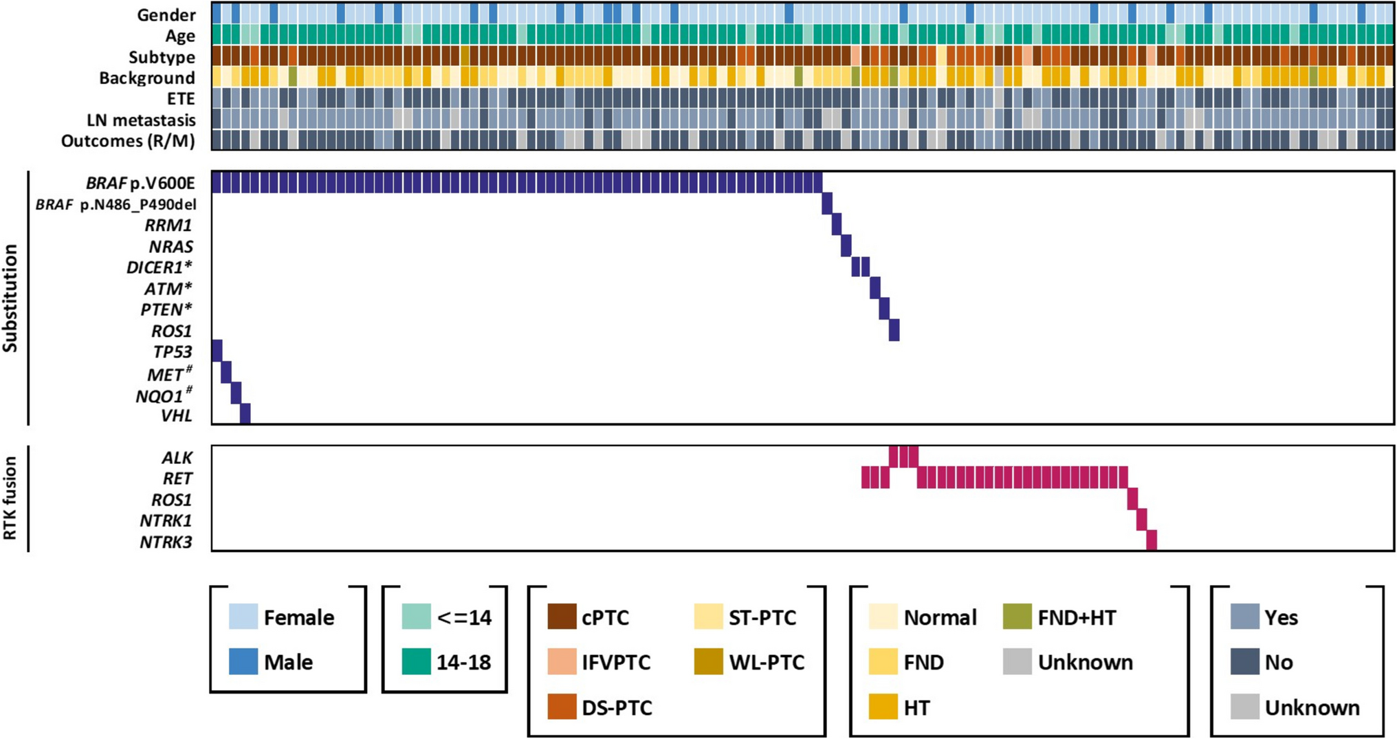
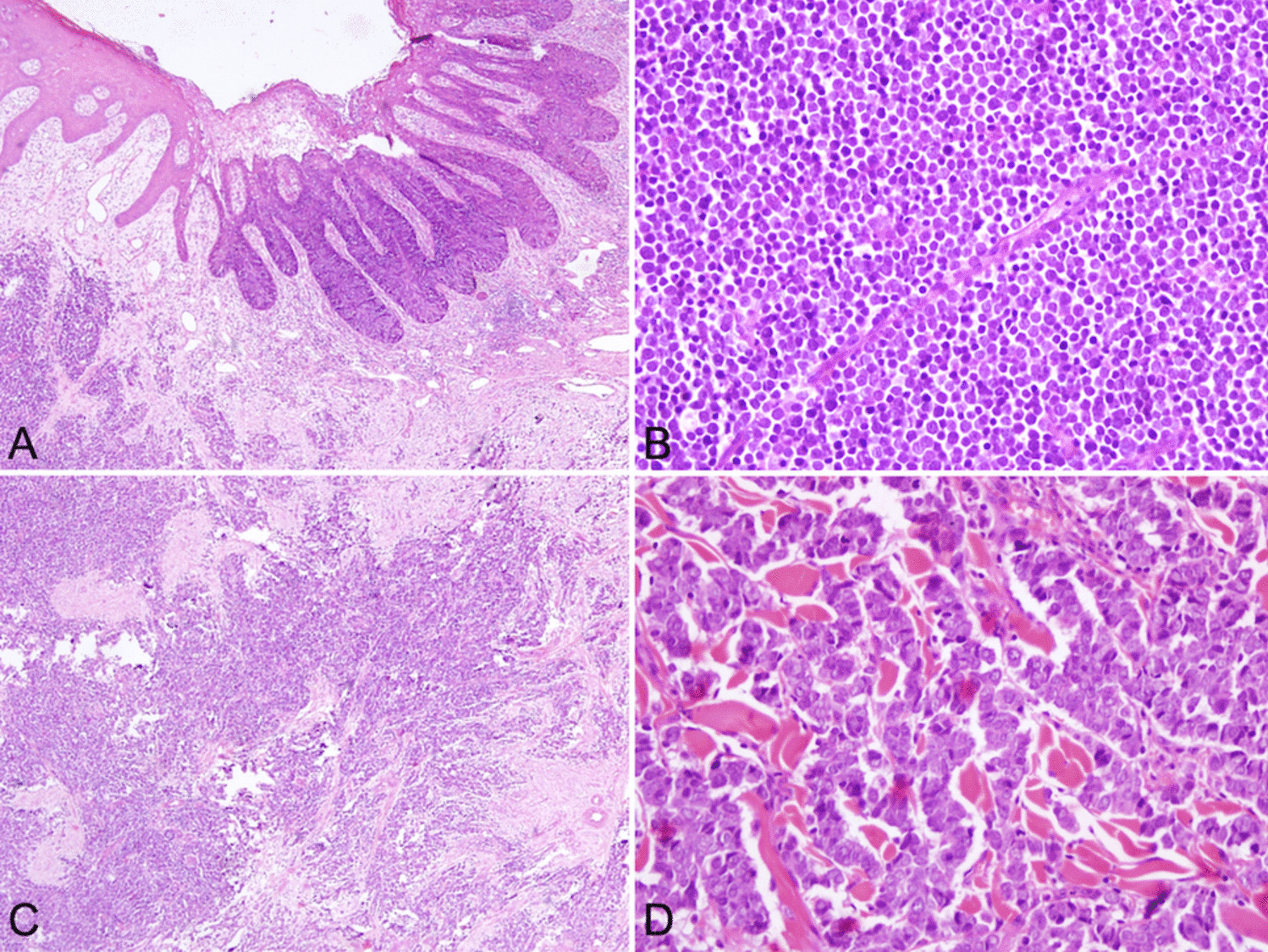
Finally, the significance of using lymph node metastases specimens for genomic analysis should be highlighted. When lymph node metastases are used as specimens for genomic analysis, it is important to determine whether the genetic mutations in lymph node metastases are identical to or greater than those in the primary tumor. Previous studies have reported that genetic alterations in primary tumors and lymph node metastases exhibit differences and similarities, depending on the type of cancer [23,24,25]. However, BRAF mutations and RET and NTRK fusions, major markers for molecularly targeted therapies in thyroid carcinoma [26,27,28], are considered early molecular events in thyroid carcinogenesis [1, 29,30,31], which may indicate their presence in lymph node metastases similar to their primary lesions. Therefore, if the quality of nucleic acids is equivalent to or greater than that in the primary tumors and the number of tumor cells and the tumor content ratio meet the thresholds for standard genomic analysis, it would be acceptable to consider lymph node metastases as candidates for genomic analysis in thyroid carcinoma. In addition, we discuss the characteristics of lymph node metastasis in thyroid carcinomas. Based on our clinical experience, lymph node metastases in thyroid cancer often exhibit a regional growth pattern and tend not to intermingle extensively with the background lymphocytes. This characteristic potentially facilitates the acquisition of samples with a high tumor content, especially when combined with macrodissection techniques, making them particularly suitable for genomic analysis. Our previous studies indicated that the calcification of lymph node metastases, particularly in cases of PTC with RET or NTRK fusions, is often mild or absent, even when the thyroid gland itself exhibits significant calcification (Fig. 5) [32,33,34]. Decalcification methods are known to significantly damage nucleic acids in FFPE tissue samples [32, 34, 35]. Considering that the nucleic acid quality of lymph node metastases is comparable to that of thyroid gland tumors, these metastases can be considered valuable specimens for genomic analysis, especially given their lower degree of calcification. However, our findings also revealed that the quality of nucleic acids in lymph node metastases tended to decrease with increasing metastasis size. Consequently, surgeons, pathologists, and/or laboratory technicians may be required to excise or collect separately fixed tumor samples from large lymph node metastases to preserve the quality of nucleic acids for analysis.
Fig. 5
Macroscopic and microscopic images of papillary thyroid carcinoma with ETV6-NTRK3 fusion gene. a Macroscopic findings of the cut surface of the thyroid gland in a case of papillary thyroid carcinoma with ETV6-NTRK3 fusion. Extensive calcification was observed in the tumor area, accompanied by marked sclerosis. b Microscopic findings of papillary thyroid carcinoma with ETV6-NTRK3 fusion gene. Prominent coarse calcifications are observed against a background of extensive fibrosis. Hydrochloric acid-based decalcification was required for slide preparation for diagnosis, although it is known to decrease nucleic acid quality (scale bar = 1000 µm). Note that this figure is included for illustrative purposes only and does not represent the cases analyzed in this study
In conclusion, the preparation of separately fixed tumor samples is an effective method for preserving DNA and RNA quality for genetic testing. Specimen collection using biopsy punches is feasible in many facilities, even when managing frozen specimens. These results will contribute to the establishment of a method for preserving high-quality pathology specimens that are widely used in general facilities. However, considering the burden of preparing additional samples, it is important for clinicians, pathologists, and laboratory technicians to appropriately assess the necessity for a sample. This can be facilitated by ensuring that the pathology request form includes detailed information on the potential requirements for future genetic testing, TNM classification, and any other relevant clinical observations. Clinicians and pathologists should share this information in advance. Additionally, our findings revealed that lymph node metastases often exhibit nucleic acid quality equal to or superior to that of thyroid gland tumors, underscoring their potential as reliable sources for genomic analysis.
LimitationsThis study has several limitations. First, the efficacy of negative-pressure fixation in enhancing formalin permeation and preserving nucleic acid quality has not yet been conclusively established. Additionally, the stability of nucleic acid quality in separately fixed tumor samples over time remains to be thoroughly investigated. Electrophoretic assessments, such as RIN and DV200, were used for RNA quality evaluation because of the lack of established qPCR protocols for analyzing degraded RNA in our hospital. Furthermore, this study primarily focused on papillary thyroid carcinoma, which may limit the applicability of our findings to other histological types of thyroid carcinomas, such as follicular, poorly differentiated, and anaplastic carcinomas. Additionally, non-neoplastic benign thyroid lesions were not included in this study. This study used 3–5 mm biopsy punch needles, similar to those used in another ongoing study in lung cancer research. We cannot rule out the possibility that different diameters may be advantageous in some situations. While frozen tissue samples were collected, detailed information about their quality is lacking because processing was conducted in a different division of our institution.
留言 (0)