Study population
The current study is embedded within a prospective population-based birth cohort, the Generation R Study [15]. Women from the city of Rotterdam in the Netherlands, with a delivery date between 2002 and 2006, were invited to participate in the study. Children and both their parents were followed from the gestational period, throughout childhood into early adulthood, with routine visits at certain ages including brain MRI and cognitive assessments [13]. Between 2017 and 2020, a subset of the mothers and their partners were invited for a sub-study that specifically aimed to determine brain health as a means to unravel the ORigins of Alzheimer’s disease aCross the LifE-course: the ORACLE Study [16].
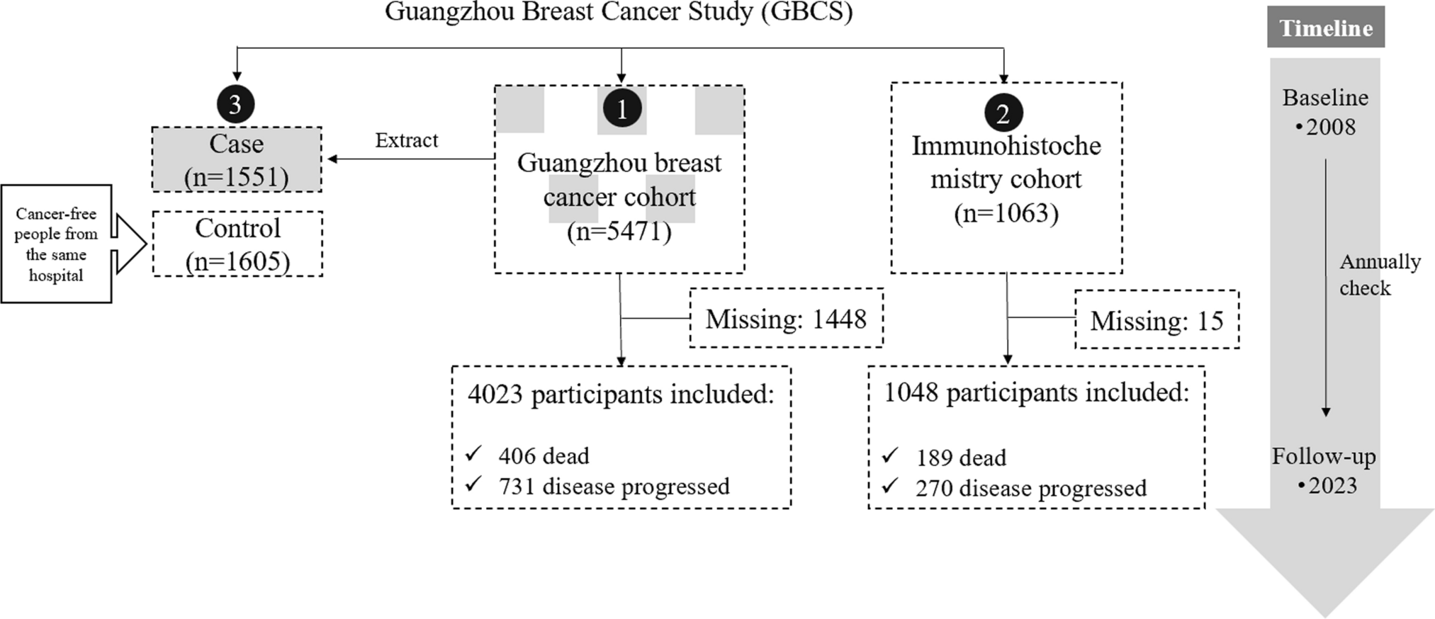
Of 3,559 parents invited for the ORACLE Study, 2,084 (58.5%) agreed to participate. They visited the study centre for one day that included questionnaires, blood pressure measurement, cognitive assessment, and brain magnetic resonance imaging (MRI). For the current study, we included all parents who completed the questionnaire on family history. The questionnaire involving family history of dementia was introduced later during the ORACLE study and was available in 1,511 of 2,084 participants (72.5%). We excluded participants who had missing cognitive tests (n = 162) and missing sequences on the MRI (n = 59). We additionally excluded 31 parents because of motion artefacts or incidental findings on brain MRI (e.g., brain tumour, large cortical infarct), leaving 1,259 parents eligible for the study.
The children of these parents were included if they had undergone brain imaging during their research visit at age 9 (2011–2015), and cognitive testing during their visit at the age of 13 (2016–2020). Of the 1259 parents eligible to participate in this study, 1332 of their children had complete information on brain MRI and cognition available. We excluded 197 children because of low image quality (e.g. motion artefacts) or incidental findings and an additional 269 children were excluded to ensure only one child per family was included in the analysis, leaving 866 (65.0%) children for analysis.
The study was designed in accordance with the guidelines set by the World Medical Association Declaration of Helsinki. The study has been approved by the Medical Ethics Committee of the Erasmus Medical Center, University Medical Center, Rotterdam, the Netherlands. Written informed consent was obtained for all participants. Children aged between 12 and 16, both the child and legal guardian gave consent.
Data availability
Datasets generated during the current study are not publicly available due to legal and ethical regulations. However, requests for access to the data reported in this paper can be directed to the secretary office of the Generation R Study (secretariat.genr@erasmusmc.nl), in accordance with local, national, and European Union regulations.
Family history
Family history of dementia was acquired through structured questionnaires administered during an interview [16]. Parents were asked ‘Has your mother or father been diagnosed with dementia?’. The question could be answered with yes, no, or uncertain. If answered yes, parents were asked to provide details about the affected parent, including age at the time of the diagnosis. For the current study, we considered family history positive if a parent reported a diagnosis of dementia in at least one of their parents. For the children, this meant their grandparental family history was positive if either of their parents reported a diagnosis of dementia in at least one grandparent.
Image acquisition and processingParent and child structural imaging
For both parent and child, structural magnetic resonance images (MRI) were obtained on a 3T GE Discovery MR750w MRI System (General Electric, Milwaukee, WI, USA) with an 8-channel head coil [16, 17]. The complete procedure has been previously described [16, 17]. In short, we collected T1-weighted images with an inversion recovery-prepared fast spoiled gradient recalled sequence (Tr = 8.77 ms, Te = 3.4 ms, Ti = 600 ms, flip angle = 10 0, Field of view = 220 × 220 mm, acquisition matrix = 220 × 220, slice thickness = 2 mm (1 mm for the children), number of slices = 230). The T1-weighted images were processed through the FreeSurfer analysis suite, version 6.0.0 [18]. Non-brain tissue was removed and the voxel intensities were normalized for B1 inhomogeneity. Next, the images were segmented and all segmentations were manually inspected. The brain measures of interest were the volume of total brain, grey matter, white matter, hippocampal, entorhinal cortex, the middle temporal gyrus, and the parahippocampal gyrus [12, 19, 20]. Volumes across left and right hemispheres were averages, as we did not expect lateralized effects. Intracranial volume (ICV) encompasses the total volume within the skull, including the brain tissue and cerebrospinal fluid spaces.
Assessment of cerebral small vessel disease
For the parents, volume of white matter hyperintensities (WMH) was acquired from FreeSurfer segmentations. Trained researchers rated all scans for the presence of lacunes and cerebral microbleeds, blinded to familial history of dementia. Lacunes were defined following the STRIVE criteria as focal lesions between ≥ 3 and < 15 mm within the white matter, cerebellum, basal ganglia, or thalamus, as seen on a 2D axial fluid-attenuated inversion recovery (FLAIR) sequence (0.8 × 1.1 × 2.5 mm3) and the T1-weighted sequence [21]. Cerebral microbleeds were defined as small hypointense foci with a maximum size of 10 mm on T2*-weighted sequence (0.8 × 1.1 × 1.0 mm3).
CognitionParents
Parents completed a cognitive test battery as part of the ORACLE study [16]. The battery consisted of six tests, assessing different domains of cognition. A detailed description of the complete test battery can be found elsewhere [16]. Briefly, the assessment included the 15-word learning test [22], the Stroop task [23], the letter-digit substitution test [24], a word fluency test [25], the Purdue pegboard test for manual dexterity [26], and the design organization test [27]. As the latter two tests were introduced later during the study course, they were not offered to 4.5% (design organisation test) and 18.5% (Purdue pegboard) of participants, respectively. We imputed these two cognitive tests (missing completely at random), using a single imputation based on age, sex, education, and other available cognitive tests. To summarize the tests into a single score for global cognition, we computed the g-factor [28] isolating the first component of a principal component analysis, using all six cognitive tests as indicators. The g-factor explained 64.1% of the variance amongst the cognitive tests.
Children
For children, we assessed the Intelligence Quotient (IQ) as an indicator of cognitive function. During the research centre visit at age 13, children were administered four subtests from the Wechsler Intelligence Scale for Children-Fifth Edition (WISC-V) [29]. Matrix reasoning was used to assess fluid reasoning and was administered digitally. Digit Span (forward, backwards & ranking from high to low) was administered verbally to assess working memory. Symbol substitution was administered digitally, and used to measure processing speed. Finally, Vocabulary was administered verbally, measuring verbal comprehension. All subtest scores were age-normed according to the manual [29]. IQ scores were derived by summing the normed subtests and then converted into IQ scores using a conversion table specifically created for these four subtests by Pearson [30].
APOE genotype
Genotyping in the parents was done by sequencing DNA from blood samples collected during early pregnancy. APOE genotype for the parents was determined with a biallelic TaqMan assay (rs7412 and rs429358), and classified in APOE-ε4 carriers and non-carriers. Of the total 1259 parents, 1071 (85.0%) had information on APOE and of these, 296 (27.6%) had at least one APOE-ε4 allele. Genotyping in children was done by sequencing DNA from blood samples obtained from the umbilical cord or with blood samples collected at the age of 6 [31]. APOE-ε4 carrier status was determined based on the genotype data. Out of the 866 children, 584 (67.4%) had information on APOE and 142 (24.3%) had at least one APOE-ε4 allele.
Other measurements
Information on ethnicity and education was obtained through self-reported questionnaires administered at study inclusion. Because of the relatively small sample size within subgroups, country of origin was categorized for this analysis into Western which includes European, North-American, and Oceanian (as well as Japanese) and non-Western (South America, Central-American, Asia (other than Japan), and African). At the visit of the MRI, height, weight, medication use, and smoking (current smoking, yes or no) were self-reported. BMI was computed from height and weight (kg/m2). Systolic and diastolic blood pressure was measured using an automatic sphygmomanometer Omron 907 (OMRON, Matsusaka Co., Ltd., Japan) [32]. Blood pressure was measured two times over a 60-second interval, and individuals with an average systolic blood pressure > 140 mmHg or diastolic blood pressure > 90 mmHg, or the use of blood pressure-lowering medication were classified as hypertensive.
Statistical analysis
Missing data on non-genetic covariates were imputed through multiple imputation procedures using the package mice [33]. We had 16.7% missing data on education, 13.6% on ethnicity, and 2.1% on hypertension information. As for smoking and BMI, we had less than 0.01% missing data. Data were imputed 20 times (20 iterations) using chained equations and the model estimates for each imputed data set were subsequently pooled using Rubin’s rules [34]. The distribution of covariates was similar in the imputed and non-imputed datasets. All analyses were done in R 3.6.3 [35].
Among the parents, we determined the association of parental history of dementia with brain volumes, using linear regression. Although we expected a low prevalence of small-vessel disease in our population, in an exploratory analysis the association of parental history with the presence of lacunes (yes/no) and microbleeds (yes/no) was determined using logistic regression. All models included age, sex, ethnicity, and ICV (model 1), with further adjustment for educational attainment, hypertension, smoking, BMI (model 2), and APOE genotype (model 3). Similarly, for the children, we determined the association of grandparental history of dementia with the same structural brain measures using linear regression. Markers of small-vessel disease were absent in childhood, and therefore not further assessed. All models included age, sex, ICV, and ethnicity (model 1), with further adjustment for APOE genotype (model 2).
Next, we determined the association of parental history of dementia with the g-factor and the underlying cognitive tests. Among the children, we determined the association of grandparental history of dementia with IQ and the tests that encompassed IQ, using linear regression models. Models included the same covariates as those for the imaging outcomes.
We performed various sensitivity analyses. First, we stratified the participants by APOE ε4 carrier status. Second, we stratified parents at age 50, to account for the fact that some of their parents may still be relatively young for developing dementia. Third, we stratified by age—(grand)parent under or over the age of 80 at diagnosis— based on previously reported increased genetic risk, particularly with younger age at onset [1]. Fourth, we stratified by maternal vs. paternal family history, because previous research has suggested effects may be more profound for maternal than for paternal family history [7, 36].


留言 (0)