
記住我


With age, all organs of the human body, including the brain, undergo a series of physiological and pathological changes, which are manifested in the decline of language ability, visuospatial ability, cognitive ability, and the decline of concentration and memory (Mattson and Arumugam 2018). At the cellular and molecular level, brain aging is characterized by the following features: increased inflammatory response, impaired mitochondrial function, oxidative folding of proteins, endogenous formaldehyde (FA) accumulation, and DNA damage (Cheng et al. 2023; Tong et al. 2011a). Gaseous FA is a common small molecule substance with strong metabolism and active chemical properties, and has been known for its strong mutagenicity and carcinogenicity for over a century (Farooqui 1983). Endogenous FA accumulation in the aging brain can be confirmed by measuring FA concentrations in humans by gas chromatography-mass spectrometry. More than 40 years ago, researchers used this method to obtain FA concentrations of 0.08 mmol/L in human blood and 0.2–0.4 mmol/L in brain tissue (Heck et al. 1982). The above FA levels are at normal levels and can be metabolized by the body normally. However, the concentration of FA in the body of healthy people tended to increase with increasing age and induce decline in memory as well as cognitive disorders. The neuromolecular mechanism of FA-induced memory loss has been elucidated, that is, it can spontaneously react with the α/ε amino groups of proteins, leading to protein aggregation and loss of activity; while it can react with the cysteine sulfhydryl group on the NR2B subunit of the NMDA receptor, affecting the function of the NMDA receptor (Tong et al. 2011a). Moreover, the mechanism of FA-induced cognitive impairment is closely related to norepinephrine depletion. Hippocampal norepinephrine plays a very important role in learning and memory, which can enhance hippocampal long-term potentiation and memory formation (Gelinas and Nguyen 2007). It was found that FA in vitro and intrahippocampal injections can significantly reduce norepinephrine levels, which becomes a key factor in cognitive ability and memory decline (Mei et al. 2015).
FA induces genotoxicityFA is characterized by its distinctive molecular structure and physicochemical properties, such as a limited steric hindrance, reactive carbonyl electrophilicity, cellular permeability, and temperature-dependent stability of methylene bridge adducts (Hoffman et al. 2015). Its elevated genotoxicity primarily manifests in the cells of organisms through DNA damage. The diverse forms of DNA damage caused by FA have been identified in current research, including various types of DNA breaks, DNA-DNA cross-linking, DNA-RNA cross-linking, DNA protein cross-linking (DPC), and DNA adducts. The specific mechanisms of this damage can be categorized into two aspects: direct damage to DNA through a series of chain chemical reactions, and indirect damage to DNA through impairment of the damage recognition and excision repair system (Ortega-Atienza et al. 2016; Tan et al. 2017). Recent studies have shown that endogenous FA, rather than exogenous sources, is the main cause of FA genotoxicity, challenging previous beliefs about the dangers of inhaling FA (Lu et al. 2010a). It can contribute to disease development by causing DNA damage and impairing repair processes through various pathways.
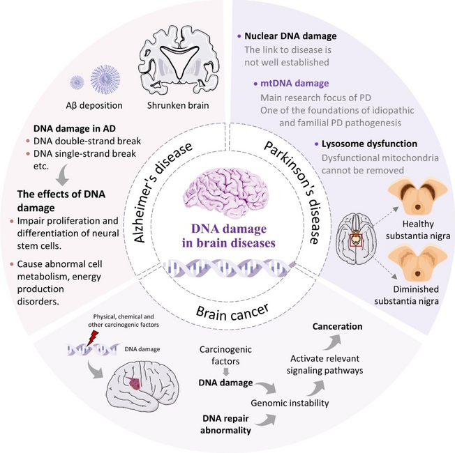
DNA damage is involved in the brain diseasesNumerous studies have linked cellular DNA damage buildup and decreased repair ability to various diseases, often resulting in impaired cell repair and proliferation in regenerative cells (Fang et al. 2016; Lautrup et al. 2019; Shiloh and Lederman 2017). In contrast, the narrow sense of nerve cells, i.e., neurons, as a kind of non-renewable cells, their DNA damage often manifests degenerative lesions, such as Alzheimer’s disease (AD) and other neurodegenerative diseases (Misiak et al. 2017; Sykora et al. 2015). Moreover, due to the presence of a large number of glial cells in the brain, their DNA damage can manifest as primary tumors of the central nervous system, such as Li-Fraumeni syndrome, in which approximately 2%-10% of patients develop brain tumors in childhood (Orr et al. 2020), and the complex interaction of different DNA damages in glioblastoma results in redundant repair mechanisms, potentially causing tumor resistance to drugs (Rominiyi and Collis 2022). Various evidence indicates that DNA damage is closely associated with brain diseases, including cancer. The traditional Aβ hypothesis is still prevalent in understanding neurodegenerative diseases like AD, but the connection between these diseases and DNA damage requires further exploration.
This paper reviews how endogenous FA production in the aging brain leads to DNA damage and contributes to the development of brain diseases. It offers a novel perspective on the development of AD and FA-related diseases, along with new diagnostic markers and treatment options.
Endogenous FA accumulation in the ageing brainDisorders of FA metabolizing enzyme systems during ageingThe brain can produce FA through a variety of pathways, the most prominent of which are enzymatic reactions, for example, semicarbazide-sensitive amine oxidase (SSAO) (Obata 2006), cytochrome P450 (CYP450) (Hasemann et al. 1995), lipid oxidases, demethylase, endoplasmic reticulum demethylase (Kalász 2003; Yu et al. 2003) and other demethylase (Trewick et al. 2002). Of course, FA can be degraded to maintain homeostasis of FA levels in brain tissues by some enzymes, such as: glutathione-dependent FA dehydrogenase (FDH or named ADH3) and alcohol dehydrogenase 1 (ADH1) (Martínez et al. 2001), and aldehyde dehydrogenase-2 (ALDH2) (Fei and Tong 2020). It has been found that abnormalities in the content and activity of these enzymes, particularly ALDH2 and dehydrogenase 5 (ADH5), can lead to FA accumulation and memory impairment. This was demonstrated in ADH5 and ALDH2 knockout mice. Among them, FA was degraded more significantly by ALDH2 compared to ADH5 when FA levels are too high (Kou et al. 2022). During the aging process, there is a decline in the function of FA metabolism enzyme system in the brains. For instance, brain FA levels were higher in 3-month-old senescence accelerated mouse-prone 8 strain compared to controls. Enzyme levels were examined and found to be elevated in SSAO expression levels, while enzyme activity of ADH3 were reduced (Qiang et al. 2014). This results in a situation where the rate of FA generation exceeds the rate at which the brain is able to degrade FA, leading to the accumulation of FA in brain tissue and brain diseases (Kou et al. 2022) (Fig. 1).
Fig. 1
Multiple metabolic pathways of endogenous FA. Red arrows: FA-generating pathways; blue arrows: FA-degrading pathways (Fei and Tong 2020). Abbreviations: ADH1, alcohol dehydrogenase 1; ADH3, alcohol dehydrogenase 3; ALDH2, aldehyde dehydrogenase 2; CAT, catalase; ER, endoplasmic reticulum; FA, formaldehyde; LSD1, lysine special demethylase 1; MeOH, methanol; MIT, mitochondria; MMA, monomethylamine; SA, sarcosine; SARDH, sarcosine dehydrogenase; SSAO, semicarbazide-sensitive amine oxidase; TET1, TET methylcytosine dioxygenase 1. Adapted with the permission of ref. (Fei and Tong 2020), copyright@Acta Physiologica Sinica, 2020
FA causes DNA damageTypes of DNA damage caused by FAFA-induced mutagenesis FA is well known as a very common mutagen, which is particularly effective for deletion mutagenesis (Anderson 1995). As we have mentioned above, ALDH2 and ADH5 are the most important FA-degrading enzymes. Significant FA accumulation was present in the serum of Aldh2-/-Adh5-/- mice obtained by hybridization. At the same time, an increase in FA-modified DNA in tissues and mutational signatures was found including single-nucleotide substitutions, double-base substitutions, insertions and deletions, which is similar to patterns observed in human cancers (Dingler et al. 2020). It seems clear that FA in peripheral tissues can lead to genetic mutations and thus cause diseases. However, in contrast, few studies have addressed the relationship between FA-induced mutations and brain diseases.
FA-induced DNA breakage DNA breakage occurs when one or both strands of the DNA molecule are broken, either as single-strand breaks (SSBs) or double-strand breaks (DSBs). Different cell types show varying levels of sensitivity to FA's toxic effects (Jimenez-Villarreal et al. 2017). FA-induced DNA breaks have also been confirmed at the cellular experimental levels (Bedford and Fox 1981). FA caused DNA damage at specific concentrations (5, 7.5, 10, 15 μM) that could be quickly repaired. This indicates that FA may cause single-strand breaks at lower concentrations and DNA-DNA cross-links at higher concentrations, but the exact biological mechanism is unclear (Liu et al. 2006). In addition, FA also causes mitochondrial DNA double-strand breaks (Miwa and Brand 2003) via reactive oxygen species (ROS) (Nadalutti et al. 2020).
FA-caused DNA-DNA cross-linking There are two types of DNA-DNA cross-linking: intrastrand and interstrand. Intrastrand occurs within the same DNA strand, while interstrand occurs between two DNA strands. Helicases unwind the DNA double helix for replication and transcription, but interstrand crosslinking hinders these processes. Therefore, interstrand cross-linking is one of the most consequential DNA damages. At the same time, it is the only form of formaldehyde-induced DNA-DNA cross-linking in the available studies. FA preferentially forms dA—dA cross-links at dinucleotide sequence 5'-d(AT) in certain AT-rich double-stranded DNA sequences but the mechanism is not clear (Huang et al. 1992). Exposure of human peripheral blood lymphocytes to different concentrations of FA induced DNA-DNA cross-linking when FA concentration was greater than 25 μM (Liu et al. 2006).
FA-induced DNA-RNA cross-linking DNA-RNA cross-links were found in NNK-treated mouse lung DNA in 2022. The authors propose that these cross-links may be DNA-RNA hybrids formed during transcription, replication, or gene expression, and that FA reacts with the base portions of DNA and RNA to induce these cross-links on the R-loop (Dator et al. 2022). In addition, mechanisms of FA-induced DNA-RNA cross-linking are unclear, which need to be further investigated.
FA-caused DPCs The DPCs, complexes formed when DNA and protein interact through a series of non-covalent forces, are considered to be the most consequential type of DNA damage and are widely found in FA-induced DNA damage (Weickert and Stingele 2022). FA, a common cross-linking agent, can cause various damages by inducing excessive DPCs and interfering with normal cellular physiological activities such as DNA replication and transcription. However, FA induces the production of DPCs only at higher concentrations, because the induced DPCs could be slowly repaired within the cell and would not accumulate for a long period of time (Liu et al. 2006).
FA-induced DNA adducts The DNA adducts are a piece of DNA covalently bound to some chemical substances (Parthiban et al. 2015). In order to distinguish between DNA adducts induced by endogenous and inhaled FA exposure, rats were exposed to 10 ppm [13CD2] FA for 1 or 5 days (6 h/day) using a nose-only chamber. The N2 -HO13CD2-dG produced by exogenous [13CD2]-FA exposure is formed only in DNA at the nasal inlet; endogenous N6 -hydroxymethyl-dA is present in all tissues (Lu et al. 2010a). Tissue samples from rats exposed to 1,30 and 300 ppb FA were analyzed by the same method as above. Endogenous adducts were detected, while exogenous adducts were not found in any groups (Leng et al. 2019). FA can activate certain drugs to bind covalently to DNA, in addition to directly binding and forming DNA adducts. In the process of forming certain drug-DNA adducts, FA plays a crucial role, particularly in the case of anthracyclines and anthracenediones. Mitoxantrone, an anticancer anthracenedione, was identified as the initial anthracenedione capable of generating substantial DNA crosslinks facilitated by FA (Pumuye et al. 2020) (Fig. 2).
Fig. 2
Types of DNA damage induced by FA. The types of damage induced by FA include DNA break, interstrand crosslink, DNA-RNA crosslink, DPCs, and DNA monoadducts. Among the several types of DNA damage induced by FA, relevant studies have clarified the specific chemical structures of DNA interstrand crosslink, reproduced with the permission of ref. (Hu et al. 2019), copyright@American Chemical Society, 2019 and DNA–protein crosslink, reproduced with the permission of ref. (Lu et al. 2010b), copyright@American Chemical Society, 2010, including 7 types of ICLs and 8 types of DPCs respectively
FA-induced RNA–protein crosslinking In addition to the above widely studied types of FA-induced DNA damage, a 2023 study demonstrated for the first time FA-induced RPCs, which stall ribosomes and thus inhibit the translation process (Suryo Rahmanto et al. 2023). The newly established modeling system photoactivatable ribonucleoside-enhanced crosslinking can help to confirm this process (Zhao et al. 2023). Such RPCs significantly activate K6-linked ubiquitination while marginally increasing K33-linked ubiquitination, ultimately resolved by the ubiquitin-dependent remodeler valosin-containing protein (Suryo Rahmanto et al. 2023). More issues remain to be investigated, such as the levels of RPCs in physiologic and certain disease states and whether they cause cytotoxicity other than the translational process.
Mechanisms of DNA damage caused by FAMolecular structures of the complex of FA/nucleic acidsFA is an active alkylating agent with its own unique molecular structure and physicochemical properties: 1) simple and small-sized molecular structure leading to a small spatial site resistance; 2) active carbonyl group electrophilicity; 3) cell permeability; 4) temperature-dependent stabilization of methylene-bridge-containing adducts etc. (Hoffman et al. 2015). These properties make FA easy to cross-link with various macromolecules and form a variety of adducts (Fraenkel-conrat et al. 1945). It is commonly used as a cross-linking agent in various biological assays due to its strong cross-linking properties (Hoffman et al. 2015; Kim and Dekker 2018; Klockenbusch et al. 2012; Sutherland et al. 2008), and the more commonly used techniques are chromatin immunoprecipitation (ChIP) and chromosome conformation capture (3C) analysis (Kim and Dekker 2018). In living cells, high levels of macromolecules and FA can cause significant DNA damage. Research has been done to understand how FA interact with DNA and other molecules in a controlled setting. (Feldman 1973; McGhee and von Hippel 1975a; McGhee and von Hippel 1975b; Metz et al. 2004), which are summarized in a review (Hoffman et al. 2015). This difference poses challenges in detection, yet it is essential to acknowledge that FA's distinctive molecular structure and physicochemical properties confer a significant advantage in inducing DNA damage (Fig. 3).
Fig. 3
Reaction properties of FA and the basic process of reaction with biological macromolecules. FA is an active alkylating agent with its own unique molecular structure and physical and chemical properties, which is shown in the upper part of the figure. Existing studies have clarified the specific steps of the reaction between FA and biological macromolecules such as DNA and proteins through in vitro experiments, which are shown in the lower part of this figure, adapted with the permission of ref. (Hoffman et al. 2015), copyright@Journal of Biological Chemistry, 2015
FA induces oxidative stressFA-damaged antioxidant system Living organisms have both oxidative and antioxidant systems, with enzymes and non-enzymatic substances playing a role in these reactions. Among them, the oxidative system includes superoxide radicals (-O2-), hydrogen peroxide (H2O2), and hydroxyl radicals (-OH) in ROS, xanthine oxidase (XO), and the lipid peroxidation end-products malondialdehyde (MDA), 4-hydroxynonenal (4- HNE), etc.; in the antioxidant system, glutathione (GSH) is the most important antioxidant in the body, in addition to the three classical enzymatic antioxidants: superoxide dismutase (SOD), glutathione peroxidase (GSH-Px) and catalase (CAT) (Sies 1997). In addition to the above substances there are glutathione reductase (GR) and glutathione s-transferase (GST) involved in the system. Under physiological conditions, the oxidative and antioxidant systems are in a balanced homeostasis. However, FA exposure leads to dysregulation of the oxidative and antioxidant systems associated with a decrease in the levels of SOD, CAT, GSH-Px, XO, GSH and MDA, while an increase in the level of ROS in the cerebellar cortex (Zararsiz et al. 2011).
FA-impaired mitochondria The mitochondria are the main site of ATP generation by aerobic respiration in most eukaryotic cells and are closely associated with various redox reactions (Zorova et al. 2018). In living cells, mitochondria are the main source of ROS, a central substance of oxidative stress (Miwa and Brand 2003). However, FA can lead to mitochondrial dysfunction through a variety of pathways. For example, FA can reduce in mitochondrial membrane potential levels and an increase in Cyt-c transfer from mitochondria to the cytoplasm (Tang et al. 2012), and inhibit the respiratory chain by inactivating the phosphate transport system in the mitochondrial membrane, blocking ATP production in the mitochondrial respiratory chain (Tyler 1969).
FA-induced oxidative stress associated with brain diseases FA-induced oxidative stress is associated with brain diseases. Although there is no definitive conclusion on the involvement of FA-induced oxidative stress as a dominant factor in brain diseases, it is certain that oxidative stress plays an important role. Using multiple bioinformatics analyses, provided important guidance on candidate genes and possible pathways of FA leading to brain cancers and neurodegenerative diseases (NDD). Through toxicogenomic databases, it was found that among the FA exposure-related genes, the antioxidant enzyme SOD2 gene overlapped the most with genes related to common brain diseases, which included brain cancer, AD, PD, amyotrophic lateral sclerosis, and other NDD while the antioxidant enzyme SOD1 ranked the third, which included PD, amyotrophic lateral sclerosis, and other NDD. Indeed, 20% of the genes in oxidative stress are affected by FA exposure. In addition, among the significantly enriched pathways for FA and NDD subgroups or brain tumor-related genes, the oxidative stress pathway was highly enriched (Rana et al. 2021).
FA inhibits DNA damage recognition and excision repair in cellsFA not only directly damages DNA through various pathways, but also impairs the DNA damage recognition and excision repair system, thus preventing the repair of damaged DNA and causing irreversible DNA damage. For example, a large number of proteasome-targeted k48-linked polyubiquitin proteins were detected in the nuclei and cytoplasm of human lung epithelial cells treated with FA exposure for a short period of time and triggered a strong heat shock response, suggesting that a large number of damaged proteins accumulated rapidly in the cells (Ortega-Atienza et al. 2016). In addition, FA-induced DPCs are mainly repaired by hydrolysis through proteasomal scavenging (Ortega-Atienza et al.
留言 (0)