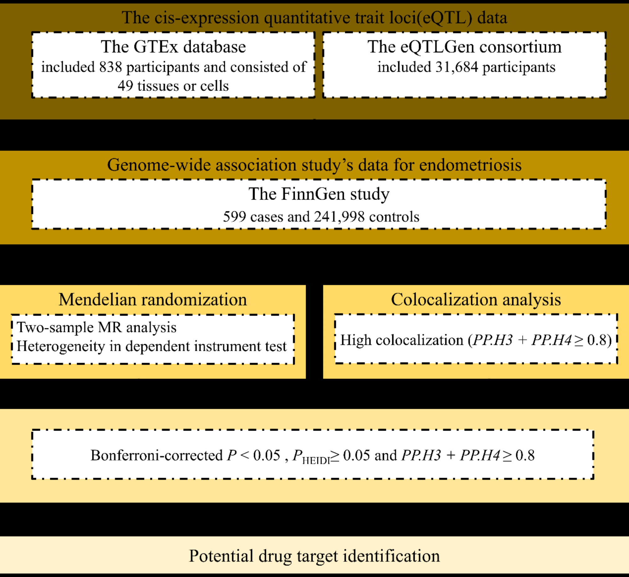
Our study employed comprehensive genome-wide MR and colocalization analyses to clarify the causal relationships between genes and POI, providing valuable insights into potential therapeutic targets. MR analysis effectively reduced confounding factors in assessing the associations between gene expression and disease. Colocalization analysis confirmed that the eQTL instrument in the MR was not incidentally associated with both traits, thereby ruling out the possibility of the MR effect stemming from alternative causal variants in linkage disequilibrium. Among the genes analyzed, only FANCE and RAB2A exhibited evidence of a shared genetic effect with POI outcomes through both MR and colocalization analyses. FANCE and RAB2A were linked to a reduced risk of POI, highlighting their potential as promising targets for POI treatment.
FANCE is a subunit of the FA pathway, which plays a key role in repairing DNA interstrand cross-links. FA pathway genes encode proteins involved in gonadal development, DNA replication, and DNA repair [27]. Mutations in the FANCE gene cause FA in humans [28]. Our study confirmed that FANCE is associated with a reduced risk of POI and is a risk locus for POI. Interestingly, clinical evidence indicates that female FA patients exhibit reduced fertility, manifesting as POI [29]. Previous studies have shown that FANCE−/− mice exhibit ovarian dysplasia and severely reduced numbers of follicles by five days after birth, resembling women suffering from POI [30]. Animal experiments have also demonstrated that FANCE defects impair the rapid mitotic proliferation of primordial germ cells (PGCs) in mouse embryos, leading to a sharp decrease in PGCs number and abnormal cell cycle distribution [31]. These findings indicate that FANCE is essential for PGC survival, with potential mechanisms involving cell cycle regulation, DNA damage repair, cell death prevention, and the regulation of lysosome and ribosome functions [32].
In FA cells, DNA damage remains unrepaired due to a dysfunctional DNA repair process, causing cells to be blocked in the G2/M phase [33]. When DNA damage occurs in the primordial follicle, it responds by phosphorylating and activating TAp63 [34]. Activation of TAp63 induces the transcription of pro-apoptotic factors such as BH3, PUMA and NOXA. The upregulated expression of these proteins facilitates their interaction with the pro-apoptotic BCL2 family members, BAX and BAK [34]. The translocation of BAX and/or BAK to the oocyte’s mitochondria causes mitochondrial dysfunction, release of apoptogenic proteins, and activation of caspase-9 and proteolytic enzymes, collectively triggering apoptosis and cell death [34]. The depletion of oocytes damages fertility and leads to POI [35]. Two rounds of meiosis are vital for oocyte maturation [36]. During the prophase of the first meiosis, fully grown oocytes are arrested in the germinal vesicle (G2) phase [37]. Meiotic resumption marks the initiation of oocyte maturation, characterized by germinal vesicle breakdown (GVBD), followed by meiotic spindle assembly and migration during metaphase I (MI)[38]. Subsequently, cytokinesis occurs, the oocyte extrudes the first polar body, and arrests in the metaphase II (MII) phase [37]. The G2/M phase and the MII phase are two major stages in oocyte meiosis [38]. G2/M cell cycle blockade leads to meiotic recovery failure, chromosome misalignment, increased aneuploidy, abnormal spindle assembly, and severe meiotic defects in oocytes [37, 39, 40]. These issues are the main causes of ovarian aging and reduced female fertility [41].
RAB2A, a member of the Rab family, is localized to the endoplasmic reticulum-Golgi intermediate compartment (ERGIC) transport complex and regulates ERGIC transport within the cell membrane [42]. Our findings indicate that genetically predicted RAB2A is inversely associated with the risk of POI. The HEIDI test and colocalization analysis further ruled out the possibility of horizontal pleiotropy. While current studies show that RAB2A is associated with sperm viability and motility function in men [43], the mechanism of premature ovarian aging in females with POI remains unclear.
Numerous studies have shown that ovarian aging is associated with mitochondria, oxidative stress, DNA damage, protein homeostasis, aneuploidy, apoptosis, and autophagy [44]. Previous research has demonstrated that RAB2A regulates autophagosome-lysosome fusion [45]. RAB2 may regulate autophagy initiation through three mechanisms: (1) Transporting Golgi-derived ATG9 + vesicles to phagophore assembly sites. (2) Recruiting ULK1 to phagophore assembly sites, as ULK1 appears soluble and forms a diffused cytosolic pattern in the absence of RAB2A. (3) Facilitating ULK1 activation to propagate signals for autophagy initiation [45]. Autophagy plays a crucial role in oocyte development. Under normal circumstances, oocytes cannot actively induce mitophagy to clear damaged mitochondria. However, oocyte mitophagy can be initiated by drugs or abnormal environmental stimuli, affecting the developmental ability of oocytes [46]. Based on our findings and literature reports, we hypothesize that high expression of RAB2A may induce abnormal autophagy in oocyte mitochondria, resulting in a decrease in the number and quality of oocytes, ultimately leading to POI. However, this hypothesis requires further study as no correlation has been reported in the literature yet.
The strength of our study lies in employing MR and colocalization analyses to evaluate gene causality in POI records through genetic variation. MR analysis reduces biases from confounders and reverse causation, thereby increasing the reliability of causality results. Colocalization analysis is instrumental in revealing the pleiotropic effects of specific loci on multiple traits, avoiding LD and identifying potential therapeutic targets. Additionally, we utilized extensive GWAS data, allowing the simultaneous examination of numerous genes and variants. This cost-effective and efficient method thoroughly investigates the gene-disease relationship. Our study integrated multiple databases, enhancing the robustness of our findings. Focusing on European populations minimized bias related to ethnic disparities.
Nonetheless, certain limitations should be acknowledged. Firstly, our colocalization analyses are constrained by the availability of outcomes from existing studies, limiting the examination of undiscovered genetic variants. Moreover, the reliance on instrumental variants in colocalization analysis can introduce bias if these variants correlate with unmeasured variables. Finally, while focusing on specific ethnicities reduces racial bias, it also limits the generalizability of our findings to diverse ethnic groups.


留言 (0)