
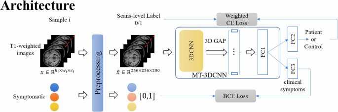
記住我

Baseline state anxiety did not differ between the placebo (33.96 ± 5.84) and OT groups (35.55 ± 7.23), t(112) = −1.29, p = 0.20, suggesting that participants of both groups showed similar baseline anxiety levels. Also, there was no significant difference for post-experiment state anxiety between the placebo (33.31 ± 8.88) and OT (35.73 ± 9.77) groups, t(112) = −1.44, p = 0.15, indicating that intranasal OT did not modulate self-reported anxiety measured after fear conditioning (see Fig. 2a).
Fig. 2: Effects of intranasal oxytocin (OT) on fear conditioning.
a State anxiety levels of the placebo and OT groups were not significantly different at baseline or post-MRI. b Skin conductance responses (SCRs) were not significantly different across groups. c Cortisol concentrations in saliva were not significantly different between groups at baseline, pre-MRI, or post-MRI. These results suggest that intranasal OT did not modulate fear acquisition.
PsychophysiologyThe task was effective to evoke conditioned fear towards the CS+ compared to CS− stimuli. A significantly larger amplitude for CS+ trials (0.19 ± 0.22) compared to CS− trials (−0.02 ± 0.08) was observed, t(98) = −11.20, p < 0.001, d = 1.12 (see Supplementary Fig. 2 for the average amplitude of each trial). It should be noted that the time window within which the SCR was calculated was prior to the occurrence of the electric stimulus, thus the SCR reflecting the anticipatory responses to pain (pain-related fear), rather than the pain itself. No significantly different SCR was observed between the placebo (0.20 ± 0.17) and OT (0.21 ± 0.20) groups, t(97) = −0.22, p = 0.82, resembling the observations in the previous rodent study [12] that found no effects of OT on freezing behavior during fear acquisition (see Fig. 2b). For visualization and analysis of the early and late phase of extinction learning, see Supplementary Fig. 3.
fMRITo examine whether the experimental task successfully activated the brain regions involved in fear acquisition, the differential BOLD responses of CS+ compared to CS− for all participants (placebo and OT groups combined) were tested. Known regions from a previous review [22] were observed including the anterior insular cortex extending to the frontal operculum, the ventral striatum including the putamen, a large expanse of the medial wall cortex including the anterior cingulate gyrus, the dorsolateral prefrontal cortex, and the lateral cerebellum. See Supplementary Fig. 4 for anatomical localization and Supplementary Table 4 for statistical and stereotaxic details. Regarding differences between placebo and OT groups, whole-brain analysis revealed no significant BOLD signal for the differential contrast of CS+ versus CS− (see Supplementary Table 5 for details). This result suggests that intranasal OT did not significantly modulate the BOLD responses during fear acquisition.
HormonesBaseline hormones including OT, estradiol, progesterone, testosterone from blood plasma as well as cortisol from saliva samples were not significantly different between the placebo and OT groups (all ps > 0.13), suggesting that the participants in both groups had similar hormonal profiles. For details, see Supplementary Table 2. The pre-MRI (placebo: 1.91 ± 0.55; OT: 2.08 ± 0.55; t(110) = −1.57, p = 0.119) and post-MRI (placebo: 1.93 ± 0.42; OT: 2.09 ± 0.50, t(110) = −1.86, p = 0.067) cortisol levels between the placebo and OT groups were not significant although a trend towards significance emerged at post-MRI with higher cortisol levels in the OT group (see Fig. 2c).
Resting-state fMRI (day 1)The connectivity of the anterior insula was altered in the OT group, compared to the placebo group. Specifically, seed-to-voxel analysis revealed a stronger negative connectivity of the left anterior insula with an area predominantly (99%) located in the left postcentral gyrus (peak voxel at x = −56, y = −14, z = +28), t(110) = −4.72, FDR-corrected p < 0.001 (see the blue region in Fig. 3a). Moreover, a stronger positive coupling for the OT group, compared to the placebo group, was observed between the left anterior insula and an area (peak voxel at x = +4, y = +64, z = +30) predominantly comprising the right ventromedial prefrontal cortex (vmPFC; 57%) and the right superior frontal gyrus (33%), t(110) = 4.37, FDR-corrected p < 0.001 (see the red bulb in Fig. 3a). The brain-behavior associations showed significant negative correlations between the insula-postcentral gyrus connectivity and baseline cortisol levels on the next day, r(107) = −0.27, p = 0.004, and baseline state anxiety levels on the next day, r(109) = −0.24, p = 0.01. See Fig. 3b, c for scatter plots. Controlling for sex hormones (estradiol, progesterone, and testosterone) using partial correlation did not affect the relationship, all ps < 0.007.
Fig. 3: Effects of intranasal oxytocin (OT) on fear memory consolidation following fear conditioning.
a Seed-to-voxel resting-state functional connectivity after fear conditioning revealed that in the OT compared to the placebo group the left anterior insula showed a stronger negative correlation with the left postcentral gyrus (blue bulb) as well as a stronger positive correlation with the right ventromedial prefrontal cortex (vmPFC) and the right superior frontal gyrus (red bulb). b Significant negative correlation between the anterior insula-postcentral gyrus connectivity and baseline cortisol levels on the next day prior to extinction learning. c Significant negative correlation between the anterior insula-postcentral gyrus connectivity and the baseline state anxiety on day 2 prior to extinction learning. The red line represents the fitted line and the gray areas are the confidence intervals at 95%.
Extinction learning (day 2)BehaviorNo significant state anxiety differences were observed between the PP, PO, OP, and OO groups at baseline, F(3, 110) = 1.2, p = 0.32, or post-MRI, F(3, 110) = 0.82, p = 0.49. See Supplementary Table 6 for details. Participants were asked post-MRI to rate the intensity and unpleasantness of electric shock pain that they received on day 1 in the scanner. No significant difference was observed between the placebo (4.98 ± 1.00; PP and PO groups pooled) and OT (5.30 ± 1.09; OP and OO groups pooled) groups for the memory of pain intensity, t(110) = −1.56, p = 0.122, suggesting that the application of intranasal OT prior to fear acquisition (day 1) did not change the recollection of pain intensity after extinction learning on day 2. In contrast, a significant difference was observed between the placebo (5.02 ± 1.76) and OT (5.67 ± 1.67) groups for the memory of pain unpleasantness, t(110) = -2.01, p = 0.047, indicating that intranasal OT applied on day 1 increased the memory of pain unpleasantness on day 2. Participants were not able to guess better than chance which substance they had received when they were asked on day 1, on day 2, and on day 2 about day 1 (see Supplementary Table 7 for details).
PsychophysiologyThere was a significant between-group difference in SCR, F(3, 95) = 3.09, p = 0.031. Post-hoc analyses revealed that groups PO (0.07 ± 0.10; p = 0.004), OP (0.05 ± 0.09; p = 0.047), and OO (0.05 ± 0.10; p = 0.043) had significantly higher amplitudes compared to the PP group (−0.01 ± 0.09; see Fig. 4a).
Fig. 4: Effects of intranasal oxytocin (OT) on extinction learning.
a Compared to participants who received placebo on both days (PP), other groups who received OT on day 1 (OP), on day 2 (PO), or on both days (OO) showed a greater skin conductance response (SCR). b Salivary cortisol was elevated in the OP group compared to three other groups post-MRI. c Whole-brain analysis revealed that the BOLD activation for the CS+ versus CS− contrast was stronger in the left anterior insula of the OP compared to the PP group. d Whole-brain analysis showed a stronger differential BOLD signal (CS+ > CS−) in the bilateral middle temporal gyrus for the PO versus PP group. These results suggest that intranasal OT (regardless of being administered prior to fear conditioning or extinction learning) increases anxiety responses towards extinguished conditioned stimulus.
fMRIWhole-brain analysis revealed increased BOLD responses in the OP group, compared to the PP group, towards CS+ versus CS− in the left anterior insula (peak voxel at x = −42, y = 3, z = −4.5), z = 4.72, pFWE = 0.040, suggesting that the administration of intranasal OT prior to fear conditioning (day 1) led to a hypermetabolism of the anterior insula during extinction learning (day 2). See Fig. 4c for anatomical localization. No other cluster passed the significance threshold. See Supplementary Table 8 for statistical and stereotaxic details. For the PO compared to the PP group for the CS+ versus CS− contrast, increased BOLD activations were observed in the bilateral middle temporal gyrus. See Fig. 4d for localization and Supplementary Table 9 for statistical and stereotaxic details. For the group that received intranasal OT on both days (group OO) compared to the PP group towards CS+ versus CS−, no significant difference was observed for the contrast of CS+ versus CS−. See Supplementary Table 10 for details.
HormonesAt baseline, no significant differences in cortisol levels were observed across groups, F(3, 108) = 1.94, p = 0.13 (Fig. 4b). At pre-MRI, a significant between-group difference emerged, F(3, 108) = 2.97, p = 0.035. Post-hoc analysis showed significantly higher cortisol levels for the OP (2.04 ± 0.58) compared to the PO group (1.65 ± 0.46), FDR-corrected p = 0.025. At post-MRI, the cortisol levels in OP participants (2.28 ± 0.60) were higher compared to the other groups: PP (1.92 ± 0.43; FDR-corrected p = 0.025), PO (1.88 ± 0.42; FDR-corrected p = 0.025), and OO (1.94 ± 0.47; FDR-corrected p = 0.035).
留言 (0)