In the presented case of resective epilepsy surgery, for effective and safe elimination of the epileptic focus without deterioration of the motor function, particularly caused by damage of the anterior choroidal artery, it was crucially important to attain both intraoperative ECoG recordings with induction of the epileptogenic activity and MEP monitoring. Therefore, a novel technique of intermittent switching TIVA and sevoflurane was applied.
Upon opening of the dura mater and interruption of the propofol administration, the baseline ECoG recordings were attained with the end-tidal sevoflurane concentration of 2.5% (corresponding to 1.5 minimum alveolar concentration [MAC]). It revealed induced synchronized high-amplitude epileptiform activity within the wide area of both the frontal and temporal cortex. Such an effect, which may resemble ECoG findings during an epileptic seizure, was specifically analyzed by Kurita et al. [5], who also demonstrated that a decrease of sevoflurane concentration diminishes the area of spikes’ occurrence and may better define their location at the onset of seizure. On the other hand, an increase of the end-tidal sevoflurane concentration up to 3.1–3.4% results in the rise of both the number of induced spikes and the frequency of their appearance [6]. It may be presumed that if a sufficiently large amount of propofol remains in the body, it will result in a more or less prominent suppression of epileptic spikes since their significant reduction was observed even after a single propofol dose of 2 mg/kg [7] and such anticonvulsant effects are seemingly mediated by GABA and NMDA receptors [8]. Although exact blood concentrations of propofol during surgery in our patient were not available, dynamic estimation of its effect-site concentrations using simulation software demonstrated a drop from 2.30 to 1.65 μg/ml within 5 min after interruption of administration and switching to sevoflurane, which allowed for informative ECoG recordings.
Nevertheless, sevoflurane is not suitable for effective MEP monitoring. It was shown that administration of this inhalational anesthetic at 0.3, 0.5, and 0.7 MAC results in corresponding decreases of the MEP amplitude to 66.2%, 41.3%, and 25.3% of the control [9]. It was clearly confirmed in our patient since administration of sevoflurane resulted in flattening of MEP. Such an effect was gradually reversed nearly to the baseline level within approximately 30 min after switching the anesthesia back to TIVA when the end-tidal sevoflurane concentration was 0%. It allowed for effective MEP monitoring during subsequent brain resection.
In full accordance with the contemporary practice of neuroanesthesia with TIVA, continuous infusion of remifentanil 0.25 μg/kg/min was attained in our patient during the entire procedure. In fact, the effects of opioids on induction of seizures may be comparable to those of sevoflurane and may be realized through inhibition of the GABAergic hippocampal neurons. McGuire et al. [8] analyzed ECoG during epilepsy surgery performed under general anesthesia attained by means of isoflurane and nitrous oxide with administration of droperidol and found that upon interruption of those anesthetics (with the end-tidal isoflurane concentration of zero), remifentanil at the highest plasma concentration of 4 ng/ml effects in noticeable increase of the spikes’ number. In our patient, according to the Minto pharmacokinetic model effect-site, concentrations of remifentanil during switching from TIVA to sevoflurane and vice versa were rather stable (range, 6.12–7.28 ng/ml).
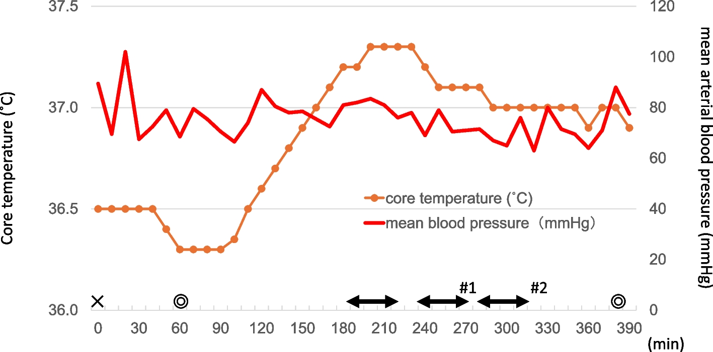
There were concerns that switching from TIVA to inhalational anesthetic and vice versa during surgery may cause instability of circulatory dynamics and/or the depth of anesthesia. Therefore, during the entire procedure, the mean arterial pressure was monitored constantly and kept stable around the baseline level by means of the vasopressor administration. On the other hand, we observed a decrease of BIS value, which was most prominent during approximately 15 min after the change of sevoflurane to TIVA, while it returned to baseline values thereafter. Seemingly, such fluctuations of BIS values might be caused by temporary additive effects from the combination of anesthetic drugs [10]. Changes of the depth of anesthesia may have an important impact on postoperative recovery. Specifically, significantly higher incidence (28% vs. 19%) of postoperative delirium and worse cognitive function at 1-year follow-up were found in a subgroup of patients who were operated on under deep anesthesia (BIS value, 35) than under more superficial (BIS value, 50) one [11]. Nevertheless, despite the transient decrease of BIS values during surgery in our patient, he demonstrated an uneventful postoperative course and stable condition during subsequent follow-up.
Our technique presenting herein closely resembling those previously described by Fukamachi et al. [12], who used a similar intraoperative switch of TIVA to sevoflurane during callosotomy for intractable epilepsy in a pediatric patient. However, in difference with those authors, we have applied such a strategy for resective epilepsy surgery with an objective to localize precisely the epileptic focus and to guide its resection with the preservation of eloquent and non-affected cerebral cortex. To attain these goals, during surgery, we have switched anesthetic agents several times. Currently, we are collecting similar cases for their further detailed analysis. In any case, described intraoperative switch of anesthetic agents according to specific intraoperative requirements seems rather useful for cases of brain surgery requiring both ECoG recordings and MEP monitoring.


留言 (0)