SIT is a rare congenital deformity [6], in which the thoracic and abdominal viscera are arranged as a complete mirror image of the normal configuration in the right-left axis in the patient. This anomaly is usually found in conjunction with Kartagener syndrome in approximately 20–25% of patients, which is characterized by primary ciliary dyskinesia [7]. Our patient did not have Kartagener syndrome. However, if present, the surgical procedure could be more challenging due to strong adhesions in the connective tissue and increased and irregular vascularity due to chronic and recurrent infections of the bronchopulmonary tree [8]. Radiologic images of bronchial dilation and the presence of Kartagener syndrome-related symptoms, such as hemoptysis and pneumonia, could help surgeons in diagnosis.
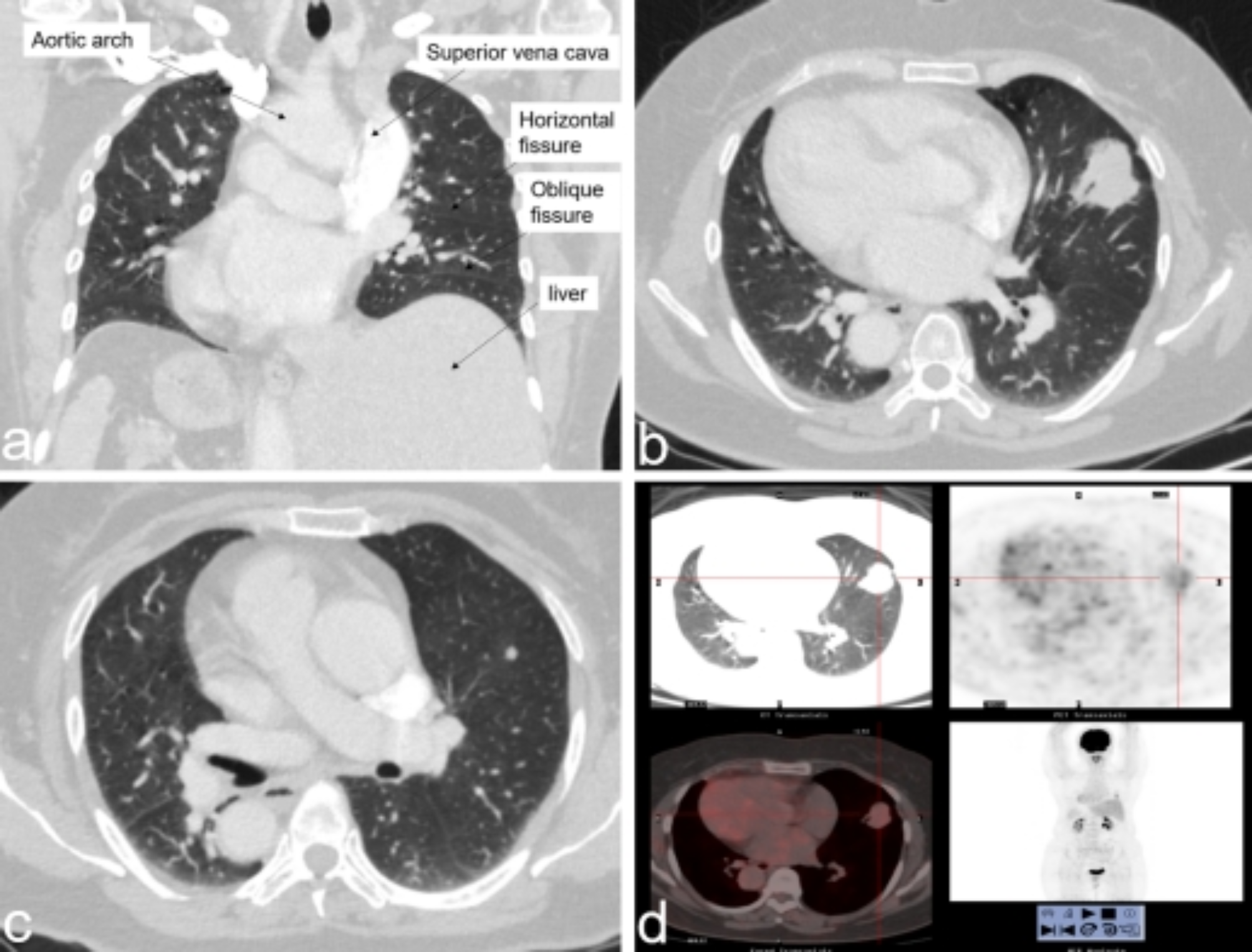
Compared to normal anatomy, the risks and challenges during surgery are high in SIT patients. Previous studies suggest that patients with SIT present with inversed but regular lung and vessel anatomy, which enables the use of the VATS technique [9, 10]. However, the research on thoracoscopic lobectomy is limited, and it is mostly performed by the multiportal approach. Due to the relative fixation of the camera in the uniportal approach and reduction in the operating space, UVATS requires advanced surgical skills and proficiency of both the operator and assistant, especially in cases with difficulty in diagnosis and orientation and dissection during surgery due to the anatomical variations in SIT. In SIT, the organs are sagittally reversed, and the courses of the pulmonary vessels and bronchi also show some variations. Accurate identification and appropriate handling of the vascular and bronchial branches in the hilum of the lung is crucial. Therefore, individual preoperative anatomical simulations and accurate preoperative evaluation are conducive to the safe and efficient performance of UVATS of anatomical lobectomy in SIT patients. The 3D-CTBA could meticulously display the mirror-imaged anatomy and vascular patterns for further analysis of the regional anatomy and reasonable preoperative planning, which finally helped us avoid blind dissection of the vessels and unexpected complications during the procedure. Preoperative imaging can also help to select the appropriate surgical incision for different vascular variations [11].
Based on our experience and relevant literature, the optimal option is inverting the tube, if a DLT is required during surgery in SIT patients [12]. In this case, the left bronchial anatomy was situated on the right, and a conventional right-sided DLT was inserted in the left main bronchus. This method allowed us to perform one-lung ventilation safely without a specially designed instrument for SIT cases. We believe that this method can provide some references and ideas for thoracic surgeons when they encounter similar situations. Moreover, considering that the neurological structures may not be clearly imaged preoperatively, particular vigilance is necessary to avoid intraoperative injury to the recurrent laryngeal nerve.
The etiology of SIT has not been elucidated; however, evidence suggests that it could be associated with improper extra-embryonic fluid flow and false heart tube rotation by disturbed ciliary motility during embryogenesis [1, 5, 7]. Moreover, the correlation between SIT and neoplasms is controversial and requires further clinical and epidemiological studies, which is of great relevance for the prevention, diagnosis, and treatment of the disease.


留言 (0)