
記住我


In contrast, we report a 3.7-year-old girl with prenatal diagnosis of complete AR STAT1 LOF (homozygous STAT1 c.1011_1012delAG; p.Val339ProfsTer18) (Fig. 1A) with development of atypical disease patterns triggered by viral infections after early and successful allogeneic HCT (Fig. 1B). The patient is the third child from healthy consanguineous parents of West African origin. The prenatal diagnosis was made due to her older brother who was diagnosed with complete AR STAT1 LOF by identifying a novel homozygous STAT1 frameshift variant after a clinical history of severe viral infections. The parents were identified to be heterozygous carriers (Fig. 1A). [4] After birth the patient was put into protective isolation and received antimicrobial prophylaxis using cotrimoxazole, azithromycin, nystatin, and IV immunoglobins (IVIG) every four weeks. The conditioning regimen consisted of fludarabine (40 mg/m2/d on days -7 to -4), busulfan (4.8 mg/kg/d on days -7 to -4; targeted area under the curve 87.876 ng*h/ml) and anti-thymocyte globulin (Grafalon®; 10 mg/kg/d on days -4 to -3). Allogeneic HCT was performed at the age of 15 weeks using 6.9 × 10⁸/kg nucleated cells of erythrocyte depleted marrow from her HLA-identical father (5.8 × 10⁶ CD34+/kg, 74.1 × 10⁶ CD3+/kg). Engraftment was observed on day +13. Under GvHD prophylaxis using cyclosporine A and mycophenolate mofetil there was no development of acute GvHD. At the age of four months (day +33) the patient was admitted to hospital for treatment of CMV reactivation. The patient received IV ganciclovir, CMV hyperimmunoglobulin and letermovir. The patient received valganciclovir prophylaxis until day +120 when immunosuppression was also discontinued. On day +58 complete donor chimerism was observed in peripheral blood (Supplementary Table S1). On day +132 the patient again presented with CMV reactivation treated with IV ganciclovir, and valganciclovir prophylaxis was reestablished. On day +180 chimerism and immune reconstitution were complete (Supplementary Table S1). One year after allogeneic HCT phosphorylation of STAT1 in PBMCs was tested, showing normal STAT1 expression and phosphorylation upon interferon stimulation in CD14 monocytes (Supplementary Figure S1) machting simultaneous chimerism testing. On day +240 the patient was readmitted presenting with fever and vesicular exanthema. The exanthema spread from the head to the whole body including the oral mucosa and the conjunctivae followed by the development of bullae (Fig. 1C). Enterovirus/Coxsackievirus A16, that has a tropism for keratinocytes and is a known cause of hand, foot, and mouth disease in immunocompetent individuals, was isolated from cerebrospinal fluid, blood, stool, and skin samples. The affected epidermis nearly completely detached in up to 10% body surface. Histopathology showed toxic epidermal necrolysis with inflammation and edema (Fig. 1D). Dermatologists diagnosed Stevens-Johnson syndrome (SJS) and/or toxic epidermal necrolysis (TEN). The critically ill patient with epidermolysis, hypovolemic renal failure, and hepatitis was treated on the PICU like a severe burn. Under local treatment with dexpanthenol and polihexanide she recovered over a period of eight days with scar free healing. 29 months after HCT, the patient was readmitted to hospital presenting with fever, abdominal pain and a papulous exanthema on the hands, inguinal region, and feet. A throat swab multiplex PCR detected human Rhino-/Enterovirus. The patient developed vomiting, diarrhea, and recurring fever unresponsive to antibiotics. Acute-phase proteins were elevated with C-reactive protein 14.6 mg/dl (reference range < 0.8), procalcitonin 0.3 ng/ml (< 0.1) and fibrinogen 702 mg/dl (160–400) without signs of HLH. Assuming hyperinflammatory immunodysregulation, IVIG 2 g/kg and methylprednisolone 2 mg/kg/d were administered, and the skin rash and fevers resolved. From this time onwards the patient´s chimerism showed to be mixed, but stable over time until 2.9 years after allogeneic HCT (Supplementary Table S1). Regularly performed immunophenotyping of peripheral blood of the patient showed stable numbers within the reference ranges (Supplementary Table S1). At the age of 39 months the patient was again admitted to hospital presenting with pneumonia with need for oxygen supplementation testing positive for influenza A virus. Laboratory abnormalities showed a HLH-like hyperinflammatory immunodysregulation (ferritin > 8000 ng/ml (10–500), triglycerides 372 mg/dl (25–119), thrombocytes 22 G/l (195–464), hemoglobin 10.2 g/dl (10.7–13.4), GPT 1374 U/l (< 59), fibrinogen 94 mg/dl (160–400), sCD25 5810 U/ml (158–623)). Steroid treatment was initiated (prednisolone 1.3 mg/kg/d). Within one week the respiratory distress resolved and the inflammatory parameters normalized.
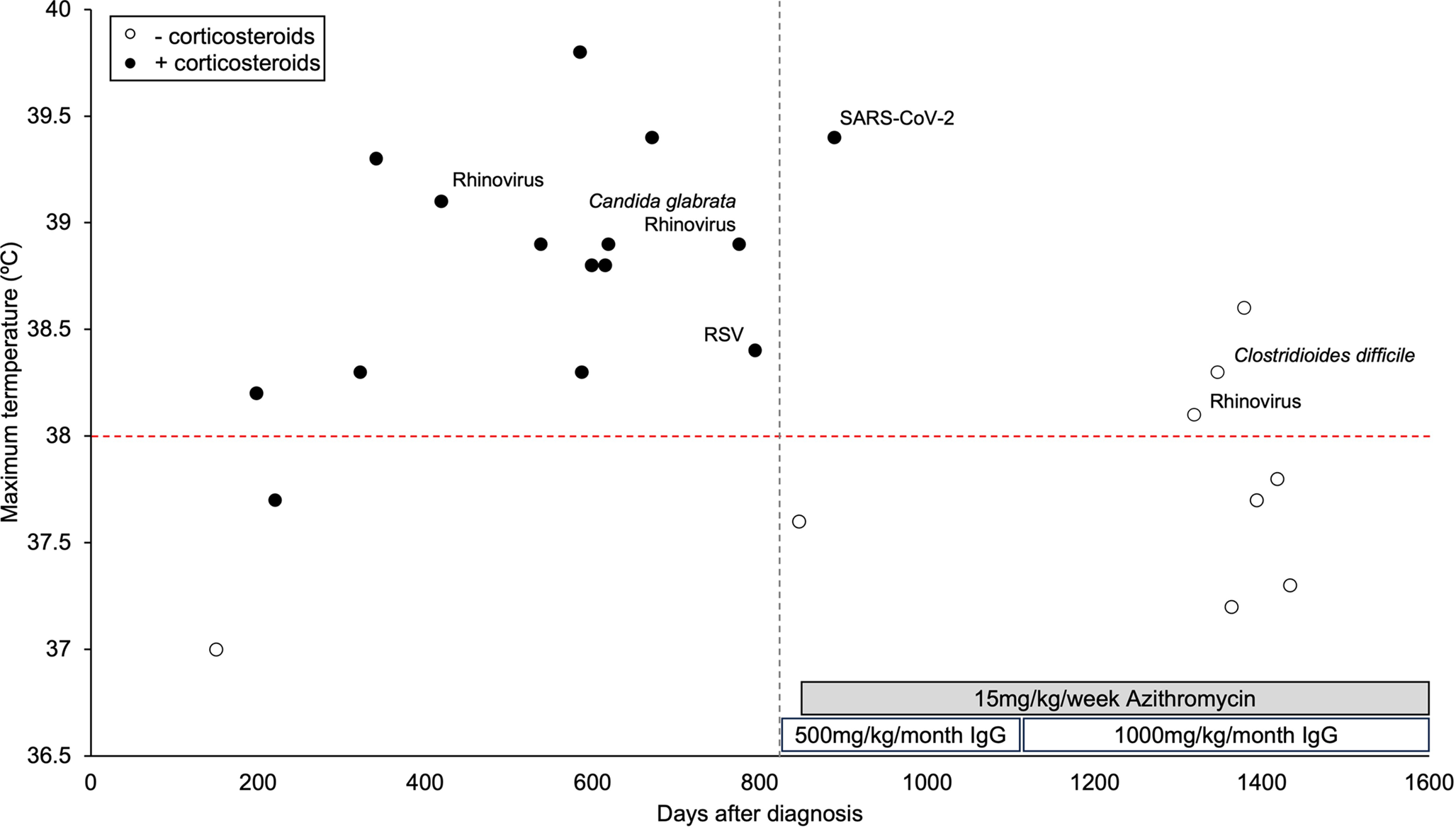
Fig. 1
(A) Electropherogram of parents and patient (P). (B) Timeline of the patient´s clinical course. (C) Enterovirus/Coxsackievirus A16 infection leading to epidermolysis at the age of 12.25 months. (D) Histopathology of patient´s skin showing degenerative interface dermatitis and single cell necrosis on the base of the blister and complete necrosis with eosinophilic and neutrophilic granulocytic infiltration on the roof of the blister. AlloHCT, allogeneic hematopoietic cell transplantation. CMV, cytomegalovirus. STAT1, signal transducer and activator of transcription 1
留言 (0)