
記住我


Pediatric acute liver failure (PALF) is potentially fatal and may lead to rapid deterioration of hepatic function and synthetic liver failure. The preceding period may last for days or weeks; however, once PALF is established, the clinical course is unpredictable and often rapidly progressive. Large multicenter data from the west reported native liver survival (NLS) of around 50% about 2 decades ago, whereas recently the PALF study group (PALFSG) re-analyzed 1144 cases and reported an increase in NLS from 47.5% to 68.5% in the present era [1, 2]. Data from the PALF study group reported 60% NLS among young infants less than 90 days old [3]. On the other hand, data from India report NLS of 47.7%, a greater proportion of deaths (42.2%), and only 10% of liver transplantation (LT) [4]. The proportion of patients with NLS is even lower in younger Indian children and infants [5].
PALF is characterized by coagulopathy and hepatic encephalopathy (HE) in children without an underlying chronic liver disease (CLD). It results from rapid and massive hepatocyte necrosis or injury, leaving behind a liver parenchymal mass insufficient to sustain liver functions, resulting in high rates of mortality. PALFSG has defined PALF in an infant or child according to the following criteria: (i) no known evidence of CLD, (ii) biochemical evidence of acute liver injury, and (iii) corrected (6 h post1–10-mg of parenteral vitamin K) coagulopathy defined as an international normalized ratio (INR) ≥ 1.5 in the presence of clinical HE or INR ≥ 2, regardless of the presence or absence of clinical HE [1].
Age-based protocols are required in the diagnostic workup of PALF to reduce the number of indeterminate cases. Indeterminate PALF accounted for about half of the cases in older studies, but the proportion is now decreasing due to improved diagnostic work-up [6]. Although there has been a significant reduction in mortality over time in the West, the mortality rate in PALF remains close to 50% in India. This could be due to a delay in diagnosis, a poor referral system, and a limited number of formally trained pediatric hepatologists, pediatric intensivists, and pediatric LT programs in the country. Cerebral edema and systemic inflammation continue to be the major pathophysiologic mechanisms driving multi-system organ failure in PALF. There are newer non-invasive diagnostic modalities, such as optic nerve sheath diameter (ONSD) and transcranial Doppler (TCD), to detect elevated intracranial pressure (ICP) in PALF. Moreover, the use of newer therapeutic modalities such as therapeutic plasma exchange (TPE) and continuous renal replacement therapy (CRRT) has improved outcomes. Guidelines are needed to direct the appropriate use of these new modalities in the management of PALF. The ideal prognostic models and LT listing criteria for PALF have not been well established. There are no consensus guidelines on the diagnosis and management of PALF, although few review articles or position papers have been published previously [6, 7].
Hence, a consensus meeting was planned on February 25, 2024, at the Department of Pediatric Hepatology, Institute of Liver and Biliary Sciences, under the aegis of the Indian Society of Pediatric Gastroenterology, Hepatology, and Nutrition (ISPGHAN) to develop recommendations on the diagnosis and management of PALF. The scientific committee invited international and national experts to participate in a consensus meeting. Before the meeting, questions were identified by a subgroup of experts and allotted to participants to review and analyze the published literature. Literature search, level of evidence (LOE), and grade of recommendations for each question were reviewed by a subgroup of experts based on the Grading of Recommendations Assessment, Development, and Evaluation (GRADE) system (Table 1) [8]. A 1-day meeting was held on February 25, 2024, in New Delhi, India, to discuss and finalize the consensus statements, recommendations, and guidelines. A large group of experts deliberated on each question, reviewed the literature, discussed the contentious issues, and deliberated to prepare the consensus statement deciding the LOE and grade of recommendations on the particular question. The final statements and recommendations were circulated to all experts for their suggestions. The manuscript with consensus recommendations was prepared by a subgroup of experts, and the final manuscript was re-circulated for comments. The experts’ suggestions were used to revise and finalize the recommendations. A brief background of each question has been included, providing recommendations and evidence from the published literature.
Table 1 Level of evidence and strength of recommendations used for the Indian Society of Pediatric Gastroenterology, Hepatology and Nutrition guidelines on pediatric acute liver failure (adapted from GRADE recommendations) [8]Pediatric ALF definitionAll adult definitions of acute liver failure (ALF) have three common features: (i) the presence of HE and coagulopathy, (ii) a specified interval (from onset of illness to HE), and (iii) no pre-existing liver disease [9]. Although initially meant to be used only as study entry criteria (and not primarily for diagnosis), the PALFSG criteria have now gained almost universal acceptance for defining PALF [1]. However, even though any known evidence of CLD is an exclusion criterion, patients with Wilson disease, autoimmune liver disease, etc. (with underlying cirrhosis in the majority) are commonly included in the PALF groups considering them as an acute presentation of underlying CLD [6, 10]. There is a need to differentiate between pure PALF and PALF associated with CLD since they are discrete in their natural course of illness and outcome. The negative impact of including patients with cirrhosis in PALF studies is further evident from the placebo-controlled clinical trial on the use of intravenous N-acetylcysteine (NAC) in patients with non-acetaminophen (APAP) PALF [11]. The negative outcome of the study, particularly among those aged < 2 years, formed the basis of non-usage of NAC in PALF patients worldwide. However, closer scrutiny of the study’s methodology revealed that more than half of the patients in both study groups (with known etiology) had underlying potential cirrhosis (including metabolic liver diseases such as Wilson disease, tyrosinemia, galactosemia, etc.). While a similar study in adult ALF majorly including patients with potentially non-cirrhotic etiologies such as drug-induced liver injury and hepatitis B showed a beneficial effect of intravenous NAC in non-APAP ALF [12]. The results of this trial led to NAC being recommended as standard medical management in non-APAP ALF in adults [10]. The regeneration potential differs depending on the underlying hepatic fibrosis/cirrhosis [13]. Thus, any study evaluating therapies (e.g., extracorporeal liver support systems (ELSS) like TPE and CRRT) is likely to be confounded if patients with underlying CLD are included. Therefore, all efforts should be made to include only ‘pure ALF’ cases, especially in studies on therapeutic modalities in PALF patients.
HE is one of the most important prognostic markers in patients with ALF. Therefore, all adult ALF definitions include HE as a mandatory diagnostic criterion [9]. However, the difficulties in incorporating HE in PALF patients are well known including suboptimal assessment in early stages of HE and it may not be apparent until terminal stages in infants/young children [1]. Almost all children with ALF have HE, however, it is difficult to detect early HE in younger children. All possible efforts should be made to identify early HE in suspected PALF cases using the modified HE assessment scale by the PALFSG for optimal management [1]. The modified HE assessment scale in children as proposed by the PALFSG is depicted in Table 2. There is an urgent need to evaluate potential biomarkers for the early detection of HE in children.
1.Recommendations:
1.1.Pediatric acute liver failure should be defined as per pediatric acute liver failure study group criteria:
Children with no known evidence of chronic liver disease,
Biochemical evidence of acute liver injury, and
Hepatic-based coagulopathy defined as an INR ≥ 1.5 not corrected by Vitamin K with clinical hepatic encephalopathy or an INR ≥ 2, regardless of the presence or absence of clinical encephalopathy (LOE 5, strong recommendation).
1.2.Efforts should be made to suspect and diagnose hepatic encephalopathy early, even in younger populations using the modified encephalopathy assessment scale (LOE 5, strong recommendation).
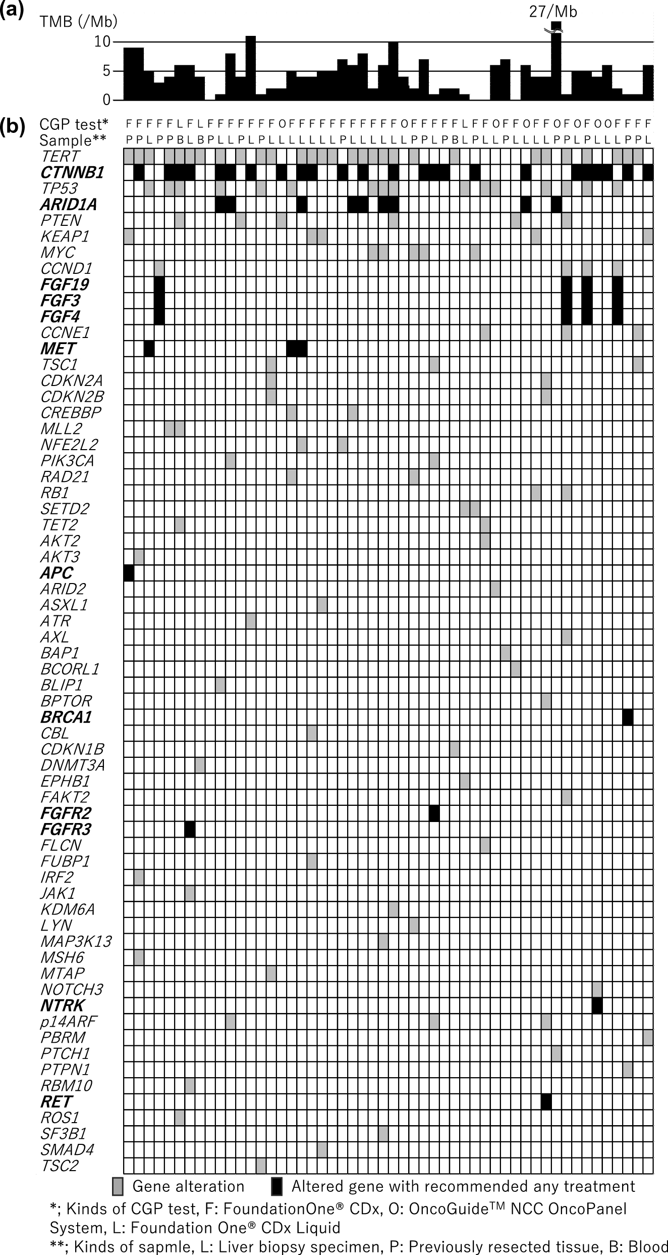
Table 2 Modified hepatic encephalopathy assessment scale for children upto 3 years of age as recommended by the pediatric acute liver failure study group [1]Etiology of PALFThe etiology of PALF is diverse and varies across different age groups and geographical locations. As per the PALFSG cohort, three different age groups were defined to segregate etiologies—0–90 days, 91 days to 3 years, and 3 years–18 years [2]. APAP, hepatotropic viruses, non-APAP drug-induced liver injury, and autoimmune hepatitis are common in older children [2, 4, 14,15,16,17,18,19,20,21,22,23,24]. In contrast, infants commonly have cardiac or ischemia-induced liver failure, non-Wilsonian metabolic liver disease (MLD), non-hepatotropic viruses (herpes simplex and enterovirus), gestational alloimmune liver disease or hemophagocytic lymphohistiocytosis [2, 3, 5, 25,26,27,28]. Etiological distribution of PALF also appears to vary based on the incidence of hepatitis-A virus (HAV) infection in different regions of the world [29]. Figure 1 summarizes the etiologies of PALF in children among various age groups.
Fig. 1
Etiologies of pediatric acute liver failure in older (≥ 3 years age) (A) and younger children (< 3 years age) (B)
Metabolic liver diseases presenting as ALFMLD constitutes approximately 28% of PALF in children less than 5 years of age with galactosemia, mitochondrial respiratory chain defects, tyrosinemia, ornithine transcarbamylase deficiency, Niemann–Pick disease being the common ones [30]. Red flags for MLD in PALF include infantile presentation, failure to thrive, hepatosplenomegaly, disproportionate synthetic dysfunction, consanguineous parents, history of miscarriages or sib-death, family history of similar illness, abnormal body fluid odors, extrahepatic features in the form of rickets, diarrhea, developmental delay or regression, nystagmus, seizures, hypotonia, bone marrow suppression, cardiomyopathy, or recurrent PALF with infection or catabolic stress [5, 30].
Indeterminate PALFAround 9–52% of children with PALF above 3 years of age and 6–52% under 3 years of age have indeterminate etiology [2,3,4,5, 14,15,16,17,18,19,20,21,22,23,24,25,26,27,28]. With the application of etiology-specific algorithms on a set of 303 ‘indeterminate’ labeled adult ALF patients, it was shown that nearly half (46.9%) got a diagnostic label, thus decreasing the prevalence of indeterminate ALF to 5.5% [31]. A similar concept was utilized by PALFSG, where a collaborative learning approach for age-specific diagnostic tests was applied using an electronic medical record admission order set at hospital admission in three phases from 1999 to 2014. It was found that the diagnosis of indeterminate PALF decreased significantly from 48 to 30.8%, along with a decrease in the 21-day cumulative incidence rate for LT from 34.6 to 20.2%. This decline was present in all three age groups, but most significantly in the older (above 3 years) age group [2]. A modified version of the age-based stepwise algorithm is presented in Table 3.
Table 3 Step-wise age-based diagnostic evaluation of pediatric acute liver failure* (Modified from reference [3])Extended viral testing in PALFTesting for an extended panel of viruses in the patients of the PALFSG cohort showed a positive viral (IgM, PCR or antigen) test in 166 (20.2%) of the 820 children tested for a causative or an associated virus. In the under-6 months age group, herpes simplex virus and Epstein–Barr virus were the common causative viruses, and cytomegalovirus and human herpes virus-6 were commonly associated viruses. In the older age group, herpes simplex virus, adenovirus, and parvovirus were the commonly detected causative viruses, and human herpes virus-6, Epstein–Barr viruses, and cytomegalovirus were the commonly associated viruses [32]. The study emphasizes that testing for viruses is often incomplete, and it should be more comprehensive to detect treatable viral etiologies of PALF.
There has been a sudden surge in cases of severe acute hepatitis/PALF since 2021. There are speculations of their association with adenovirus type 41, adeno-associated virus-2, and COVID-19. In a recent systematic review of 1643 children presenting with severe acute hepatitis, 120 (7%) required LT, and 24 (1%) died. There were inconsistent results for serological testing or testing of explants for these viruses, and considering the existing evidence, it was concluded that no definite explanation for causality could be made [33]. Another report from King’s College Hospital (KCH), London mentioned that the presence of indeterminate PALF had a similar prevalence and association with adenovirus over the previous 5 years, although with a severe phenotype but similar survival to native liver in the year 2022 [34]. This again emphasizes the need for extensive viral testing of PALF cases.
Genetic testing for PALFGenetic testing for PALF is important for identifying MLDs with a diverse or unrecognized phenotype. In a study from KCH, a next-generation sequencing (NGS) panel of 64 candidate genes identified homozygous or compound heterozygous variants in 12 (26.7%) out of 45 children tested by targeted and whole exome sequencing [35, 36]. A recent multicenter data on whole exome sequencing of 260 children with indeterminate PALF (22.7% recurrent PALF) from 19 countries showed the establishment of genetic diagnosis in 37%, with the highest diagnostic yield in infants (46%) and with recurrent PALF (64%). Common defects were linked to 36 genes related to mitochondria, vesicular trafficking, and cytosolic t-RNA synthetase and the common ones were NBAS (21%), MPV17 (8%), and DGUOK (7%), followed by TRMU, SCYL1, DLD, SERAC1, YARS1, LARS1 and ZNFX1 [36]. A prolonged turnaround time for genetic analysis is currently a potential limitation for any timely therapeutic intervention.
2.Recommendations:
2.1.Etiological evaluation of pediatric acute liver failure should be undertaken based on the age and geographic location of the patient (LOE 3, strong recommendation).
2.2.Genetic testing using whole-exome sequencing should be considered for children with pediatric acute liver failure of indeterminate etiology (LOE 4, strong recommendation).
Diagnosis of cerebral edemaThe intracranial volume is contributed by three compartments: the brain, blood, and cerebrospinal fluid. According to the Monroe-Kellie doctrine, the intracranial volume is constant, and any increase in the volume of one of the compartments would lead to a compromise of the other 2 compartments. Hence, cerebral edema caused by ALF leads to compromised cerebral perfusion (ischemia). Cerebral edema can be present in as high as 55%–74% of adult ALF and is associated with high mortality [37, 38]. Factors associated with raised ICP are hyperacute ALF, younger age, higher grade HE, serum ammonia > 150 mmol/L, systemic inflammatory response syndrome, concurrent infection, and requirement for vasopressors [37, 39]. To reduce the mortality associated with PALF, all possible attempts should be made towards early detection and appropriate management of cerebral edema.
Clinical assessment of HE and cerebral edemaHE assessment is best performed using the West Haven Criteria in older children and using a modified HE assessment scale in children up to 3 years of age [1, 40]. The classical symptoms of cerebral edema include headache, blurred vision, vomiting, and alterations in mental status (somnolence to coma). Assessment of these classical signs and symptoms in PALF may be difficult due to HE, ventilation, and sedation. Physical signs that suggest significant cerebral edema are unilateral or bilateral false localizing sign i.e., 6th nerve palsy, Cushing response (hypertension, bradycardia), irregular respiration, spasticity, and decerebrate posture. Neurological examination including pupil size should be regularly monitored as per the grade of HE (q2 hourly in grade I, q60 min in grade II, q30 min in grade III–IV) [6]. Clinical signs of raised ICP are frequently late in emergence, hence all attempts should be made to detect raised ICP early by using a range of non-invasive techniques [
留言 (0)